
Mr imaging guided therapy

a guided therapy and magnetic resonance technology, applied in the field of magnetic resonance (mr) imaging, can solve the problems of inability to measure absolute proton resonance frequency, prolong treatment time, and cell death immediate,

Active Publication Date: 2012-03-22
PROFOUND MEDICAL
View PDF25 Cites 37 Cited by
  • Summary
  • Abstract
  • Description
  • Claims
  • Application Information

AI Technical Summary

Benefits of technology

[0020]The invention provides the ability to continuously obtain MR thermometry data in the regions required for therapy monitoring. The invention uses a sparse set of temperature data collected in only a few two-dimensional image planes. The approach of the invention allows the position and orientation of the image plane used for temperature monitoring to follow the transducer without having to wait for the tissue to cool down to the baseline level. Moreover, the invention overcomes problems arising from local magnetic field changes due to the transducer movement.
[0023]In accordance with a preferred embodiment of the invention, the baseline thermographic MR image used for subsequent image reconstruction after a change of the focus of the thermal treatment is computed from MR signals that are selectively acquired from the previously chosen static second image region prior to moving the focus of the thermal treatment. In this way a sparse set of temperature data is collected continuously during treatment, thereby enabling temperature monitoring in regions which have not been pre-chosen by the user.

Problems solved by technology

As the temperature rises above 55° degrees centigrade, coagulative necrosis of the tissue occurs resulting in immediate cell death.
Because of a non-homogeneous magnetic field within the MR imaging apparatus used, absolute proton resonance frequency measurements are not possible.
Problems arise in applications in which the ultrasound transducer is moved to apply therapy at different locations.
The drawback of this scheme is that the duration of the treatment is much longer than would actually be necessary.
The presently used MR thermometry sequences do not allow the acquisition of volumetric temperature information in three-dimensional space and for different instances in time.

Method used


Image

  • Mr imaging guided therapy
  • Mr imaging guided therapy
  • Mr imaging guided therapy

Examples


Embodiment Construction

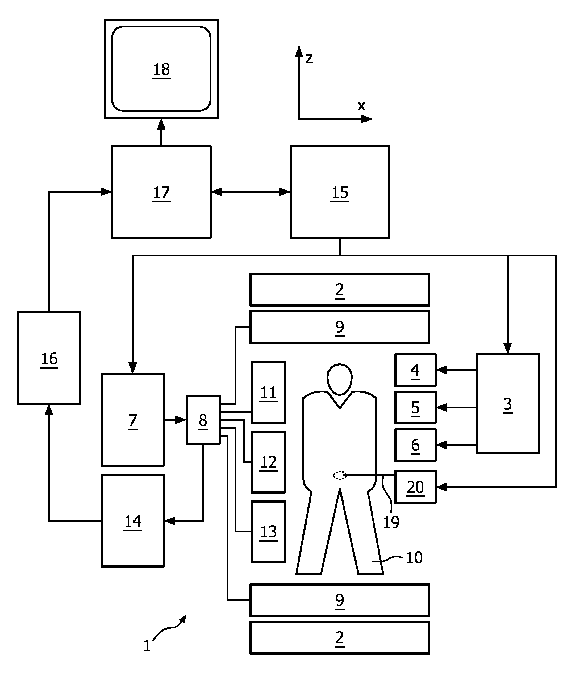
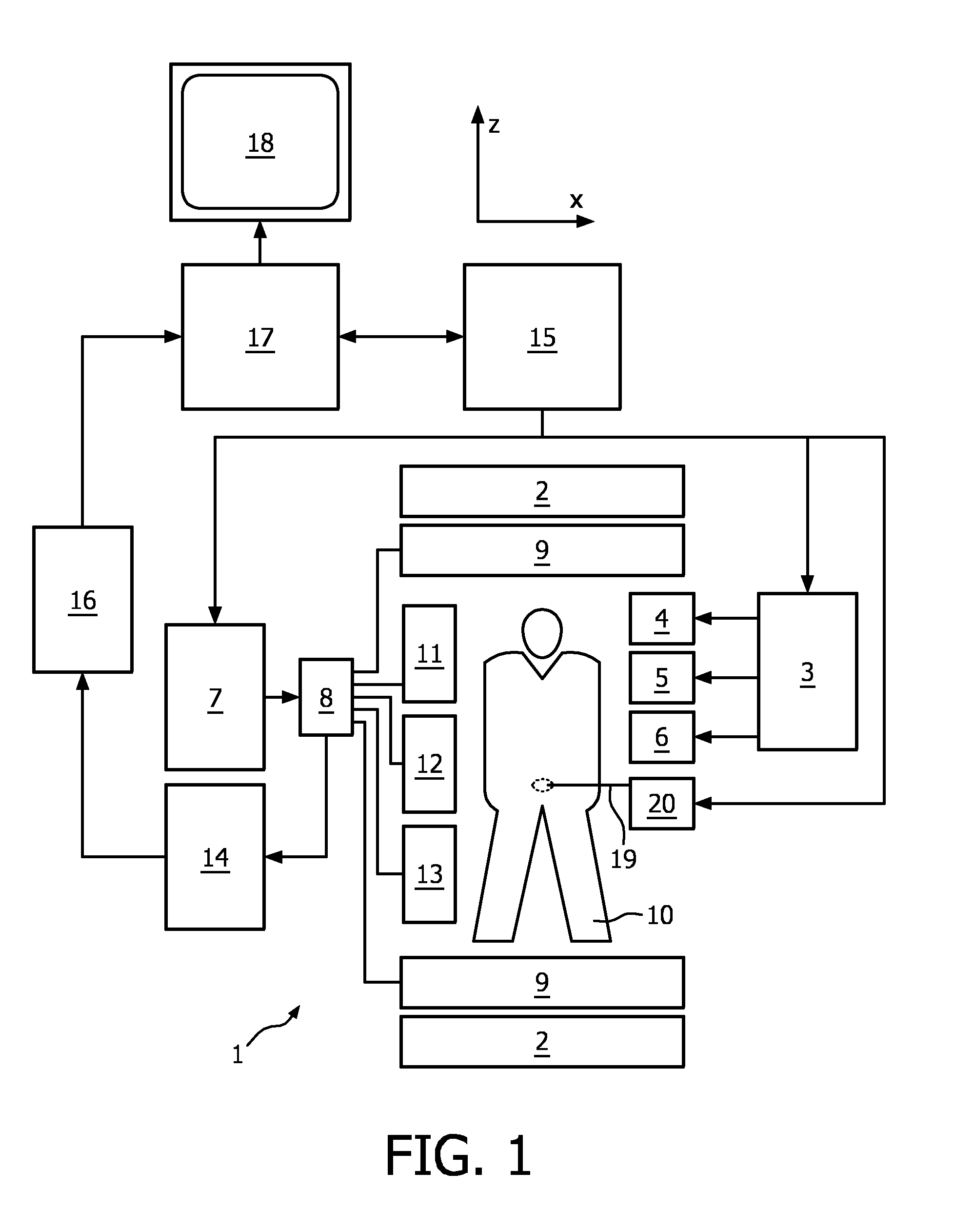
[0041]With reference to FIG. 1, a therapeutic system 1 is shown. The system comprises superconducting or resistive main magnet coils 2 such that a substantially uniform, temporally constant main magnetic field is created along a z-axis through an examination volume

[0042]A magnetic resonance generation and manipulation system applies a series of RF pulses and switched magnetic field gradients to invert or excite nuclear magnetic spins, induce magnetic resonance, refocus magnetic resonance, manipulate magnetic resonance, spatially and otherwise encode the magnetic resonance, saturate spins, and the like to perform MR imaging.

[0043]More specifically, a gradient pulse amplifier 3 applies current pulses to selected ones of whole-body gradient coils 4, 5 and 6 along x, y and z-axes of the examination volume. A digital RF frequency transmitter 7 transmits RF pulses or pulse packets, via a send- / receive switch 8, to a whole-body volume RF coil 9 to transmit RF pulses into the examination vo...


PUM


Abstract

The invention relates to a therapeutic system comprising: a MR imaging unit arranged to acquire MR signals from a body (10) of a patient positioned in an examination volume, a thermal treatment unit (19, 20) for the deposition of thermal energy within tissue of the body (10). It is an object of the invention to enable continuous temperature monitoring based on MR thermometry even in a situation in which the focus of the thermal treatment is moved. To this end, the invention proposes that the system is arranged to perform the steps of: a) initiating thermal treatment by heating the tissue of the body (10) at the position of a focus within the examination volume, b) selective acquisition of MR signals from a first image plane (24), the position of the focus of the thermal treatment being located within the first image plane (24), c) reconstruction of a thermographic MR image from the MR signals acquired from the first image plane (24), d) computing a baseline thermographic MR image from the temperature distribution within at least one second image plane (25, 26, 27) being different from the first image plane (24), e) moving the focus of the thermal treatment to a new position within the examination volume, f) changing the position and / or orientation of the first image plane (24) corresponding to the new position of the focus of the thermal treatment, g) repeating steps b) and c), wherein the baseline thermographic MR image computed in step d) is used for thermographic image reconstruction in subsequent step c).

Description

FIELD OF THE INVENTION[0001]The invention relates to the field of magnetic resonance (MR) imaging. It concerns a therapeutic system comprising a MR imaging unit and a thermal treatment unit for the focused deposition of thermal energy within tissue of a body of a patient. Moreover, the invention relates to a computer program and to a method of monitoring a therapeutic thermal treatment.BACKGROUND OF THE INVENTION[0002]As described more detailed herein below, thermal energy deposition is increasingly used in medicine as a means of necrosing diseased tissues. The present invention is disclosed in the following in the context of therapeutic thermal treatment by high intensity focused ultrasound (HIFU). In HIFU a phased array of piezoelectric transducers is used for generating a focused ultrasound beam. It has to be noted, however, that the technique of the invention can be applied equally well in connection with any type of device for the targeted deposition of thermal energy. Examples...

Claims


Application Information

Patent Timeline
Patent Type & Authority Applications(United States)
IPC IPC(8): G06K9/00
CPCA61B2017/00084A61N7/02A61B2019/5236A61B2090/374
Inventor RAJU, BALASUNDARAANAND, AJAYEHNHOLM, GOSTA JAKOB
Owner PROFOUND MEDICAL
Learn More