ROTICAM: An Orotracheal Guide with Camera

Inactive Publication Date: 2012-04-12
HANU SURGICAL DEVICES
View PDF10 Cites 19 Cited by
  • Summary
  • Abstract
  • Description
  • Claims
  • Application Information

AI Technical Summary

Benefits of technology

[0013]In a preferred intubation use, an operator selects a ROTIG device with a guide tube diameter such that coaxial advancement of an endotracheal tube over a previously inserted bronchoscope will cause the lingual aspect of the guide tube to splay open and release (“eject”) the coaxial endotracheal tube and bronchoscope from the guide tube. The proximal opening of the guide tube is typically up to one (1) mm larger than the outside diameter of the endotracheal tube to be used; the preferred embodiment of the guide tube has a slight narrowing of the lumen in the distal direction. Generally speaking, the smallest inner diameter of the lumen of a guide tube is not smaller than the outside diameter of the endotracheal tube to be passed through the guide tube. Having selected the guide tube diameter, the operator inserts the ROTIG device in a patient's oral cavity, presses the superior dentition of the patient into the upper bite tray, and presses the inferior dentition of the patient into the lower bite tray. The operator slides an endotracheal tube coaxially up a bronchoscope (the bronchoscope is in the lumen of the endotracheal tube), leaving a distal length of the bronchoscope exposed, which distal length is at least equal to the intubation depth desired. The operator then inserts and advances the exposed distal end of the bronchoscope distally through the proximal opening of the guide tube of the ROTIG device and advances the bronchoscope. The operator navigates the bronchoscope down the orotracheal passage by direct visualization from the bronchoscope through the glottis and into the trachea, advancing the bronchoscope until the distal end of the bronchoscope is correctly placed (“operably positioned”). The operator then advances the coaxial endotracheal tube distally to contact the proximal opening of the guide tube, and then into the lumen of the guide tube. From the centrifugal expansion caused by the distal advance of the endotracheal tube, the lingual slit of the guide tube splays open and releases the endotracheal tube and bronchoscope from the guide tube by rupture of the slit in the lingual aspect of the guide tube. This release (“ejection”) by the ROTIG device of the coaxial endotracheal tube and bronchoscope allows the operator to easily remove the ROTIG device from the patient's oral cavity. The ROTIG device is typically removed immediately after the endotracheal tube is operably positioned.
[0016]The ROTIG device enables fast and accurate intubation by guiding an endotracheal tube, which endotracheal tube is coaxially disposed around a bronchoscope, through the oral cavity so that the tube is correctly disposed to enter, successively, the pharynx, larynx, and trachea. In addition to facilitating procedures related to endotracheal tube intubation, such as induction anesthesia, uses of the ROTIG device include facilitation of direct transoral fiberoptic esophagoscopy (“direct esophagoscopy”), direct transoral fiberoptic bronchoscopy (“bronchoscopy”), and direct transoral fiberoptic laryngoscopy (“direct laryngoscopy”).
[0017]Since the ROTIG device adjusts to the unique oral conformation of a given patient and is simple to use, it provides a much higher probability of successful intubation by a lower skilled operator. The “self-adjusting” path of the flexible guide tube is determined by the roof of a patient's oral cavity and distance between the patient's upper incisors and posterior pharyngeal wall. Importantly, the ROTIG device solves a technical problem of allowing a lower skilled operator, e.g., an operator who does infrequent intubations, a higher success rate of intubation without inducing a gag reflex in a patient and of maintaining a mid-line entry of the endotracheal tube and bronchoscope. The ROTIG device allows an operator to devote the operator's entire attention to advancing the bronchoscope or endotracheal tube down the orotracheal passage without manually holding a floating, untethered guide in position, especially a guide that rests on a patient's tongue. Since the ROTIG device does not rest on a patient's tongue, it is not destabilized by voluntary or involuntary movement of the tongue by the patient, does not trigger the gag reflex in conscious patients, and consistently provides midline approaches to the vocal cords of a patient. The ROTIG device is also cost competitive with existing intubation guides and can be packaged with intubation tubes. The ROTIG device is non-obtrusive and easily tolerated in an awake patient. So long as an awake patient in a sitting position has a functioning airway (a safe assumption), the ROTIG device is the only known intubation device that enables awake oral bronchoscopic intubation without intrusion of the intubation device distal to the distal oral cavity. The ROTIG device can be advantageously used for various procedures, including without limitation, direct esophagoscopy, direct bronchoscopy, direct laryngoscopy, and endotracheal tube intubation.

Problems solved by technology

The guide tube for visualization uses typically has a constant diameter (non-narrowing) lumen, since ejection of the bronchoscope is typically not desired in visualization uses.

Method used


Image

  • ROTICAM: An Orotracheal Guide with Camera
  • ROTICAM: An Orotracheal Guide with Camera
  • ROTICAM: An Orotracheal Guide with Camera

Examples


first embodiment

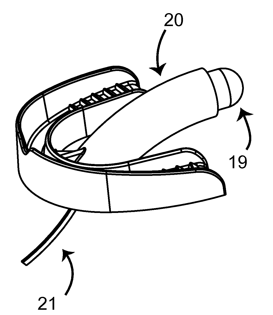
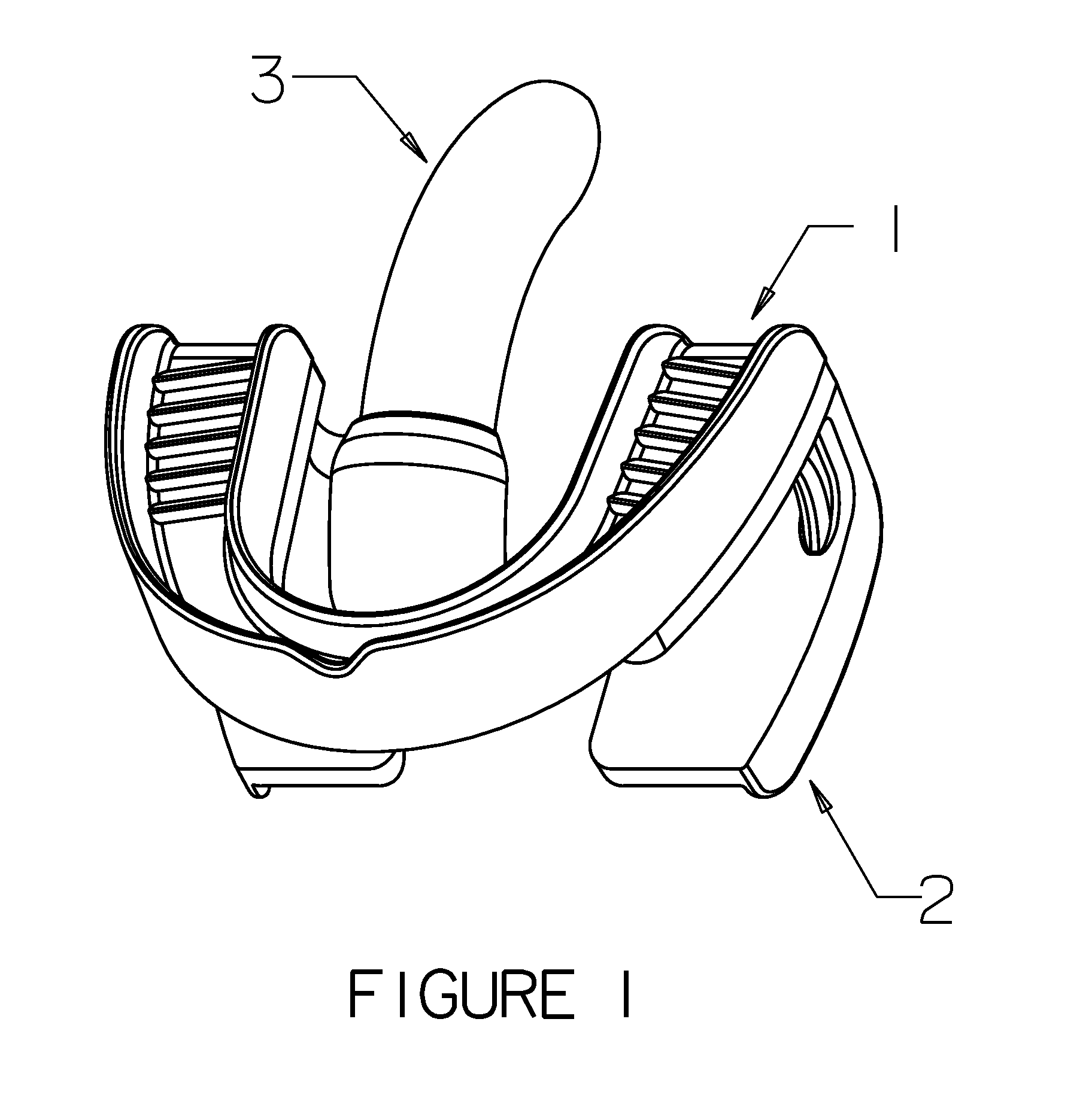
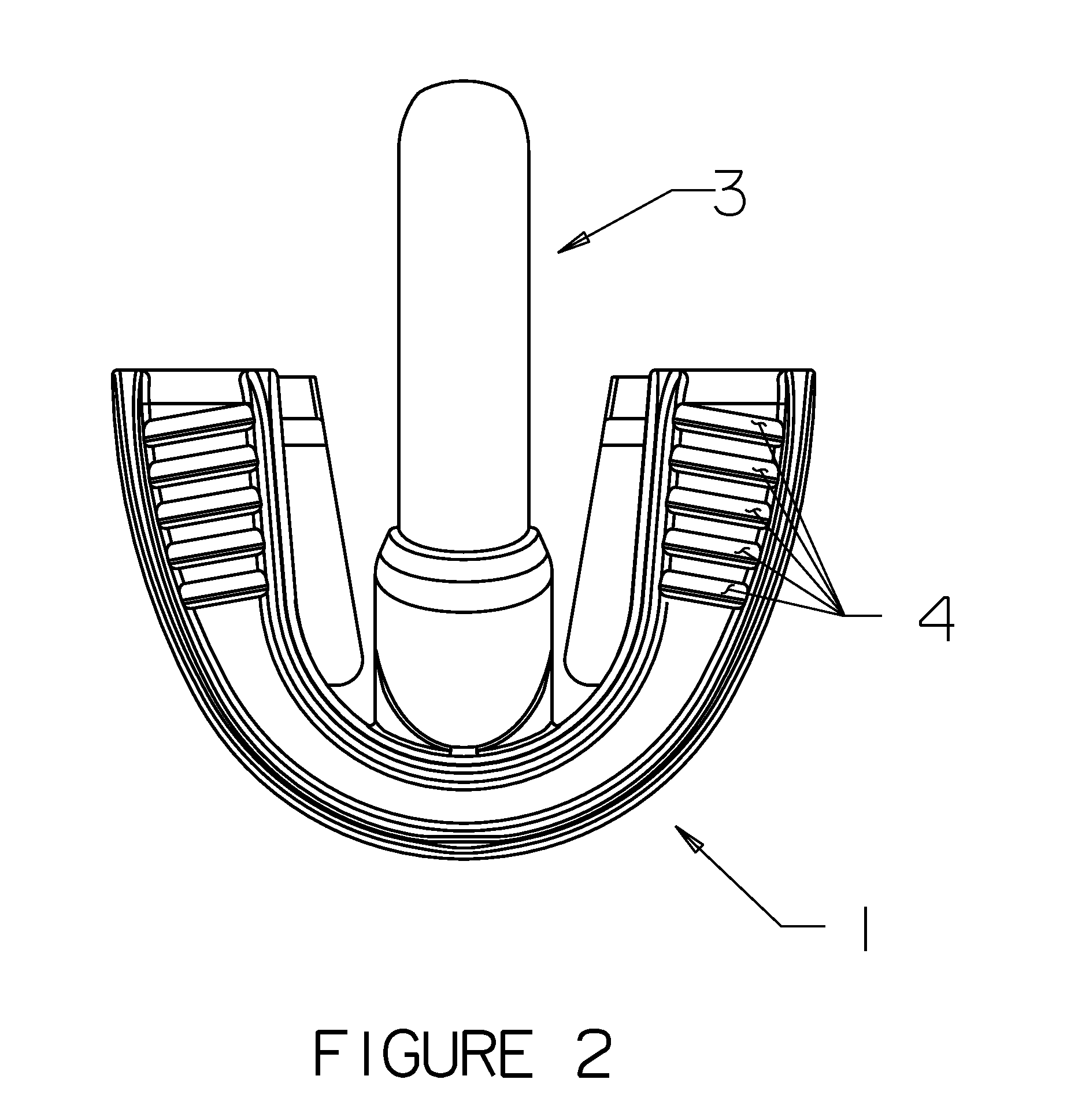
[0076]In embodiments in which the guide of a ROTIG device is replaced by a “camera mount”, as shown in FIGS. 8 to 11, the camera mount is attached to upper dental arch at the proximal end of the camera mount. A camera is attached to the camera mount at the distal aspect of the camera mount by a rigid connection or by a gimbal, Y (pitch) stage, X / Y (yaw / pitch) stage, or X / Y / Z (yaw / pitch / roll) stage, a “memory” elastomeric section (i.e., a section that retains an orientation created by manipulation of the elastomeric section), joint(s), or other means known in the art. A camera mount is attached to the distal aspect in the incisor area of upper dental arch tray, in the same manner described above for the attachment of the guide to the upper dental arch tray of a ROTIG device not equipped with a camera. In a camera mount, the camera is not separable from the camera mount without the use of tools. In this embodiment, the camera mount can house one or more cameras, sensors, one or more b...

second embodiment

[0077]In the camera mount, the camera mount is a curved cylinder into which a ROTIG camera insert is inserted. The ROTIG camera insert typically has a twist-lock or snap fit in the camera mount and can typically be removed without the use of tools. The ROTIG camera insert can house one or more cameras, sensors, one or more batteries, image storage, wireless transmission systems, etc. ROTIG camera inserts are typically reusable and have easily sterilized exteriors, while the camera mount and upper dental arch tray are typically disposable. A camera in a ROTIG camera insert is installed within the distal region of a ROTIG camera insert by a rigid connection or by a gimbal, Y (pitch) stage, X / Y (yaw / pitch) stage, or X / Y / Z (yaw / pitch / roll) stage, a “memory” elastomeric section (i.e., a section that retains an orientation created by manipulation of the elastomeric section), joint(s), or other means known in the art.

[0078]In embodiments of ROTICAMs that include an image display (typically...


PUM


Abstract

A camera system for surveillance of a patient's throat is disclosed. The camera system comprises a camera associated with an upper dental arch tray, and does not rest on the patient's tongue. The camera system can optionally include a delivery system for medication, display, and lower dental arch tray. The camera system may be configured as a guide insert in a Rapid Orotracheal Intubation Device.

Description

RELATED APPLICATION[0001]This application is a continuation-in-part of co-pending U.S. patent application Ser. No. 12 / 051,793, filed 19 Mar. 2008, and claims the benefit of said U.S. patent application Ser. No. 12 / 051,793. This application also claims the benefit of U.S. Provisional Patent Application No. 61 / 549,808, filed 21 Oct. 2011.BACKGROUND OF THE INVENTION[0002]1. Field of the Invention[0003]An orotracheal intubation guide simplifies the process of passing an endotracheal tube through a patient's mouth and larynx, and into the trachea. Orotracheal intubation is a common medical procedure that enables mechanically ventilated respiration, delivery of anesthesia to a patient's lungs, protecting patency of a patient's airway; an orotracheal guide can also be diagnostic visualizations such as bronchoscopy, operative procedures to the airway, and other diagnostic, surgical, and therapeutic procedures. When orotracheal intubation is performed improperly (e.g., producing an unrecogni...

Claims


Application Information

Patent Timeline
IPC IPC(8): A61B1/24A61B1/04
CPCA61B1/00154A61B1/05A61B1/24A61M16/0495A61M16/0488A61M16/0497A61M16/0493A61B1/267
Inventor NAPIER, BRADFORD LEE
Owner HANU SURGICAL DEVICES
Why Eureka
Try Eureka