
Self-immolative probes for enzyme activity detection

a technology of enzyme activity and probes, applied in the field of visual detection, quantification and localization of cells, can solve the problem of limited number of rationally designed probes or “signalophores” capable of detecting intracellular organic biomolecules

Active Publication Date: 2012-05-17
ENZO LIFE SCI INC
View PDF5 Cites 9 Cited by
  • Summary
  • Abstract
  • Description
  • Claims
  • Application Information

AI Technical Summary

Benefits of technology

[0013]The present invention provides several self-immolative pr

Problems solved by technology

However, only a limited number of rationally designed probes or “signalophores” capable of detecting intracellular organic biomolecules exist.

Method used


Image

  • Self-immolative probes for enzyme activity detection
  • Self-immolative probes for enzyme activity detection
  • Self-immolative probes for enzyme activity detection

Examples


example 1

Synthesis of CD-1

a). Preparation of Intermediate CD-2

[0151]

[0152]To a solution of 500 mg Rho110 in dimethylformamide, 290 μl of N,N′-diisopropylethyl-amine was added and the mixture was stirred on ice for 10 minutes. Then 160 μl of N-morpholinecarbonyl chloride was added dropwise. The reaction mixture was stirred on ice for an additional 15 minutes and then at room temperature for 2 days. The reaction mixture was evaporated to dryness and the oily residue was dissolved in chloroform containing a small amount of methanol. The product was purified on a Biotage SP4 System with a SNAP 100g column and a chloroform methanol gradient.

b). Preparation of CD-1

[0153]

[0154]To 23 mg of intermediate CD-2 dissolved in a mixture of dichloromethane (1 ml) and pyridine (1 ml), 40 mg of p-nitrobenzyl chloroformate in chloroform was added dropwise. The solution was stirred at room temperature for 2 days and the solvent was removed in vacuo. The resin was co-evaporated with toluene, dissolved in chlorof...

example 2

Synthesis of CD-3

[0155]

[0156]To 22 mg of CD-2 in the mixture of dichloromethane (0.7 ml), methanol (0.15 ml) and pyridine (1 ml), a solution of benzyl chloroformate (24 mg) in chloroform was added dropwise. The reaction mixture was stirred at the room temperature for 3 days and the solvent was removed in vacuo. The resin was co-evaporated with toluene, dissolved in chloroform and purified on a Biotage SP4 System with a SNAP 25g column and a chloroform methanol gradient, to give off-white crystals.

example 3

Synthesis of CD-4

[0157]

[0158]To 50 mg of Rho575 in dimethylformamide, 53 μl of N,N′-diisopropylethylamine was added followed by 78 mg of p-nitrobenzyl chloroformate dissolved in dimethylformamide. The reaction mixture was stirred for 3 days and the solvent was removed in vacuo. The residue was dissolved in chloroform and purified on a Biotage SP4 System with a SNAP 25g column and a chloroform methanol gradient, to give a pink product.


PUM


Abstract

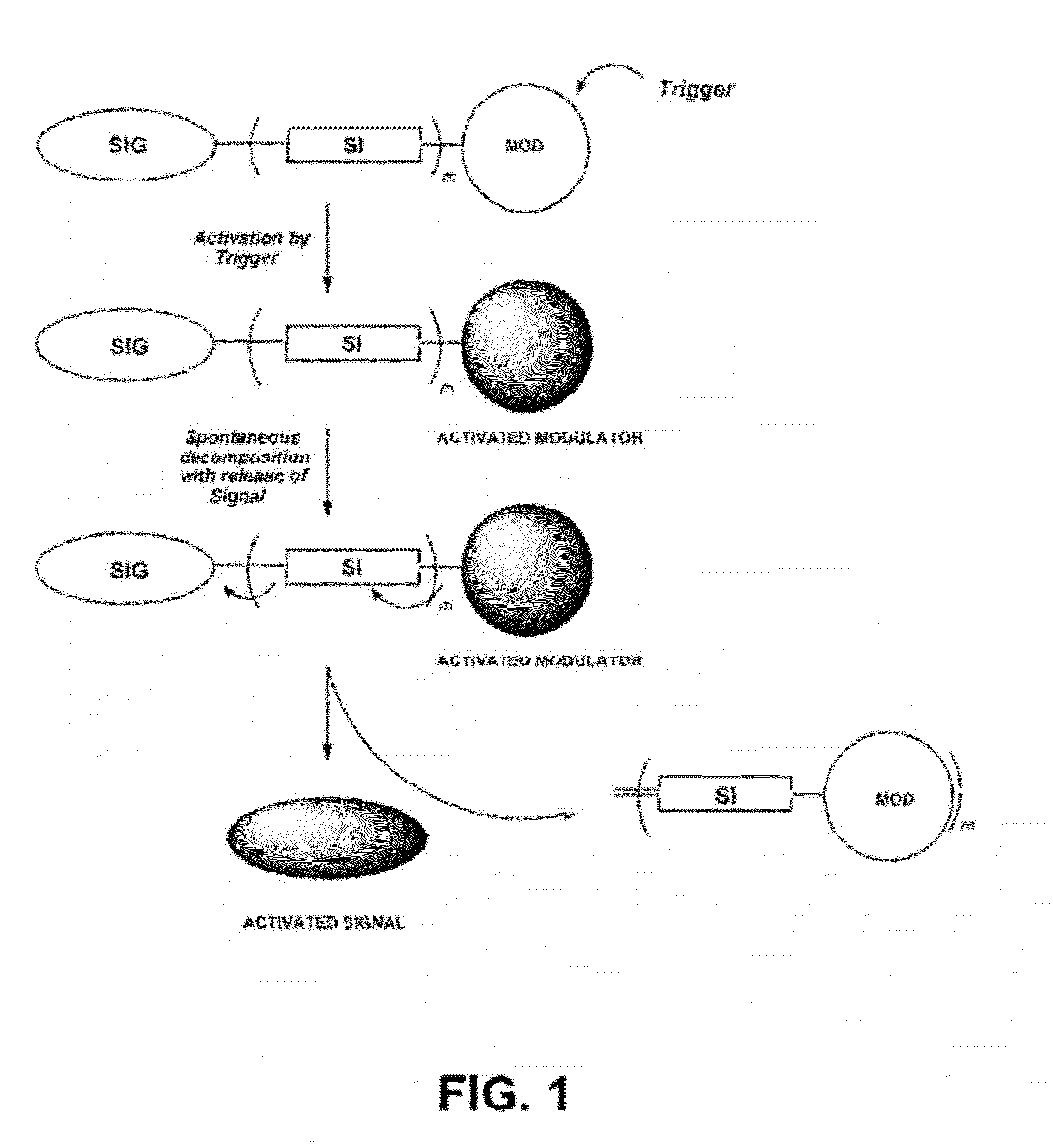
Provided is a compound comprising the structure:(SIG)-(SI-MOD)m.In this compound, SIG is a signaling molecule, SI is a self-immolative structure bound to SIG such that SIG has a reduced signal relative to the signal of SIG without SI, MOD is a moiety bound to SI that is subject to modification by an activator, and m is an integer from 1 to about 10. With this compound, when MOD is modified by an activator, SI is destabilized and self-cleaved from SIG such that SIG generates an increased signal. Also provided is a method of determining whether a sample comprises an activator, using the above-described compound. Additionally provided is a method of determining whether a cell comprises a nitroreductase using the above-described compound where nitroreductase is the activator. Further provided is a method of determining whether a mammalian cell is hypoxic using the above-described compound where nitroreductase is the activator. A method of detecting a microorganism that comprises a nitroreductase, using the above-described compound where nitroreductase is the activator, is also provided. Also provided is a method of identifying nitroreductase in a sample, using the above-described compound where nitroreductase is the activator.

Description

BACKGROUND OF THE INVENTION[0001](1) Field of the Invention[0002]The present invention generally relates to the field of reagents for visual detection, quantification and localization of cells. More specifically, probes are provided that increase their signal upon exposure to specific enzyme or chemical analyte presence.[0003](2) Description of the Related Art[0004]Signaling molecules that are responsive to the intracellular environment are indispensable tools for fast and accurate detection and measurement of both physiological and pathological processes. However, only a limited number of rationally designed probes or “signalophores” capable of detecting intracellular organic biomolecules exist.[0005]For a chemical molecule to be a suitable signalophore, it should meet several conditions. First, it should have favorable spectral properties, and be detectable by readily available sources and filter systems. Second, it should exhibit a significant signal enhancement triggered by the ...

Claims


Application Information

Patent Timeline
IPC IPC(8): C12Q1/26C07D491/22G01N21/75C07D493/10C07D413/14C07D237/32C07D311/08
CPCC09K11/06C12Q1/26Y10T436/18G01N2500/00G01N2333/902C07D311/08C07D491/052C07D491/20C07D493/10G01N2333/92C07C271/08G01N2333/918C12Q1/37C12Q1/44C12Q1/485C09K2211/1022C09K2211/1088
Inventor SZCZEPANIK, MACIEJLEBEDEVA, IRINA V.XIANG, YUEJUNPANDE, PRAVEENPATTON, WAYNE FORREST
Owner ENZO LIFE SCI INC
Learn More