
Method for deparaffinizing formalin-fixed paraffin-embedded tissue
a formalin-fixed paraffin and tissue technology, applied in the field of deparaffinizing a formalin-fixed paraffin-embedded tissue, can solve the problems of loosening and easy floating around
- Summary
- Abstract
- Description
- Claims
- Application Information
AI Technical Summary
Benefits of technology
Problems solved by technology
Method used
Image

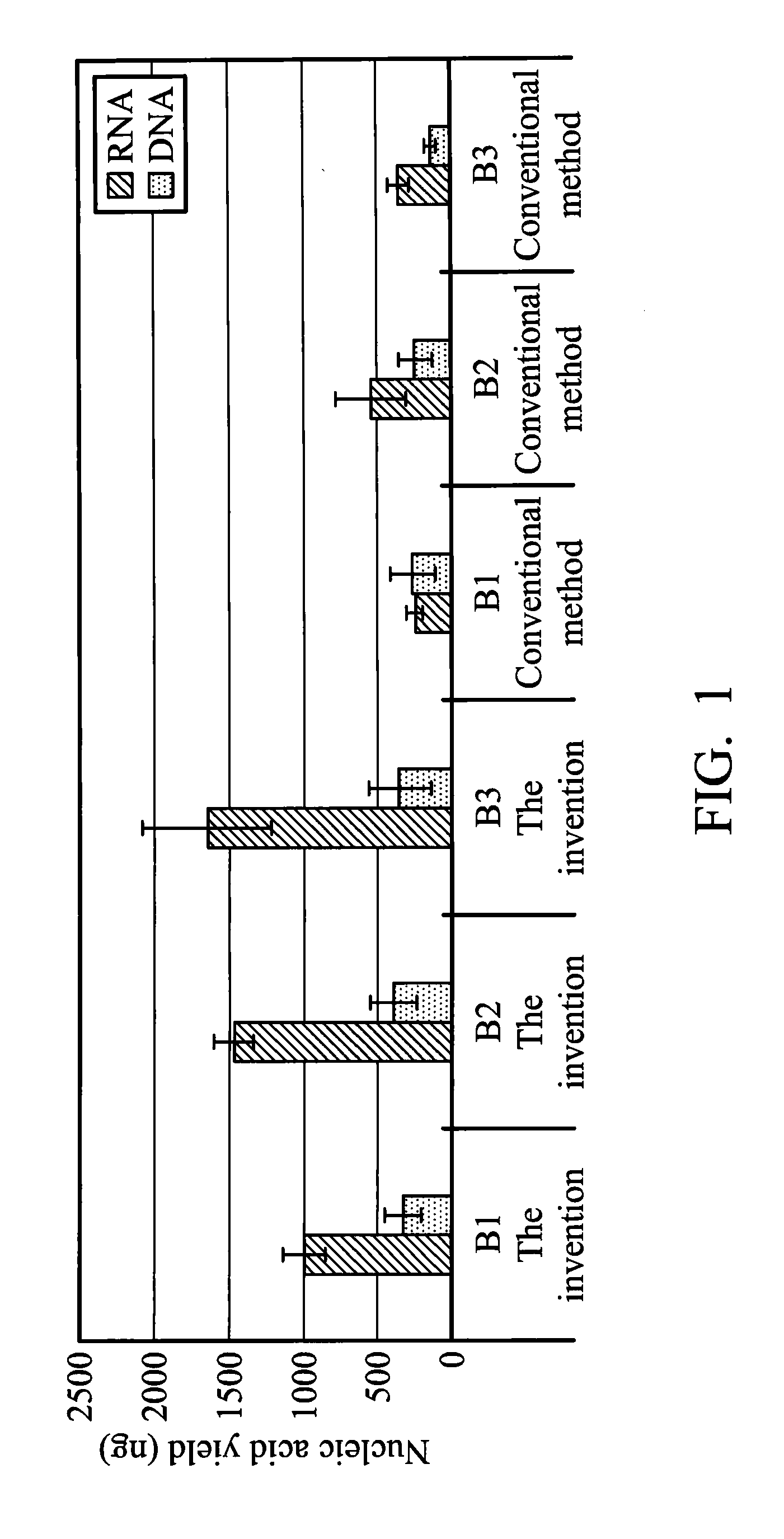
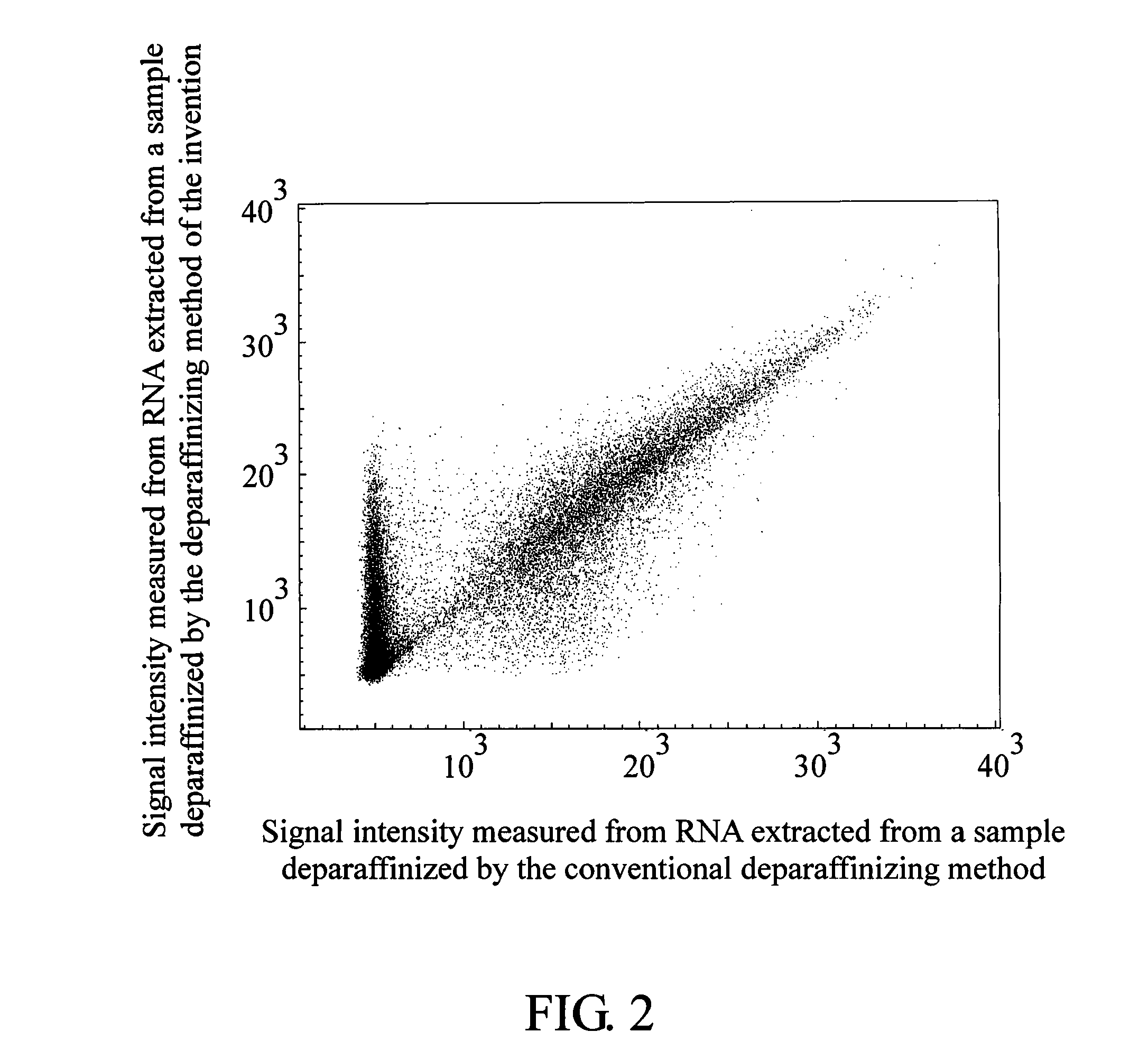
Examples
example
Example 1
[0032]Deparaffinization:
[0033]A slice of a formalin-fixed paraffin-embedded lung cancer tissue biopsy sample (tissue area: about 1 cm2; thickness: 10 μm) was placed into a microcentrifuge tube. 600 μl of 100% xylene was added into the microcentrifuge tube and mixed with the sample by vortexing for 10 seconds to form a mixture. The mixture was centrifuged to be at the bottom of the microcentrifuge tube. Then, 194 μl of RNase-free water was added into the microcentrifuge tube, and the microcentrifuge tube was placed under a temperature of 50° C. for shaking horizontally for 3 minutes. The microcentrifuge tube was centrifuged at 12000 rpm for 3 minutes. Next, 550 μl of an upper layer liquid in the microcentrifuge tube was removed. After that, 600 μl of 100% xylene was added into the microcentrifuge tube again and mixed with the remaining liquid by vortexing for 10 seconds to form a mixture. The microcentrifuge tube was centrifuged at 12000 rpm for 3 minutes. Finally, 600 μl of...
example 2
[0040]Deparaffinization:
[0041]A slice of a formalin-fixed paraffin-embedded lung cancer tissue biopsy sample (tissue area: about 1 cm2; thickness: 10 μl m) was placed into a microcentrifuge tube. 600 μl of 2,6-dimethyl-2,4,6-octatriene (Allo-ocimene), 194 μl of RNase-free water, 6.3 μl of 1 M Tris-HCl (pH 8.0) and 4.2 μl of 0.5 M EDTA (pH 8.0) were added into the microcentrifuge tube and violently mixed with the sample for 10 seconds. Next, the microcentrifuge tube was placed under a temperature of 50° C. for shaking horizontally for 5 minutes. The microcentrifuge tube was centrifuged at 12000 rpm for 3 minutes. Then, 500 μl of an upper layer liquid in the microcentrifuge tube was removed. After that, 600 μl of 2,6-dimethyl-2,4,6-octatriene was added into the microcentrifuge tube again and mixed with the remaining liquid by vortexing for 10 seconds. Afterward, the microcentrifuge tube was centrifuged at 12000 rpm for 3 minutes. Finally, 600 μl of an upper layer liquid in the microce...
example 3
[0049]Deparaffinization:
[0050]10 slices of a formalin-fixed paraffin-embedded lung cancer tissue needle biopsy sample (16G core needle biopsy) (tissue area: about 5-7 cm2 / slice; thickness: 5 μm / slice) were placed into a microcentrifuge tube. 200 μl of xylene and 185 μl of an aqueous solution (30 mM Tris-HCl, pH 8.0, 10 mM EDTA, pH 8.0) were added into the microcentrifuge tube and mixed with the samples by vortexing for 5 seconds to form a mixture. Next, the microcentrifuge tube was centrifuged at 12000 rpm for 1 minute. Finally, an upper layer liquid in the microcentrifuge tube was removed to leave a lower liquid containing a deparaffinized tissue.
[0051]DNA Extraction
[0052]15 μl of 20% SDS and 10 μl of proteinase K [Qiagen #19131] were added into the microcentrifuge tube mentioned above which contained the lower liquid containing the deparaffinized tissue and mixed with the lower liquid by vortexing for 10 seconds to form a mixture. The microcentrifuge tube was placed under a temper...
PUM
 Abstract
Description
Claims
Application Information