Tissue malignant tumor detection method and tissue malignant tumor detection apparatus

a tumor detection and tissue technology, applied in the field of tissue malignant tumor detection methods, can solve the problems of invasive injection of contrast agents, not using the functional features of microvessels, etc., and achieve the effect of accurately detecting malignant tumors

Inactive Publication Date: 2012-05-31
KONICA MINOLTA INC
View PDF14 Cites 35 Cited by
  • Summary
  • Abstract
  • Description
  • Claims
  • Application Information

AI Technical Summary

Benefits of technology

[0023]It is possible to provide a tissue malignant tumor detection method which detects a malignant tumor more accurately by detecting characteristics of cancerous tumor pulsations more appropriately.

Problems solved by technology

However, these methods require invasive injection of the contrast agent.
Furthermore, these methods only rely on the morphological structure of microcirculation at the cancerous tumor site, and do not use the functional features of microvessels, for example, blood flow and pulsations.

Method used


Image

  • Tissue malignant tumor detection method and tissue malignant tumor detection apparatus
  • Tissue malignant tumor detection method and tissue malignant tumor detection apparatus
  • Tissue malignant tumor detection method and tissue malignant tumor detection apparatus

Examples


embodiment

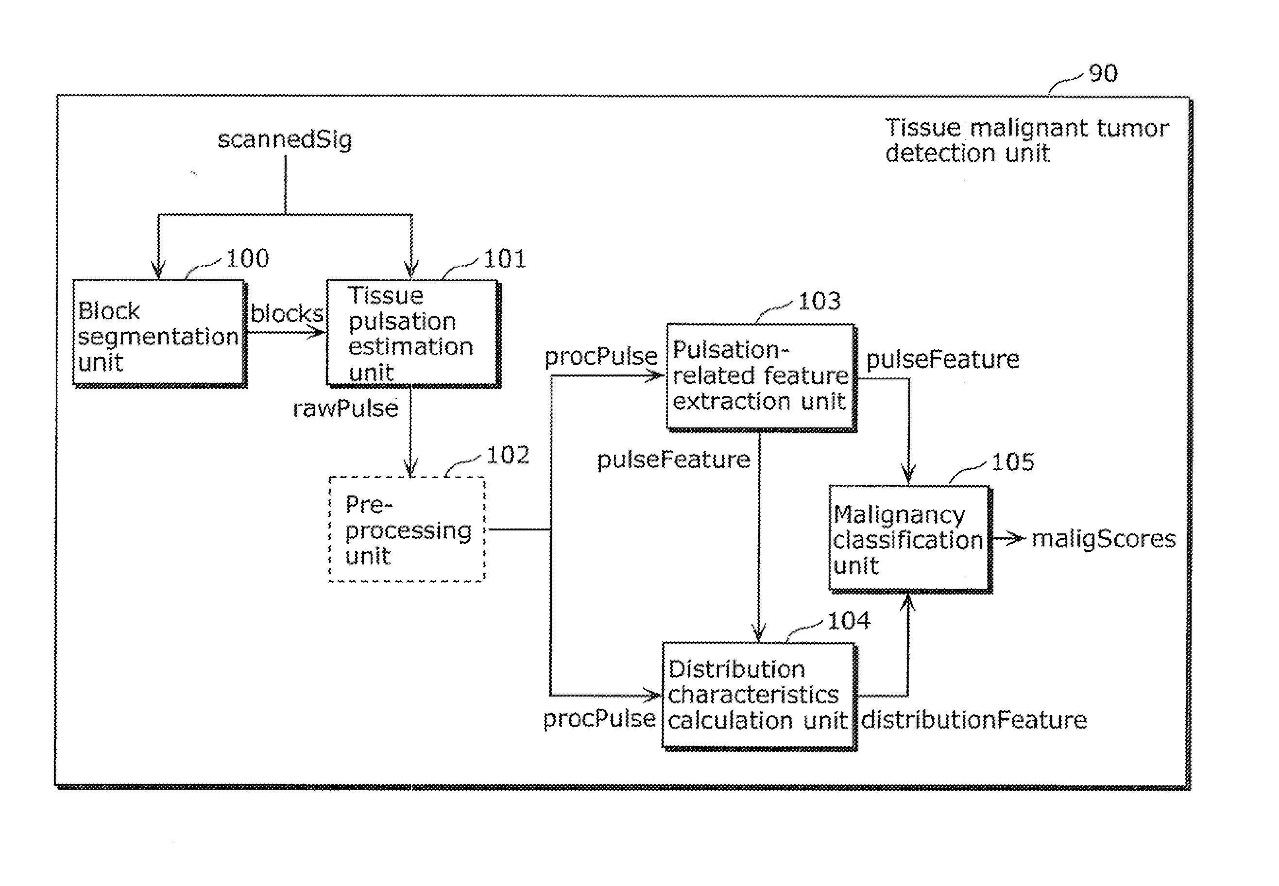
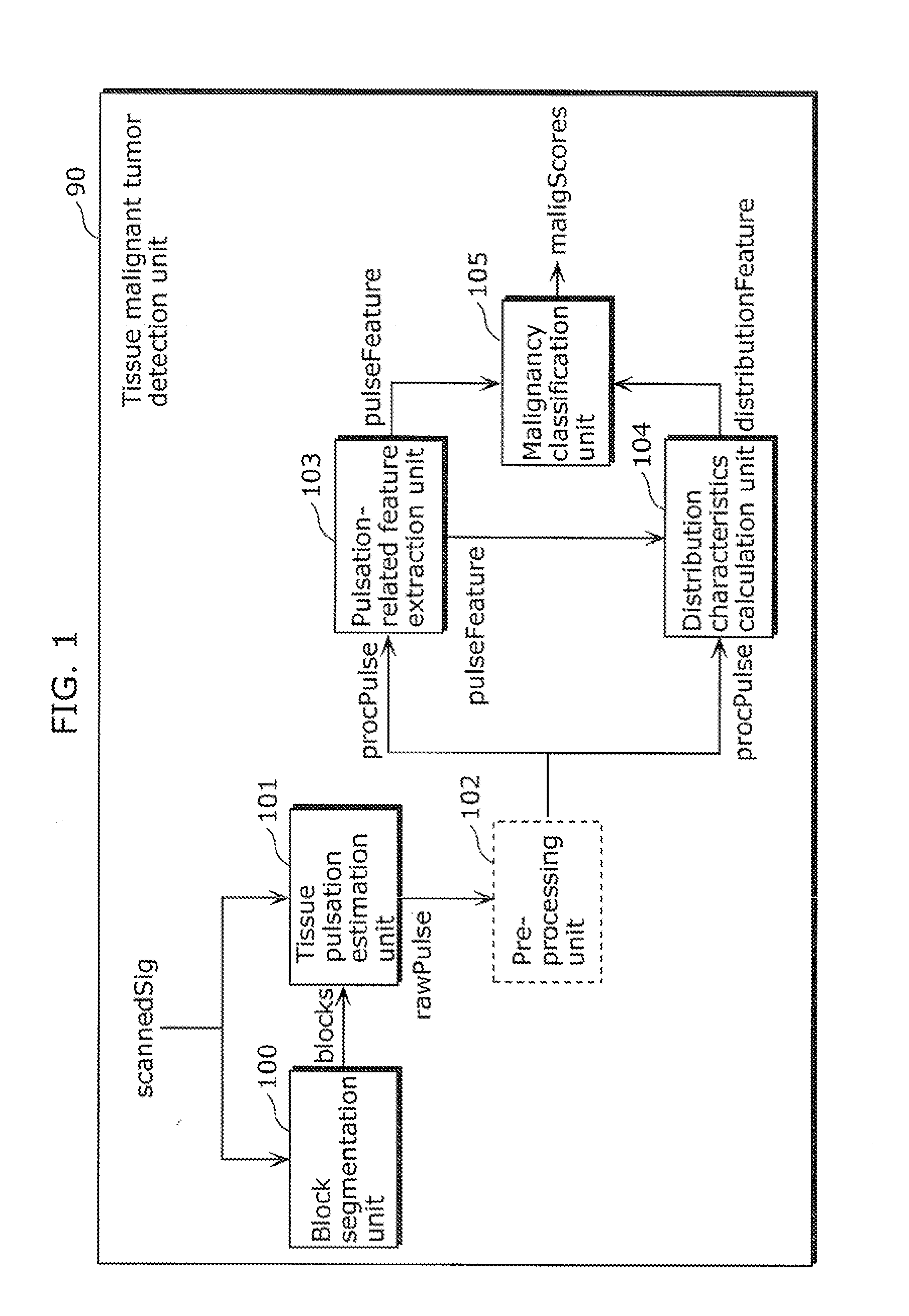
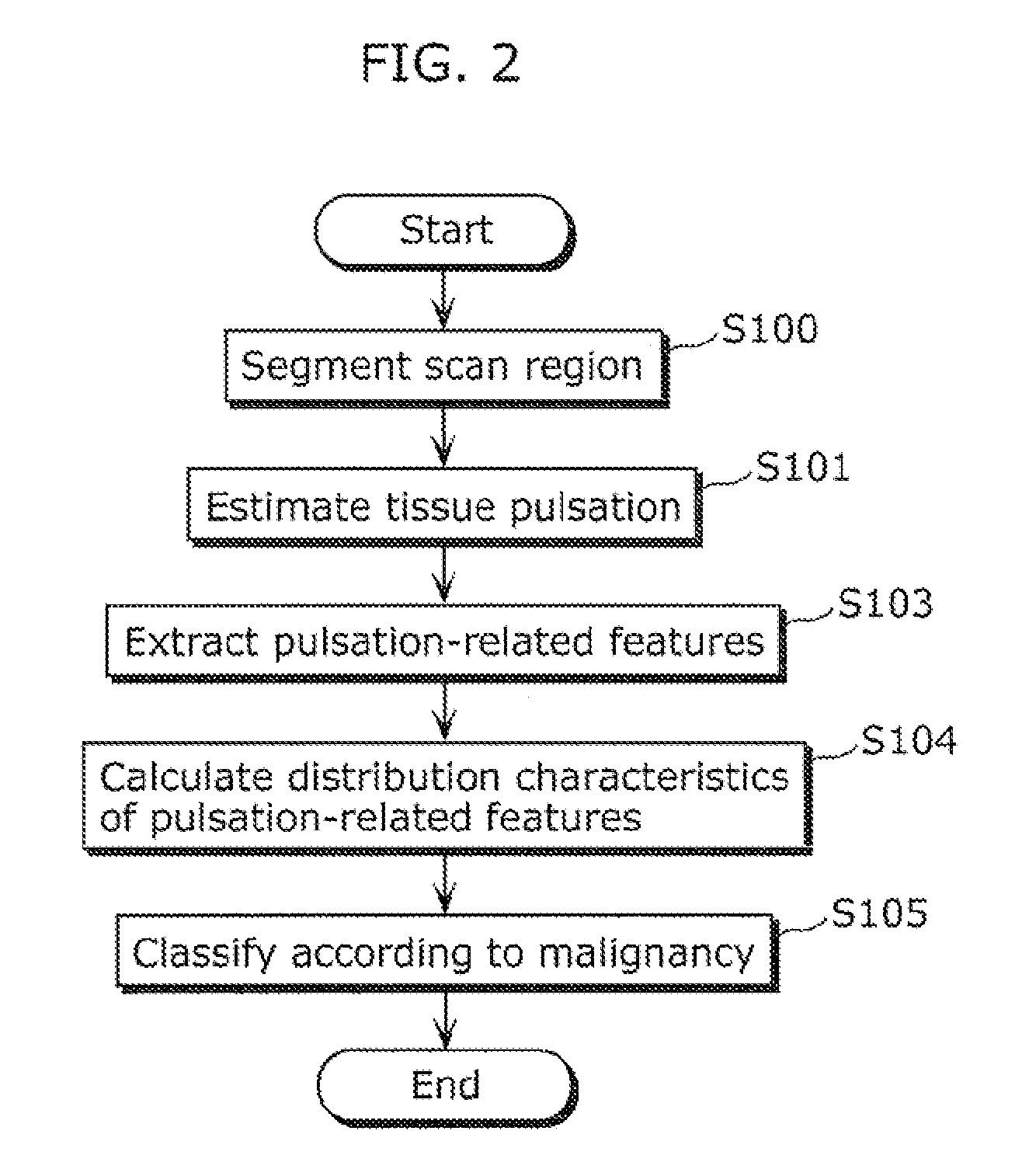
[0049]A tissue malignant tumor detection method according to an aspect of the present invention is a tissue malignant tumor detection method for detecting a malignant tumor included in a tissue, using a scan signal obtained by scanning the tissue with ultrasound, and the tissue malignant tumor detection method includes: segmenting a scanned region of the tissue into a plurality of blocks; estimating a tissue pulsation for each of the blocks, based on the scan signal, the tissue pulsation being a temporal variation in displacement of the tissue caused by pulsation of the tissue; extracting a plurality of pulsation-related features for each of the blocks, the pulsation-related features being parameters related to the tissue pulsation; calculating distribution characteristics of the pulsation-related features for each of the blocks; and classifying, based on the distribution characteristics, whether or not each of the blocks is a malignant block that is a block including a malignant tu...


PUM


Abstract

To detect a malignant tumor more accurately by detecting a feature of a cancerous tumor pulsation more appropriately, a tissue malignant tumor detection method according to the present invention includes: segmenting, into blocks, a region scanned with ultrasound (S100); estimating, based on a scan signal, a tissue pulsation that is a temporal variation in tissue displacement derived from pulsations of a tissue (S101); extracting, for each of the blocks, pulsation-related features that are parameters related to the tissue pulsation (S103); calculating distribution characteristics of the pulsation-related features for each of the blocks (S104); and classifying, based on the distribution characteristics, whether or not each of the blocks is a malignant block that is a block including the malignant tumor (S105).

Description

TECHNICAL FIELD[0001]The present invention relates to tissue malignant tumor detection methods and the like, and relates particularly to a tissue malignant tumor detection method and the like using ultrasound.BACKGROUND ART[0002]Conventionally, many techniques have conventionally been developed to detect tissue cancerous tumors (for example, see Patent Literatures 1 to 7). Examples of contrast agents for imaging tissue perfusion include: an iodine contrast agent for computed tomography (CT), a gadolinium chelated agent for magnetic resonance imaging (MRI), and radiopharmaceuticals for positron emission tomography (PET) (see Patent Literatures 4 and 5). CT and MRI typically detect cancerous tissues by taking advantage of rapid accumulation of such contrast agent in an extra-vascular space due to increased permeability of tumoral blood vessels. PET detects cancerous tissues by increased uptake of glucose molecules due to increased metabolic demands of tumor cells. Ultrasound Contract ...

Claims


Application Information

Patent Timeline
Patent Type & Authority Applications(United States)
IPC IPC(8): A61B8/08
CPCA61B8/02A61B8/5269A61B8/485A61B8/0833
Inventor FAN, SHU FENGTRAN, ANH TUANCHONG, KOK SENG
Owner KONICA MINOLTA INC
Why Eureka
Try Eureka