Eureka AIR delivers breakthrough ideas for toughest innovation challenges, trusted by R&D personnel around the world.

Photoacoustic measurement of analyte concentration in the eye

Inactive Publication Date: 2012-06-14
PEYMAN GHOLAM A
View PDF0 Cites 25 Cited by
  • Summary
  • Abstract
  • Description
  • Claims
  • Application Information

AI Technical Summary

Benefits of technology

[0019]In one aspect, the invention features methods and devices for measuring concentration of a substance in body fluids such as interstitial and other fluids that avoids the drawbacks of the above-described approaches.
[0022]This method has the advantage that the radiant energy used to stimulate the photoacoustic response need not pass through soft tissue, but rather passes through a relatively clear and optically transmissive media. Furthermore, this method has the advantage that light need not be reflected or otherwise directed to a detector for the measurement to be accomplished, as the measurement is accomplished from acoustic, rather than electromagnetic, response signals.
[0023]Although it has been known to use a photoacoustic method to measure blood glucose in opaque tissue such as through the skin, use of this method in the aqueous humor and other fluids is believed to be new, and has distinct advantages over these known applications for the reason that the aqueous humor and other fluids that are relatively transparent, and thus permit focused illumination over a larger range of tissue structure than could be achieved in opaque areas. At the same time, the glucose concentration within the aqueous humor and other fluids is reflective of the body as a whole and thus the quality of the measurement is not compromised.

Problems solved by technology

Unfortunately, these approaches all suffer from a number of drawbacks.
Specifically, light is scattered by body tissue, and thus even in the '904 patent where light is focused to a region inside the body, the location and size of the absorbing tissue region are not accurately known.
Measurements of blood glucose levels can therefore be affected by unknown variables that substantially compromise the reliability of those measurements.
However, several problems are associated with these techniques.
However, this method suffers from poor sensitivity, in part because it relies on reflecting the radiant energy off the iris.
Although this test is able to achieve high accuracy, it is difficult to administer because of the shallow angle at which the radiant energy must be passed through the eye.
All of these methods, however, measure glucose in only one area of the eye at any one time, and each relies upon optical methods for transmission and return of information.

Method used


Image

  • Photoacoustic measurement of analyte concentration in the eye
  • Photoacoustic measurement of analyte concentration in the eye

Examples


Embodiment Construction

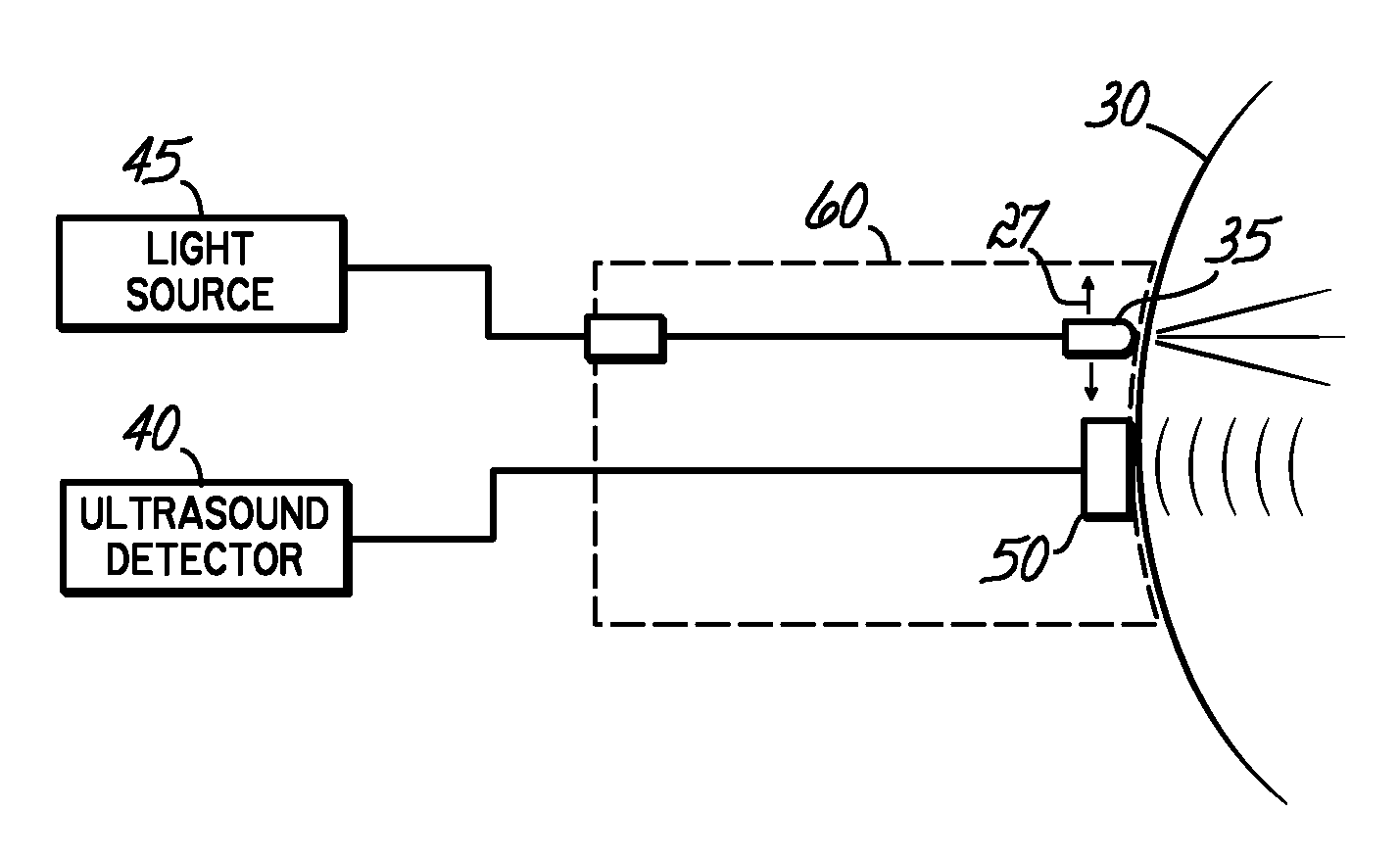
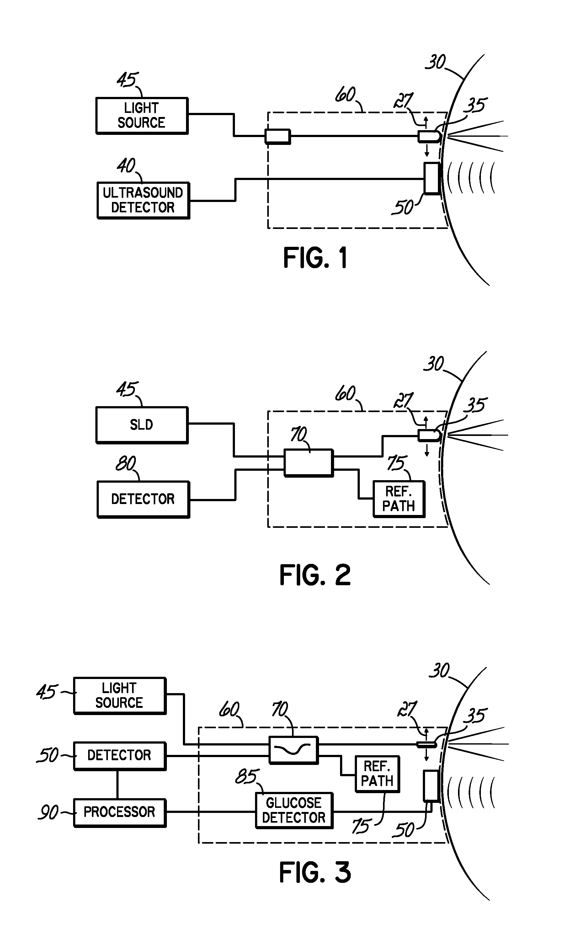
[0028]In one embodiment of the inventive system, the glucose concentration is measured in the eye, by a photoacoustic assay such as that described in U.S. Pat. No. 6,846,288 (e.g. at col. 13 line 62 to col. 18 line 49) or in U.S. Pat. No. 6,403,904, each of which is incorporated by reference in their entirety.

[0029]In one embodiment non-glucose substances, also referred to as analytes, that are present in the eye are also measured by a photoacoustic assay. For example, photoacoustic signals for methylcobalamin and methylcobinamide of the B12 family of compounds, were measured, as described in Hung and Grabowski, J. Am. Chem. Soc. 121 (1999) 1359, which is expressly incorporated by reference herein in its entirety. These substances include, in addition to glucose and the vitamin B12 family, endogenous substances such as glycosylated hemoglobin, oxygenated hemoglobin, non-oxygenated hemoglobin, oxygen, urea nitrogen (i.e., BUN), creatinine, bilirubin and its conjugates and metabolites...


PUM


Abstract

An in vivo determination of the presence or concentration of an endogenous or exogenous substance by photoacoustically assaying the substance in the eye and correlating the presence or concentration of the substance in the eye to the presence or concentration of the substance in the blood, without removing a tissue or fluid sample from the body for assay. The eye, unlike other body sites such as the skin, has a relatively constant pressure and temperature, providing an additional utility for the inventive method.

Description

CROSS REFERENCE TO RELATED APPLICATIONS[0001]This application is a continuation-in-part of co-pending U.S. application Ser. No. 11 / 870,935 filed Oct. 11, 2007; which is a Continuation of U.S. application Ser. No. 11 / 323,232 filed Dec. 30, 2005 now abandoned; which claims priority to U.S. application Ser. No. 60 / 727,078 filed Oct. 14, 2005; each of which is incorporated by reference in its entirety herein.FIELD OF THE INVENTION[0002]The invention relates to a non-invasive, in-vivo method and a system for the determination of the concentration of a substance in different areas of the eye.BACKGROUND OF THE INVENTION[0003]Many medical diagnostic techniques project radiant energy into the body of an animal for testing for the existence of a biomedical disorder or condition. For example, the integrity of the skeletal structure may be examined by passing X-rays through the body. The dense bony material substantially blocks the passage of the X-rays, permitting a doctor or other medical car...

Claims


Application Information

Patent Timeline
IPC IPC(8): A61B6/00B82Y5/00
CPCA61B5/0095A61B5/6821A61B5/1455A61B5/14532
Inventor PEYMAN, GHOLAM A.
Owner PEYMAN GHOLAM A
Why Eureka
Eureka Blog
Learn More