Endoscope apparatus

a technology of endoscope and endoscope, which is applied in the field of endoscope equipment, can solve the problems of increasing burden on the subject, difficult to slenderize the insertion portion of the endoscope, and complex structure, and achieves the effect of rapid and accurate diagnosis or observation, simple configuration, and no burden on the subj

Active Publication Date: 2012-06-21
FUJIFILM CORP
View PDF3 Cites 49 Cited by
  • Summary
  • Abstract
  • Description
  • Claims
  • Application Information

AI Technical Summary

Benefits of technology

[0018]An object of the invention is to provide an endoscope apparatus capable of switching and using a plurality of kinds of illumination depending on screening diagnosis / observation, detailed diagnosis / observation, and the like with simple configuration without causing an increase in the size of an insertion portion at the tip of an endoscope and without imposing burden on a subject, obtaining a smooth diagnostic image suitable for observation at a high frame rate in screening diagnosis, and obtaining a high-precision diagnostic image for detailed diagnosis.
[0032]According to the invention, it is possible to switch and use a plurality of kinds of illumination depending on screening diagnosis / observation, detailed diagnosis / observation, and the like with simple configuration without causing an increase in the size of the insertion portion at the tip of the endoscope and without imposing burden on the subject, to obtain a smooth diagnostic image suitable for observation at a high frame rate in screening diagnosis, and to obtain a high-precision diagnostic image in detailed diagnosis. As a result, it is possible to rapidly perform accurate diagnosis or observation constantly without imposing burden on the subject.

Problems solved by technology

However, enlarged observation is simply performed, and diagnosis of an abnormal site by special light observation is not performed.
In this apparatus, even for normal observation, it is necessary to use a complex optical system, such as an objective lens optical system, causing complexity in the structure and an increase in cost.
A confocal optical system for obtaining an enlarged microscopic image should also be provided, making it impossible to slenderize the insertion portion of the endoscope, and increasing burden is imposed on the subject.
A processor or a monitor corresponding to the confocal optical system should also be provided, causing complexity in the configuration and an increase in cost.
However, with regard to screening, bright smooth video (motion image) for observing the wide range from a distance for diagnosis while moving inside the body cavity is not acquired.
For this reason, in this endoscope system, since it is impossible to observe the wide range for diagnosis from a distance while moving inside the body cavity in the fluorescent observation mode, it is necessary to perform screening by fluorescent observation while switching the normal observation mode and the screening fluorescent observation mode, and the operation of the operator is troublesome.

Method used


Image

  • Endoscope apparatus
  • Endoscope apparatus
  • Endoscope apparatus

Examples


Embodiment Construction

[0050]Hereinafter, an endoscope apparatus according to the invention will be described with reference to a preferred embodiment shown in the accompanying drawings.

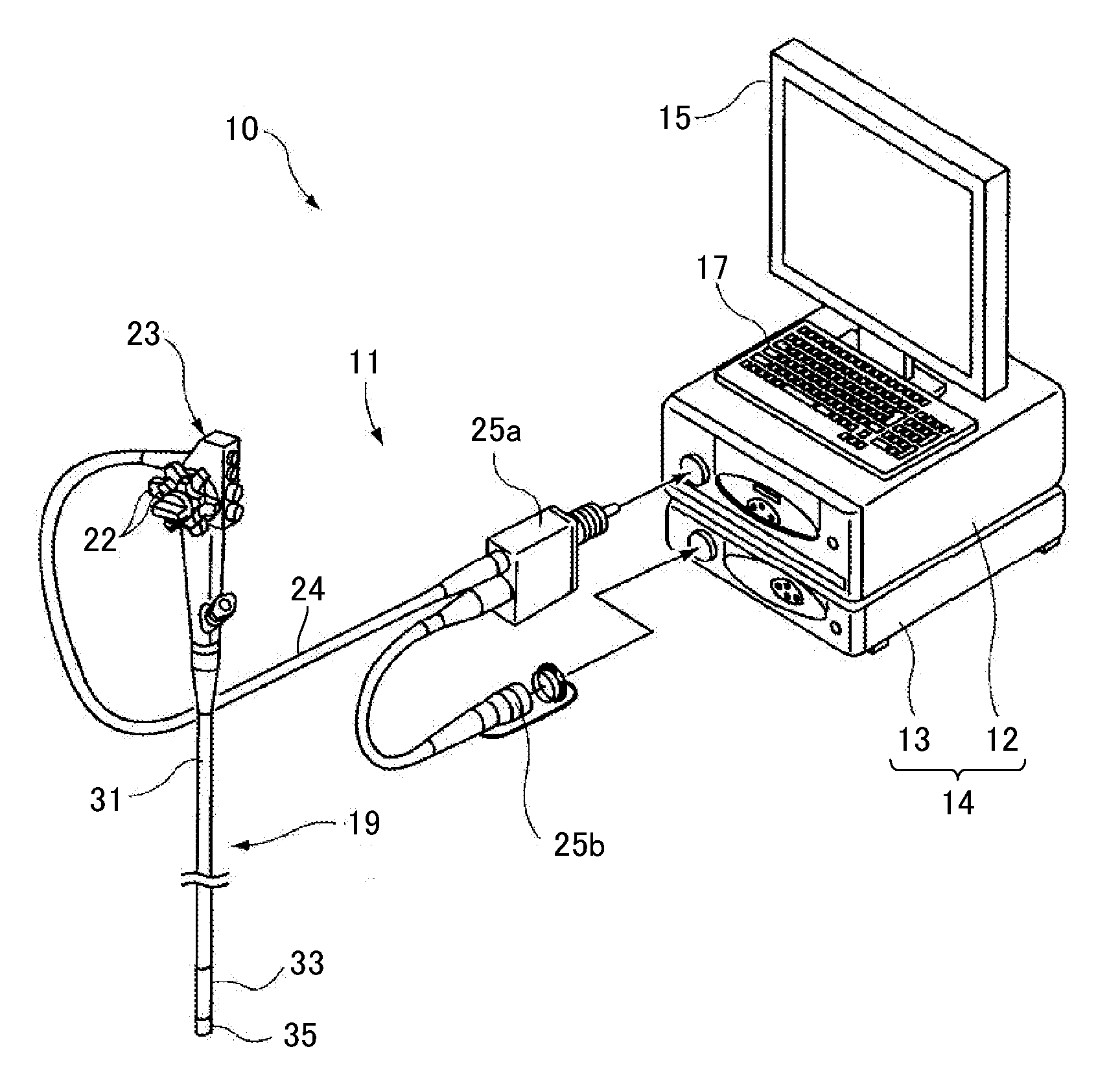
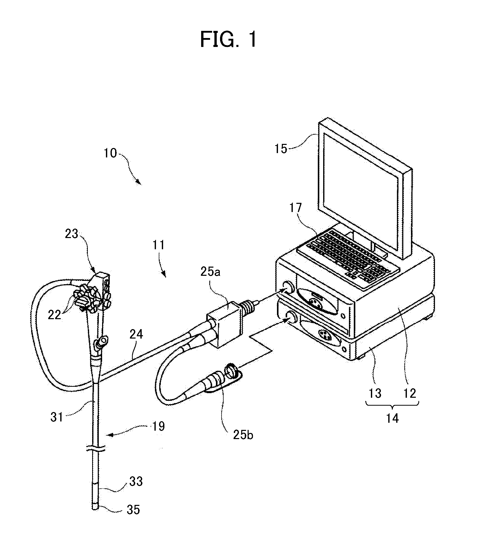
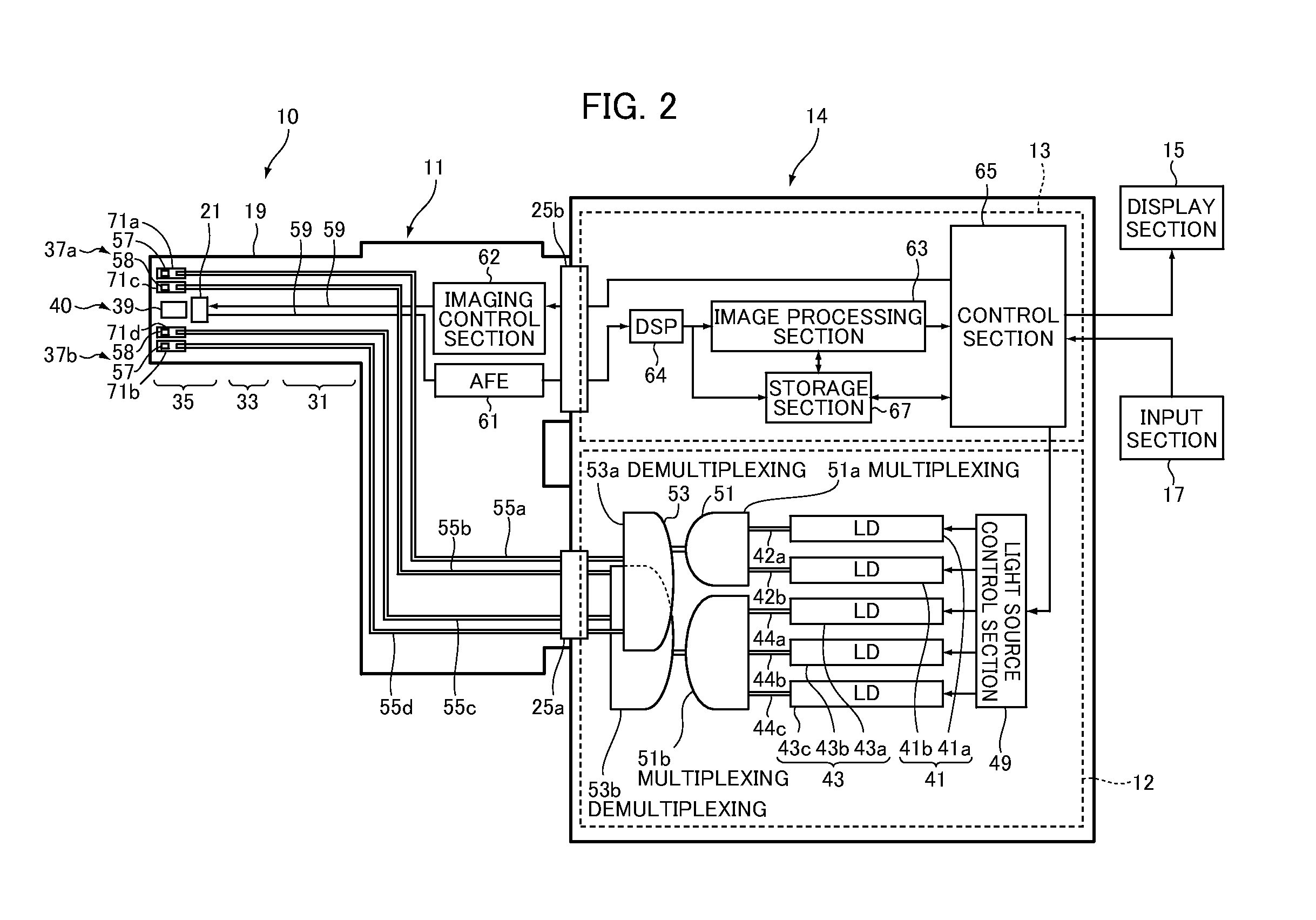
[0051]FIG. 1 is a perspective view showing the appearance of an example of an endoscope apparatus according to an embodiment of the invention. FIG. 2 is a schematic view conceptually showing the overall configuration of the endoscope apparatus shown in FIG. 1. FIG. 3 is a block diagram showing the electrical configuration of the endoscope apparatus shown in FIG. 1.

[0052]As shown in FIGS. 1 and 2, an endoscope apparatus 10 of the invention is one type of medical instrument. The endoscope apparatus 10 includes an endoscope 11 which captures the inside of a body cavity of a subject, a light source device 12 which supplies light to be irradiated into body cavity for imaging, a processor 13 which generates image information of an image including biological information, such as blood vessel information of a subject tissue inside...


PUM


Abstract

The endoscope apparatus includes a wavelength switching unit for switching emission wavelengths of a first illumination light including at least broadband light and a second illumination light including only plural kinds of narrowband light, an imaging unit for capturing an image of a subject by the first illumination light or the second illumination light having the switched emission wavelength in each imaging frame, an acquisition unit for acquiring biological information relating to form and / or function of a biological object serving as the subject and a mode switching unit for switching at least two diagnosis modes based on the biological information. The number of imaging frames by the imaging unit in which the emission wavelengths of the first and second illumination light are switched varies depending on the diagnosis mode.

Description

BACKGROUND OF THE INVENTION[0001]The present invention relates to an endoscope apparatus capable of obtaining a smooth diagnostic image suitable for observation at a high frame rate at the time of screening diagnosis and obtaining a high-definition diagnostic image at the time of detailed diagnosis by switching and using a plurality of kinds of illumination depending on purposes, such as screening diagnosis (observation) and detailed diagnosis (observation).[0002]In diagnostic imaging using an endoscope, screening observation / inspection or screening diagnosis is performed in which cancer of a body cavity, particularly, the focus site of early-stage cancer, a lesion site, or an abnormal site, such as a hemorrhage, is observed accurately, precisely, and in detail, and prior to detailed observation for diagnosis or detailed diagnosis, a site suspected of abnormality is found from each site in a wide range of the body cavity.[0003]In screening observation / diagnosis using the endoscope, ...

Claims


Application Information

Patent Timeline
Patent Type & Authority Applications(United States)
IPC IPC(8): A61B1/06
CPCA61B1/0638A61B1/0653A61B1/063A61B5/1459A61B1/00009A61B5/14551A61B1/000094A61B1/0655
Inventor YAMAGUCHI, HIROSHI
Owner FUJIFILM CORP
Why Eureka
Try Eureka