
Methods for Treatment of Muscular Dystrophy
- Summary
- Abstract
- Description
- Claims
- Application Information
AI Technical Summary
Benefits of technology
Problems solved by technology
Method used
Image

Examples
example 1
Reduced CTGF Levels Ameliorates Fibrosis in Dystophic Skeletal Muscle in an Animal Model of Duchenne Muscular Dystrophy
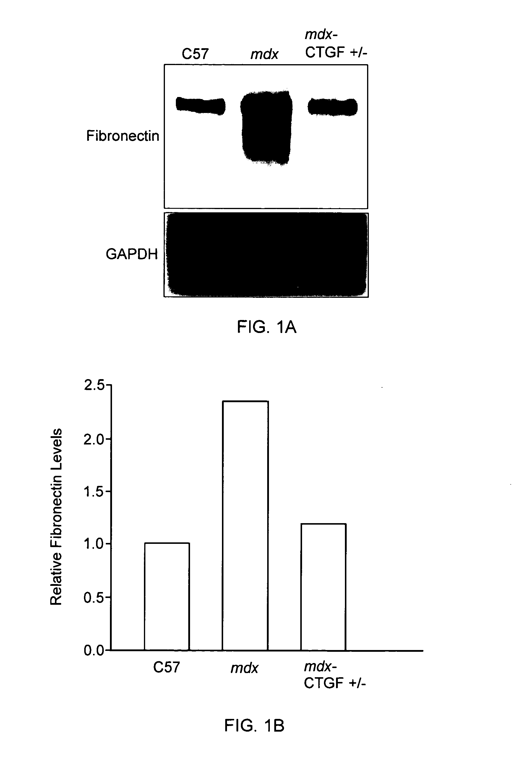
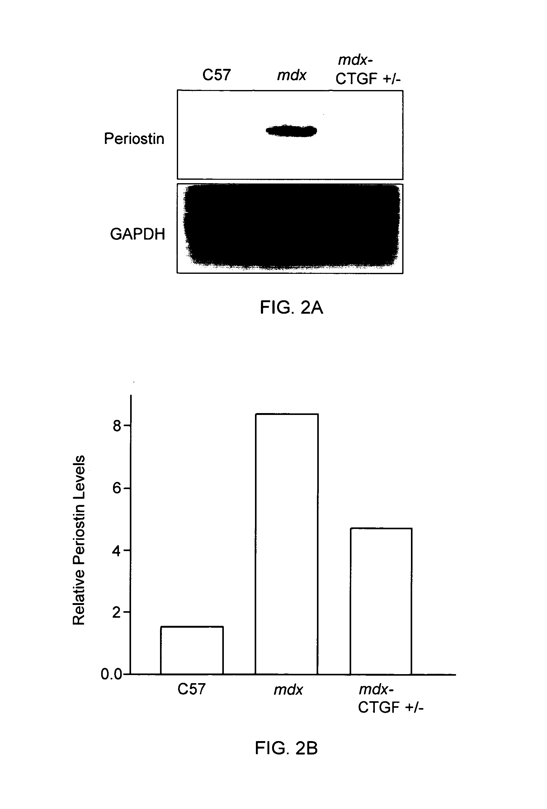
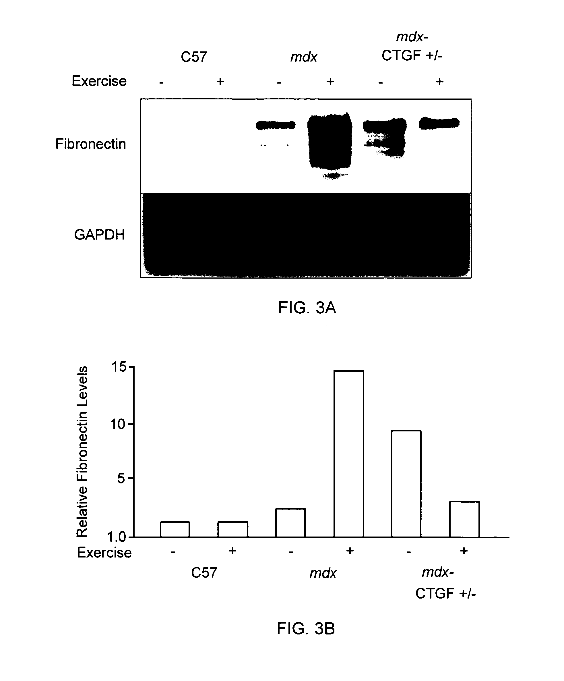
[0093]The mdx mouse is an animal model of Duchenne muscular dystrophy. In the mdx mouse, the dystrophin protein is absent in skeletal muscle and many similar pathological features seen in muscle in human subject with Duchenne muscular dystrophy are observed in these animals. (See e.g., Bulfield et al. (1984) PNAS 81:1189-1192; Stedman et al. (1991) Nature 352:536-539.) In this animal model of Duchenne muscular dystrophy, skeletal muscle fibrosis can be assessed by measuring skeletal muscle levels of extracellular matrix proteins (e.g., fibronectin, periostin). (See e.g., Mezzano et al. (2007) J Cell Commun Signal. 1:205-17.) The effects of reduced CTGF levels on fibrosis in dystrophic skeletal muscle were examined using the mdx mouse model of Duchenne muscular dystrophy as follows.
[0094]In this series of experiments, in order to reduce CTGF levels, mdx mice were mat...
example 2
CTGF Inhibition Ameliorates Dedifferentiation of Skeletal Muscle Cells In Vitro
[0101]Myoblasts are muscle precursor cells that play an important role in the growth and repair of skeletal muscle following injury. Pathological dedifferentiation of myoblasts has been observed in muscular dystrophy and contributes to disease progression. (See Vial et al. (2008) J Cell Physiol. 215:410-21.) Myoblast dedifferentiation is characterized, in part, by down regulation of MyoD, myogenin, myosin, and desmin, markers of myoblast activation and differentiation. Therefore, the effect of CTGF inhibition on myoblast dedifferentiation is examined as follows.
[0102]In a series of experiments, C2C12 myoblasts (ATTC, Manassas, Va.) are transfected with a specific siRNA for CTGF or incubated with an anti-CTGF antibody (e.g., the anti-CTGF antibody CLN-1). The myoblasts are then cultured and induced to dedifferentiate in the presence of angiotensin II, as described in Larrain et al. ((1997) Exp Cell Res. 23...
example 3
CTGF Inhibition Ameliorates Fibrosis in Skeletal Muscle Cells
[0104]Myogenic precursor cells can differentiate into myofibroblasts after muscle injury and myofibroblasts contribute to the development of fibrosis. Increased myofibroblast activation has been observed in skeletal muscle cells treated with angiotensin II. The effect of CTGF inhibition on angiotensin-induced myofibroblast activity is examined as follows.
[0105]C2C12 myofibroblasts (ATTC, Manassas, Va.) are transfected with a specific siRNA for CTGF or incubated with an anti-CTGF antibody (e.g., the anti-CTGF antibody CLN-1). Myofibroblasts are then incubated in the presence of angiotensin II as described in Larrain et al. ((1997) Exp Cell Res. 234:405-12). The expression levels in muscle of several extracellular matrix proteins (e.g., fibronectin, periostin, thrombospondin-1, decorin, and collagen III) are evaluated by real time PCR, Northern blot and / or western blot analysis. (See Caceres et al. (2000) Eur J Cell Biol 79:...
PUM
 Abstract
Description
Claims
Application Information