MR Imaging System for Discriminating Between Imaged Tissue Types

a tissue type and imaging system technology, applied in the field of mr imaging system, can solve the problems of inhomogeneity of signal variations, inability to fully compensate for phase errors introduced by and inability to detect the phase error of the first inversion puls

Active Publication Date: 2012-08-02
DUKE UNIV +1
View PDF12 Cites 21 Cited by
  • Summary
  • Abstract
  • Description
  • Claims
  • Application Information

AI Technical Summary

Problems solved by technology

2010) are susceptible to motion and blood flow resulting in signal variations known as image inhomogeneity, and in image artifacts within the imaged volume.
2010), but this requirement is problematic as it may make the method susceptible to motion and flow.
Phase errors introduced by the first inversion pulse can only be fully compensated by the second inversion in the absence of motion and flow.
With motion and flow present, the resulting tissue preparation, e.g. in the h

Method used


Image

  • MR Imaging System for Discriminating Between Imaged Tissue Types
  • MR Imaging System for Discriminating Between Imaged Tissue Types
  • MR Imaging System for Discriminating Between Imaged Tissue Types

Examples


Embodiment Construction

Definitions.

[0025]An inversion recovery (IR) pulse inverts longitudinal magnetization from the positive z-axis by 180 degrees to the negative z-axis. IR pulses are used as preparation pulses prior to a main imaging pulse sequence to achieve different kinds of MR contrast (such as T1 weighted, T2 weighted). Adiabatic IR pulses are used to give more uniform contrast throughout an imaging volume than non-adiabatic RF pulses.

[0026]iPAT (integrated Parallel Acquisition Techniques) comprises “parallel imaging”. It enables faster scanning through reduced phase encoding and addition of RF coil information. An iPAT factor of 2 enables scanning about twice as fast, iPAT factor of 3 enables scanning about three times as fast and so on.

[0027]TI=inversion time, the time between an inversion recovery pulse and the next RF excitation pulse. TI determines the image contrast.

[0028]T1=the longitudinal (or spin-lattice) relaxation time T1 decay constant.

[0029]T2=the transverse (or spin-spin) relaxatio...


PUM


Abstract

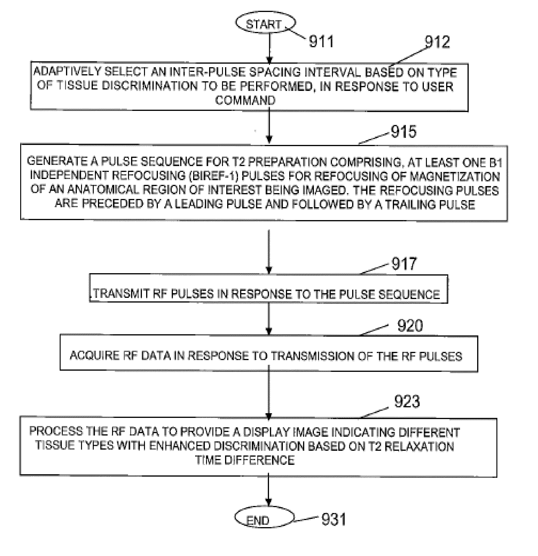
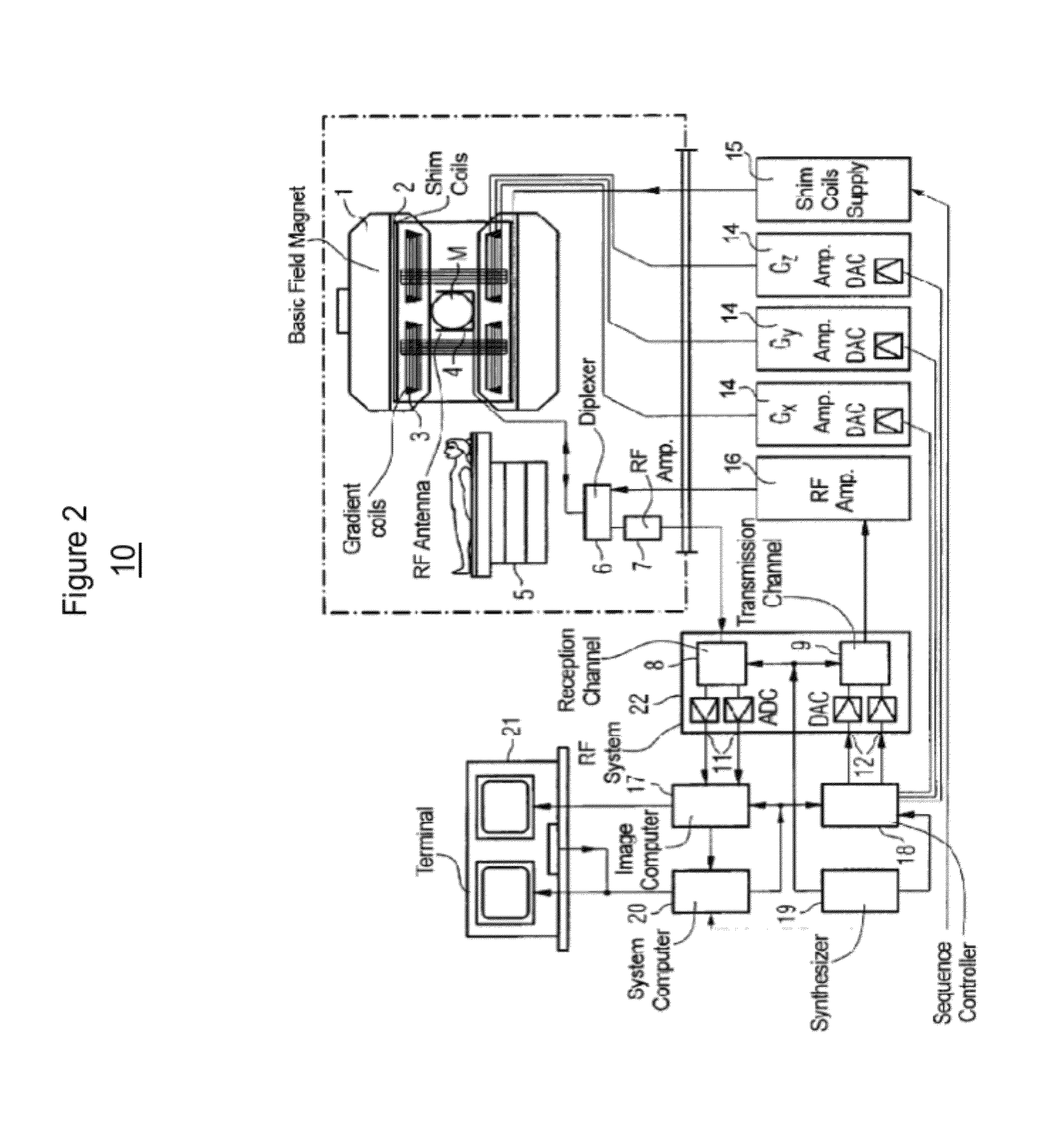
A system provides B1- and B0-insensitive, blood flow and motion-robust T2-preparation and T2-preparation combined with inversion recovery. An MR imaging system discriminates between imaged tissue types based on transverse relaxation time (T2) or transverse relaxation time combined with longitudinal recovery time (T1). A signal generator generates a pulse sequence for T2 preparation or combined T2-preparation with inversion recovery comprising one or more B1 independent refocusing (BIREF-1) pulses for refocusing of magnetization of an anatomical region of interest being imaged, and different combinations of adiabatic or non-adiabatic tip-down and flip-back pulses. Multiple RF coils transmit RF pulses in response to the pulse sequence and acquire RF data in response to transmission of the RF pulses. A processing system processes the RF data to provide a display image indicating different tissue types with enhanced discrimination based on T2 relaxation time difference or combined T2 and T1 time difference.

Description

[0001]This is a non-provisional application of provisional application Ser. No. 61 / 436,700 filed 27 Jan. 2011, by W. G. Rehwald et al.FIELD OF THE INVENTION [0002]This invention concerns an MR imaging system for discriminating between imaged tissue types based on transverse relaxation time (T2) or transverse relaxation time (T2) in combination with longitudinal recovery time (T1).BACKGROUND OF THE INVENTION[0003]In Magnetic Resonance Imaging (MRI) and in particular cardiovascular MRI, “T2-preparation” or “T2-prep” is used to magnetically prepare tissue, for example myocardium (heart tissue), to create image contrast between different tissue types due to their differences in T2. This is also known as T2-weighting. T2 is a time constant describing the decay of transverse magnetization and is a function of local tissue water content, among other parameters. The shorter the tissue T2 value in a region of interest (ROI) the darker the ROI appears in a T2-weighted MR image, and conversely...

Claims


Application Information

Patent Timeline
IPC IPC(8): G01R33/34G01R33/50
CPCG01R33/5602G01R33/50
Inventor REHWALD, WOLFGANG G.CHEN, ENN-LINGKIM, RAYMOND J.
Owner DUKE UNIV
Why Eureka
Try Eureka