
Systems and methods for producing imaging biomarkers indicative of a neurological disease state using gray matter suppressions via double inversion-recovery magnetic resonance imaging

a magnetic resonance imaging and gray matter suppression technology, applied in the field of magnetic resonance imaging methods and systems, can solve the problems of inability to capture the extent of cortical pathology in ms, limited conventional techniques, and currently unapproved clinical use of high field strengths

Active Publication Date: 2016-08-04
MAYO FOUND FOR MEDICAL EDUCATION & RES
View PDF3 Cites 6 Cited by
  • Summary
  • Abstract
  • Description
  • Claims
  • Application Information

AI Technical Summary

Benefits of technology

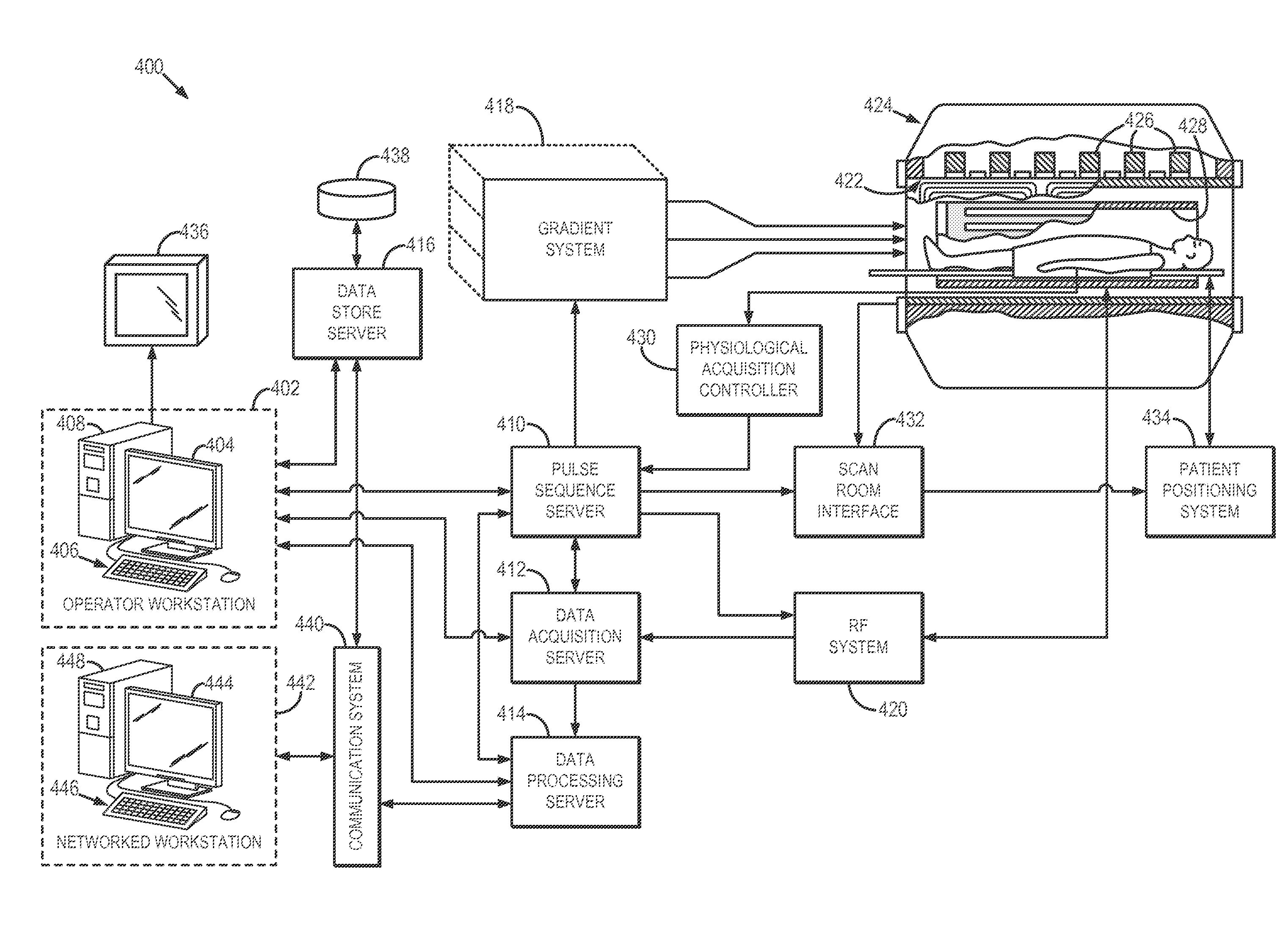
The present invention provides systems and methods for producing and utilizing imaging biomarkers indicative of neurological diseases, such as multiple sclerosis. By using double inversion-recovery magnetic resonance imaging and analyzing the resulting images, the invention can determine connectivity between abnormal lesions in the brain, which can be used as a biomarker of disease states. The invention can also identify specific regions of interest in the brain that can be used as indicators of neurological disease states. Overall, the invention provides a more accurate and reliable tool for diagnosing and monitoring the progression of neurological diseases.

Problems solved by technology

In addition, cortical demyelination is also present in the earliest stages of MS, and the acute stage of CL formation is highly inflammatory.
However, such conventional techniques are limited and typically fail to capture the extent of cortical pathology in MS.
However, application of high field strengths to patients is currently not approved for clinical use.
In addition, results achieved with standard DIR imaging techniques that are commercially and clinically available are still inferior when compared to standard histopathology.
In particular, suppression of normal-appearing WM and CSF in DIR imaging produces poor sensitivity in identifying subpial plaques, which are the most common cortical lesions observed in early MS and clinically isolated syndrome (“CIS”) cases.

Method used


Image

  • Systems and methods for producing imaging biomarkers indicative of a neurological disease state using gray matter suppressions via double inversion-recovery magnetic resonance imaging
  • Systems and methods for producing imaging biomarkers indicative of a neurological disease state using gray matter suppressions via double inversion-recovery magnetic resonance imaging
  • Systems and methods for producing imaging biomarkers indicative of a neurological disease state using gray matter suppressions via double inversion-recovery magnetic resonance imaging

Examples


example

[0049]The relationship between cortical lesions (“CLs”) and white matter lesions (“WMLs”) in multiple sclerosis (“MS”) is poorly understood. In addition to their association with disease progression, CLs may also play a role in disease initiation. Thus, it was hypothesized that white matter lesions may be present along tracts directly connected with cortical lesions. As such, an experiment was performed to demonstrate such connectivity, using double inversion recovery (“DIR”) images for CL detection coupled with DTI-based probabilistic tractography.

[0050]Study participants, which are outlined in Table 1, included patients with relapsing-remitting MS with relative mild disease (EDSS2) performed a highly conservative scoring (according to the consensus recommendation). The other rater (rater 1) took a more inclusive approach, including smaller lesions. WM lesions were obtained with consensus, all scored on DIR image, with WV lesions being confirmed with T2 lesion maps.

TABLE 1Demograph...


PUM


Abstract

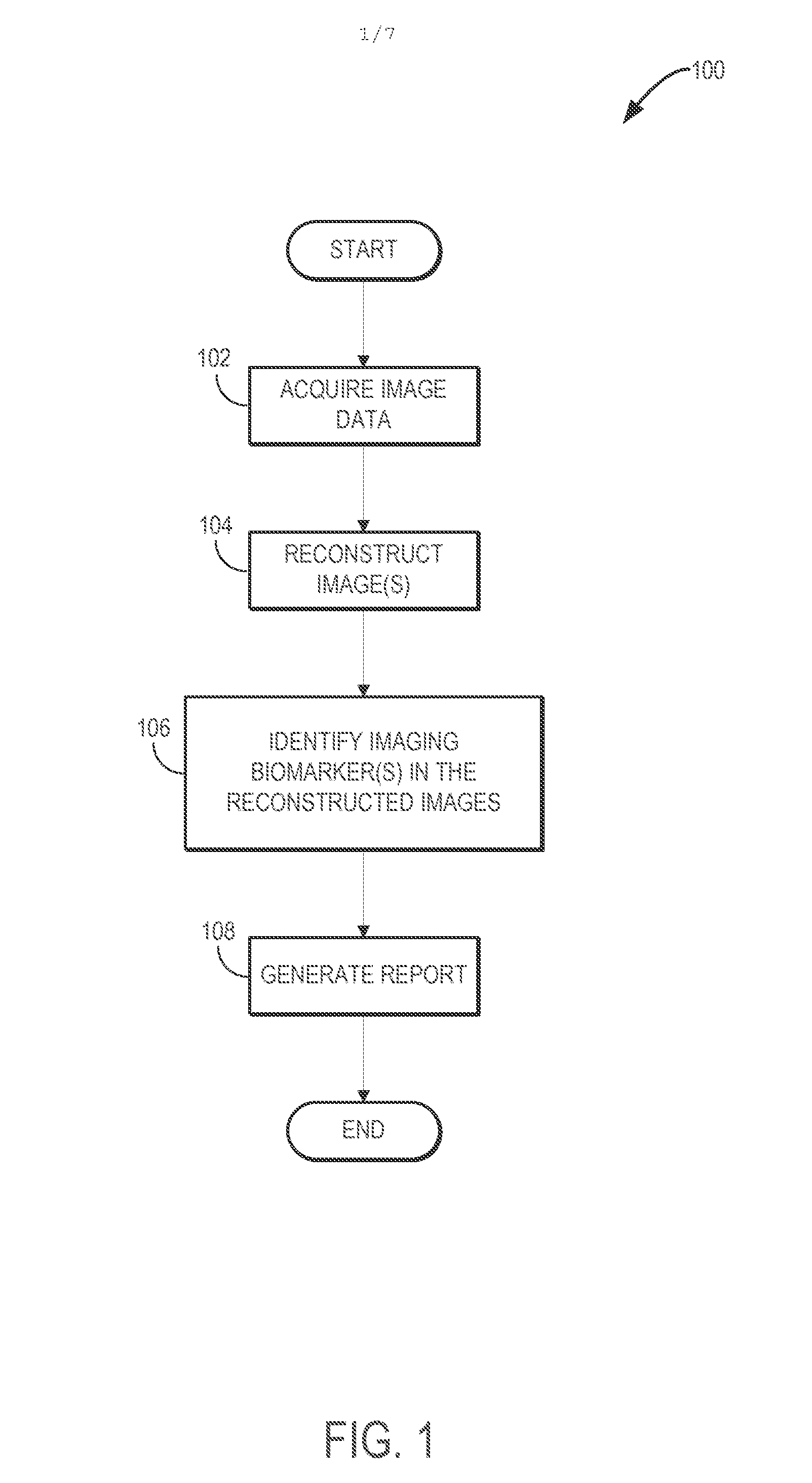
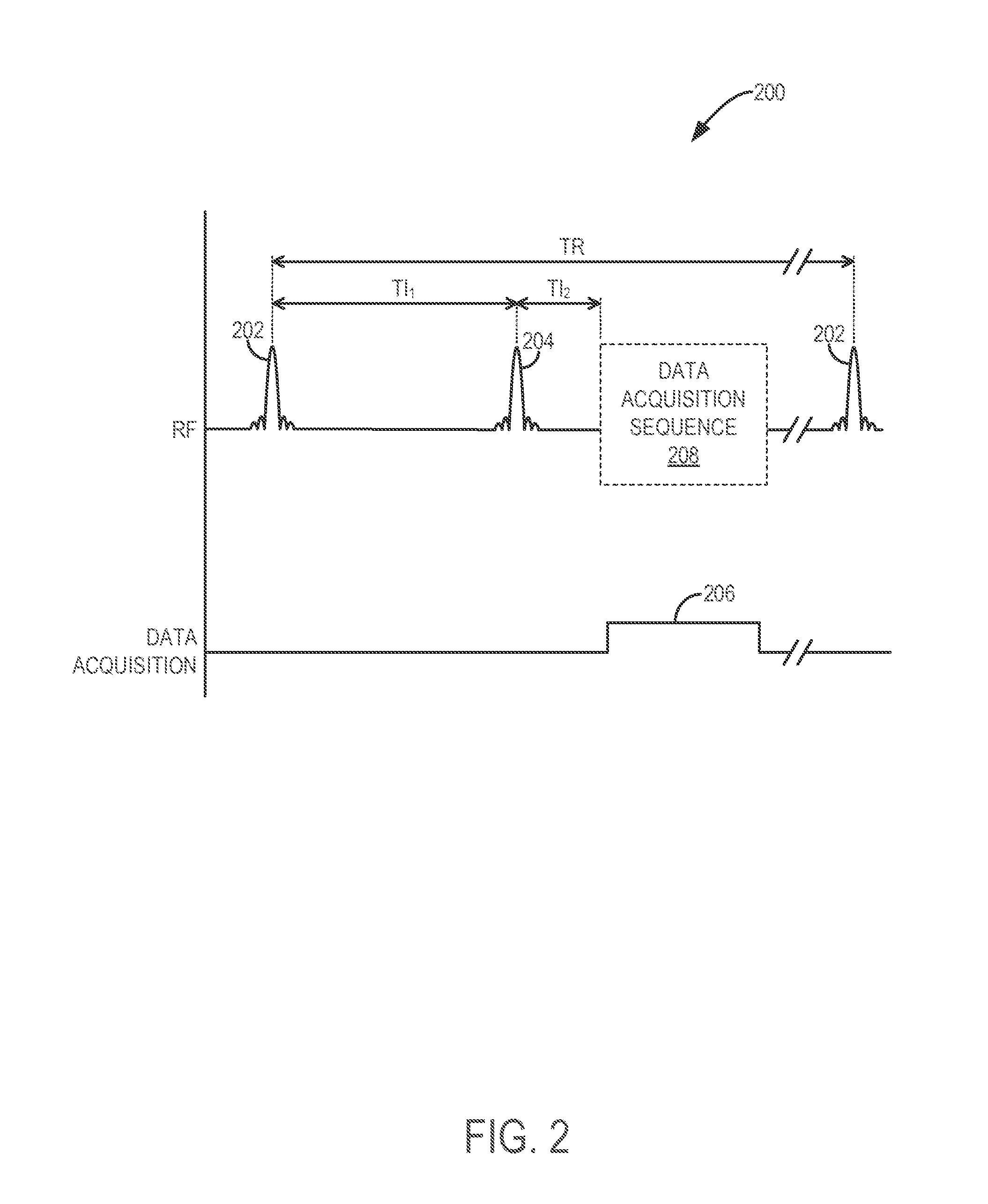
Systems and methods related to imaging biomarkers for determining neurological disease states of a subject are provided. In one embodiment, a method for producing an image indicative of a neurological disease using a magnetic resonance imaging (“MRI”) system is provided. The method includes directing the MRI system to perform a double inversion-recovery (“DIR”) pulse sequence to generate data where signals from gray matter and cerebral spinal fluid are substantially suppressed. The method also includes analyzing the DIR images, reconstructed from the acquired data, to identify cortical and white matter lesions. This includes identifying imaging biomarkers based on visual signatures of brain tissue, including white matter tissue. In some aspects, diffusion-weighted data may also be obtained using the MRI system. Diffusion-weighted data may be used in a tractography process to determine connectivities, or connectivity patterns between the identified lesions, including cortical lesions, to determine neurological disease states of the subject.

Description

CROSS-REFERENCE TO RELATED APPLICATIONS[0001]This application is based on, claims priority to, and incorporates herein by reference in its entirety for all purposes, U.S. Provisional Application Ser. No. 61 / 880,386, filed Sep. 20, 2013, and entitled “SYSTEM AND METHOD FOR PRODUCING IMAGING BIOMARKERS INDICATIVE OF A NEUROLOGICAL DISEASE STATE USING GRAY MATTER SUPPRESSION VIA DOUBLE INVERSION-RECOVERY MAGNETIC RESONANCE IMAGING.”STATEMENT REGARDING FEDERALLY SPONSORED RESEARCH[0002]N / ABACKGROUND OF THE INVENTION[0003]The field of the present invention is magnetic resonance imaging (“MRI”) methods and systems. More particularly, the present invention relates to systems methods for identifying or diagnosing multiple sclerosis (“MS”) and other neurological disorders, or individuals at risk of getting MS or other neurological disorders. Furthermore, the present invention relates to quantitative measurement of appearance and connectivity of cerebral tissues on MR images as biomarkers for...

Claims


Application Information

Patent Timeline
Patent Type & Authority Applications(United States)
IPC IPC(8): A61B5/00G01R33/56G01R33/563A61B5/055
CPCA61B5/055A61B5/4076A61B2576/026G01R33/56341A61B5/0042G01R33/5608G01R33/5602G16H30/40
Inventor PORT, JOHN D.PIRKO, ISTVANTILLEMA, JAN-MENDELTSHU, YUNHONGLUCCHINETTI, CLAUDIA F.
Owner MAYO FOUND FOR MEDICAL EDUCATION & RES
Learn More