
Method of oocyte cryopreservation using antifreeze protein

a cryopreservation method and antifreeze technology, applied in the field of oocyte cryopreservation using antifreeze protein, can solve the problems of low success rate of freezing treatment of oocytes, large amount of moisture, and damage to frozen oocytes, so as to improve the survival rate of oocytes, improve the fertilization rate, and minimize the damage of oocytes

Inactive Publication Date: 2012-09-27
SEOUL NAT UNIV BUNDANG HOSPITAL
View PDF0 Cites 5 Cited by
  • Summary
  • Abstract
  • Description
  • Claims
  • Application Information

AI Technical Summary

Benefits of technology

[0007]The present invention relates to a method for minimizing damage to an oocyte during freezing of the oocyte by using a cryopreservation liquid added with an antifreeze protein, which increases a survival rate, a fertilization rate and a blastocyst development ratio of the oocyte.
[0011]According to another aspect of the present invention, it provides a method for increasing a blastocyst development ratio of a cell, the method comprising the steps of: preparing a composition for cryopreservation by mixing 5 to 50 vol % of serum, 10 to 20 vol % of ethylene glycol (EG), 10 to 20 vol % of propylene glycol and 100 to 3000 ng / ml of antifreeze protein; suspending and cryopreserving the cell with the composition, and thawing the cell.
[0019]The inventive cryopreservation method of an oocyte minimizes damage to the oocyte, which increases the survival rate of the oocyte after freezing / thawing of the oocyte, and improves a fertilization rate, and a blastocyst development ratio of the oocyte.

Problems solved by technology

Meanwhile, an oocyte has a large size and accordingly, a large amount of moisture, and thus a frozen oocyte may be damaged.
Accordingly, it is reported that freezing treatment of oocyte has a low success rate.
Also, a matured oocyte may be subjected to a damage causing deformation of a meiosis spindle fiber, which reduces cleavage capability or blastocyst development ratio (Carrol et al., 1993; Lane et al., 1999; Hong et al., 1999, Shaw et al., 2000;).
As described above, in spite of much research on a freezing method, a significant progress in improvement for minimizing the freezing damage of an oocyte has not been achieved.

Method used


Image

  • Method of oocyte cryopreservation using antifreeze protein
  • Method of oocyte cryopreservation using antifreeze protein

Examples


example 1

Oocyte Cryopreservation Through Addition of an Antifreeze Protein

[0024]An oocyte was suspended in 20 vol % fetal bovine serum (FBS) solution in HEPES-buffered TCM-199 (Gibco) as a basic medium for 5 minutes, and was separated. 20 ml of equilibrium solution (ES) containing 7.5 vol % ethylene glycol (1.5 ml), 7.5 vol % propylene glycol (1.5 ml) and basic medium 17 ml was prepared. The separated oocyte was suspended in 700 μl of equilibrium solution for 5 minutes. Then, the oocyte was separated again. A vitrification solution (VS) containing 15 vol % ethylene glycol (3 ml), 15 vol % propylene glycol (3 ml), 0.5M sucrose (3.424 g / 20 ml) and basic medium 14 ml was prepared. The separated oocyte was suspended in 700 μl of vitrification solution for less than 1 minute. 5 to 6 oocytes on the vitrification solution (VS) were replaced on CryoTop (Kitazato, Japan), and their moisture was removed. Immediately, they were immersed within liquid nitrogen and stored in a nitrogen tank. Herein, each...

example 2

Cryopreservation of Oocyte According to the Weight Ratio of an Antifreeze Protein

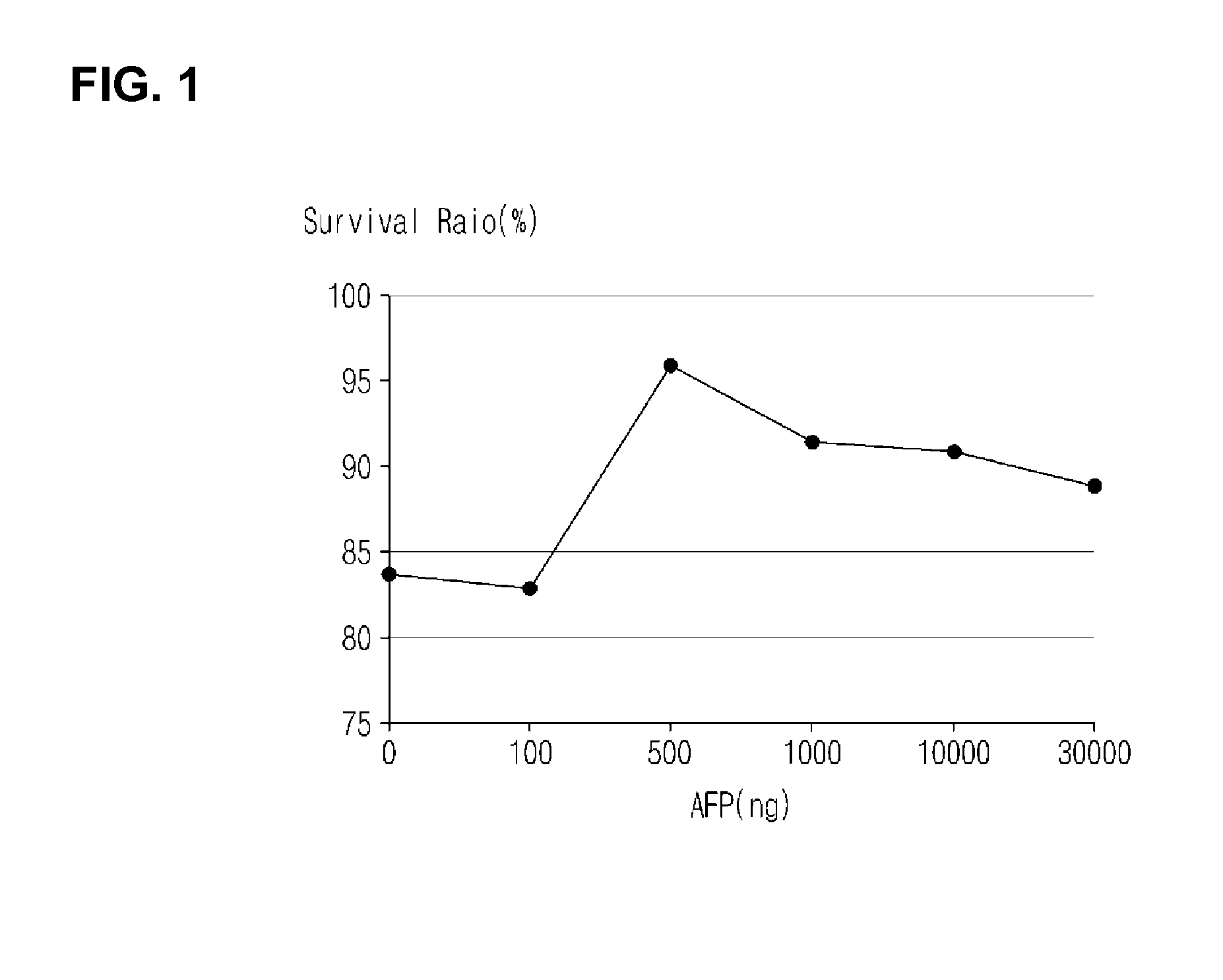
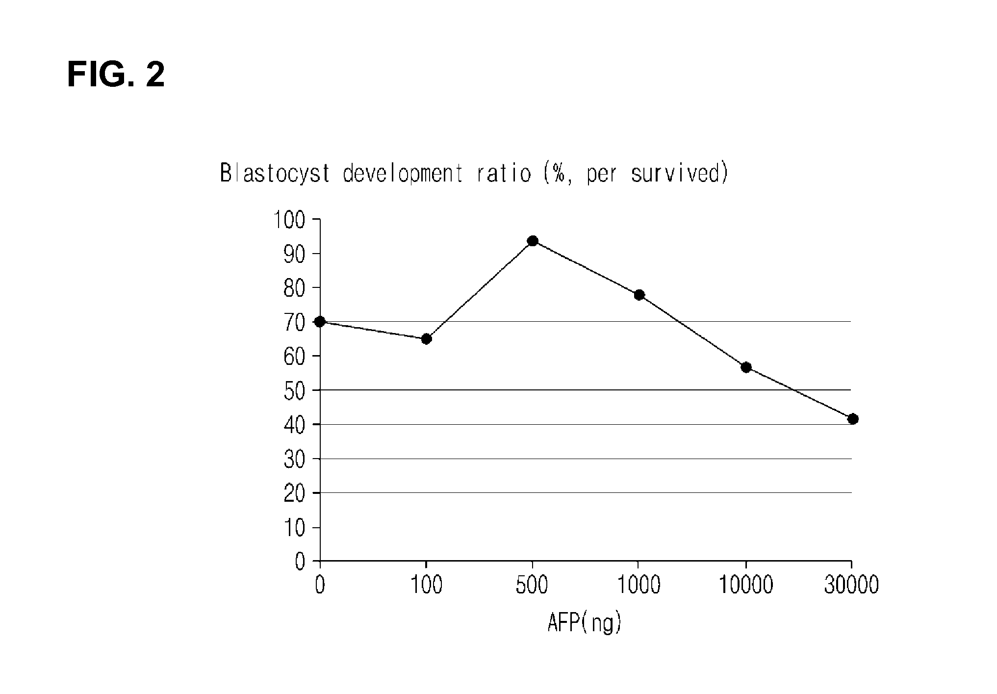
[0027]Antifreeze protein was added in different weight ratios of 100 ng / ml, 500 ng / ml, 1 μg / ml, 10 μg / ml and 30 / ml, a survival rate, a fertilization rate, and a blastocyst development ratio of an In-vivo matured MII oocyte were measured according to the weight ratio of the antifreeze protein. This experiment was carried out in the same manner as described in Example 1 except that the weight ratios of the antifreeze protein were varied.

[0028]As a result, when the antifreeze protein was added at a weight ratio of 500 ng / ml, oocyte showed very significant good results in a survival rate, a maturity rate, a fertilization rate (cleavage rate), and a blastocyst development ratio (see FIGS. 1 and 2). From this, it was identified that a specific weight ratio of an added antifreeze protein is an important factor in determining the survival rate, the maturity rate, the fertilization rate, and the blastocyst devel...

example 3

Formation of Spindle Fibers and Separation of Chromosomes According to Addition of an Antifreeze Protein

[0029]After an In-vivo matured oocyte was vitrified with addition of an antifreeze protein at a ratio of 500 ng / ml, the formation of spindle fibers and the separation of chromosomes were observed. As a result, it was found that the number of oocytes maintaining their normal status was increased (Table 4).

[0030]A normal status and an abnormal status of spindle fibers were discriminated as follows. In normal spindle fibers, a nucleus is lined up with respect to an equatorial plate, and the spindle fibers are symmetrically disposed in a spindle form with respect to the nucleus. In abnormal spindle fibers, a nucleus is lost or disposed in a loose order without being lined up, or some of spindle fiber are lost or tangled and lumped together. The normal status is graded as follows. Grade 1 of a normal status indicates that the form is partially similar or very similar to that before fre...


PUM

PropertyMeasurementUnit
vol %aaaaaaaaaa
vol %aaaaaaaaaa
concentrationaaaaaaaaaa

Abstract

Disclosed is a method for cryopreserving an oocyte by adding an antifreeze protein to a cryopreservation liquid (equilibrium solution, vitrification solution). The disclosed cryopreservation method of an oocyte minimizes damage to the oocyte, which increases the survival rate of the oocyte after freezing and thawing of the oocyte, and improves a fertilization rate, and a blastocyst development ratio of the oocyte.

Description

CROSS-REFERENCES TO RELATED APPLICATIONS[0001]This application is a U.S. Utility application, which claims priority to Korean Application No. 10-2011-0019521, filed Mar. 4, 2011. The entire contents of the aforementioned patent application are incorporated herein by this reference.TECHNICAL FIELD[0002]The present invention relates to a method for cryopreserving an oocyte by adding an antifreeze protein to each cryopreservation liquid.BACKGROUND ART[0003]Oocyte cryopreservation is a very attractive method capable of preserving fertility in the case where a large amount of oocytes are obtained during in vitro fertilization, in the case where semen collection is failed on the day of in vitro fertilization, in the case where fertility of a cancer patient is required to be preserved, or in the case where there is a concern about loss of fertility due to endometriosis, etc. An oocyte freezing method includes a slow freezing method and a vitrification method.[0004]The cryopreservation comp...

Claims


Application Information

Patent Timeline
Patent Type & Authority Applications(United States)
IPC IPC(8): C12N5/075
CPCA01N1/0221A01N1/00C07K14/00C12N5/00C12N5/0609
Inventor JO, JUN WOOSUH, CHANG SUKJEE, BYUNG CHUL
Owner SEOUL NAT UNIV BUNDANG HOSPITAL
Learn More