Supercharge Your Innovation With Domain-Expert AI Agents!

Preventive for adhesion following abdominal surgery

a technology of abdominal surgery and adhesion prevention, which is applied in the field of prevention of postlaparotomy adhesion, can solve the problems of adhesion between, obstruction or constriction of intestinal passages, and adhesive ileus

Inactive Publication Date: 2012-11-01
MEDICAL THERAPIES
View PDF0 Cites 0 Cited by
  • Summary
  • Abstract
  • Description
  • Claims
  • Application Information

AI Technical Summary

Benefits of technology

[0007]The adhesion frequency following peritoneal wall injury in midkine gene knockout mice (Nakamura, E. et al., Genes Cells, 3, 811-822, 1998) was markedly lower when compared with the control wild-type mice (FIG. 1). Since the only difference between the knockout and the wild-type mice is the presence of the midkine gene, adhesions can be prevented or treated by suppressing the expression or activity of midkine.
[0029]Cell fusion between the immune cells and myeloma cells described above can be essentially achieved by a known method, for example, the method of Milstein et al. (Galfre, G. & Milstein, C. Methods Enzymol. 73: 3-46, 1981). Specifically, the cell fusion described above can be performed, for example, in a standard culture medium in the presence of a cell fusion-promoting agent. Such fusion-promoting agents include, for example, polyethylene glycol (PEG) and the Sendai virus. Furthermore, an auxiliary substance such as dimethylsulfoxide may be added to enhance the fusion efficiency.

Problems solved by technology

Mechanical stimuli produced by laparotomy operations may cause inflammation in intraperitoneal organs, and consequently, cause adhesions between the intraperitoneal organs or between the intraperitoneal organs and the peritoneal wall.
This may cause intestinal passage obstruction or constriction and lead to adhesive ileus.
Even if adhesive ileus does not develop, chronic abdominal pain and female infertility can result.
Furthermore, such adhesions make it very difficult to perform future laparotomies.
However, this operation may result in further adhesions.
The formation of post-laparotomy adhesions is a serious problem and has been neglected as it is considered to be an inevitable side effect.
However, the membrane is only effective in preventing adhesions in regions that are separated by it, and cannot exert its adhesion prevention effect throughout the whole peritoneal cavity.

Method used


Image

  • Preventive for adhesion following abdominal surgery
  • Preventive for adhesion following abdominal surgery
  • Preventive for adhesion following abdominal surgery

Examples


example 1

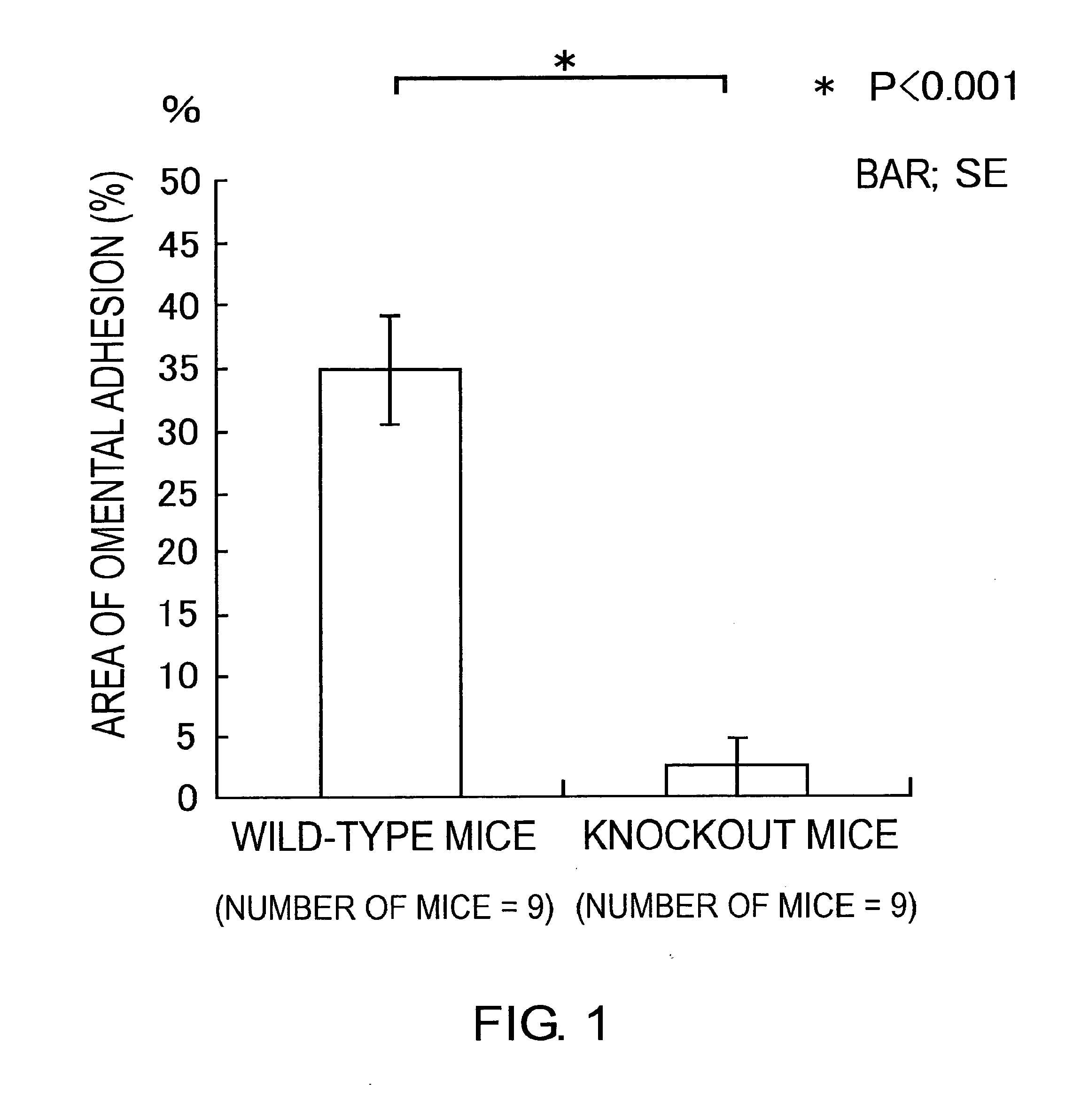
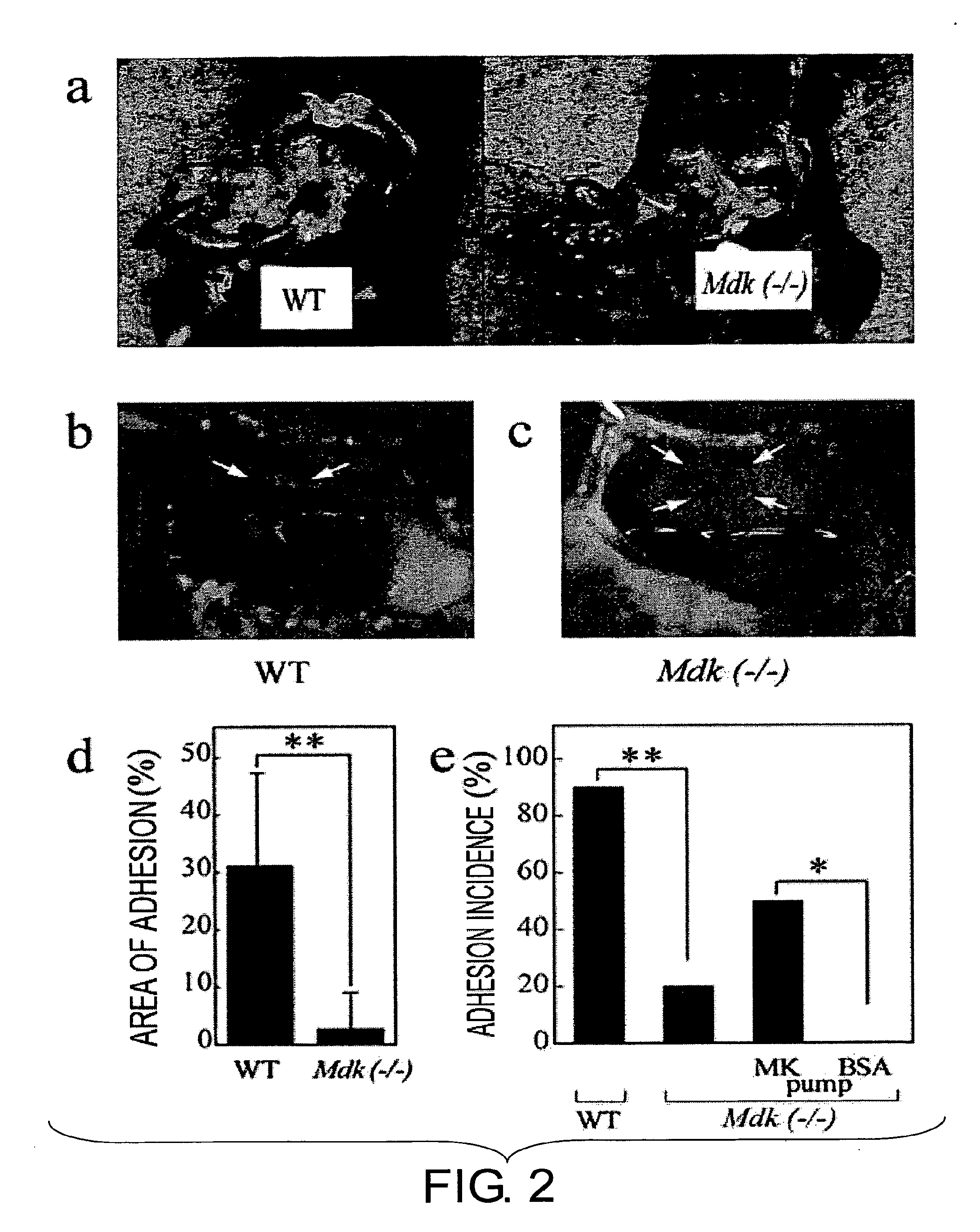
Effects of Midkine on Post-Laparotomy Adhesions

(1) Preparation of Knockout Mice

[0042]Midkine (- / -) mice were created as described (Nakamura, E. et al., Genes Cells, 3, 811-822, 1998). The heterozygote was backcrossed to C57BL / 6J mice for nine generations. Then, the backcrossed progenies were crossed with each other to produce midkine (- / -) mice having the C57BL / 6J genetic background. C57BL / 6J mice were used as the wild-type control.

example 2

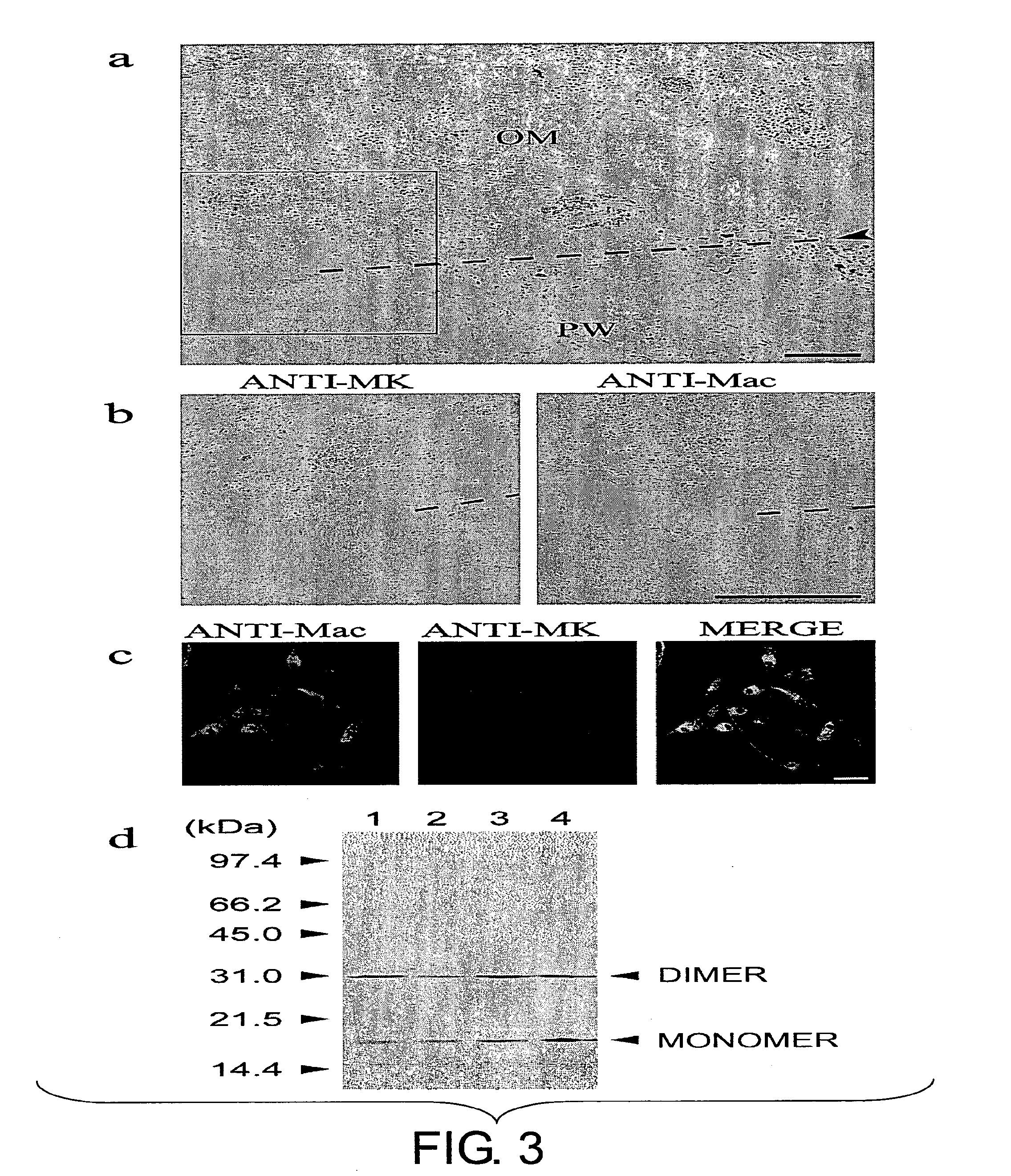
Midkine Expression in Regions of Adhesion

[0048]Midkine expression was determined by Western blotting of the heparin-binding protein derived from 1.7 mg of extract as described (Muramatsu, H. et al., Dev. Biol. 159, 392-402, 1993). The anti-mouse midkine antibody was produced as previously described (Muramatsu, H. et al., Dev. Biol. 159, 392-402, 1993). The sites of midkine expression were revealed by immunohistochemistry (Nakamura, E. et al., Genes Cells, 3, 811-822, 1998) using an affinity-purified rabbit anti-mouse midkine antibody as the primary antibody and a horseradish peroxidase-conjugated affinity-purified goat anti-rabbit IgG (Jackson ImmunoResearch Laboratories, Inc.; West Grove, Pa.) as the secondary antibody. Positive signals were visualized using diaminobenzidine tetrahydrochloride (Amersham Pharmacia Biotech; Tokyo, Japan). A fluorescein isothiocyanate-labeled goat anti-rabbit IgG (Sigma; St. Louis, Mo.) can also be used as the secondary antibody. Staining of the macro...


PUM


Abstract

The present inventors discovered that oligonucleotides which suppress midkine expression and antibodies which suppress midkine activity can be used to prevent post-surgical intraperitoneal adhesions.

Description

CROSS-REFERENCE TO RELATED APPLICATIONS[0001]This application is a divisional of U.S. patent application Ser. No. 10 / 547,011, filed 5 Jan. 2006, now allowed, which is the National Stage Application of PCT / JP2004 / 002888, filed 5 Mar. 2004, which claims priority to Japanese Application No. 2003-108428, filed 6 Mar. 2003. The contents of the above patent applications are incorporated by reference herein in their entirety.TECHNICAL FIELD[0002]The present invention relates to prevention of post-laparotomy adhesions.BACKGROUND ART[0003]Although the incidence rate of post-laparotomy adhesion varies from report to report, adhesions are reportedly formed in 90% or more of major laparotomy cases and in 55-100% of patients who undergo gynecologic laparotomy. Mechanical stimuli produced by laparotomy operations may cause inflammation in intraperitoneal organs, and consequently, cause adhesions between the intraperitoneal organs or between the intraperitoneal organs and the peritoneal wall. This...

Claims


Application Information

Patent Timeline
Patent Type & Authority Applications(United States)
IPC IPC(8): A61K31/7088A61P41/00A61P43/00
CPCC07K16/22A61K31/7088A61P41/00A61P43/00
Inventor MURAMATSU, TAKASHIINOH, KAZUHIKOMURAMATSU, HISAKOTORII, SHUHEI
Owner MEDICAL THERAPIES
Features
  • R&D
  • Intellectual Property
  • Life Sciences
  • Materials
  • Tech Scout
  • Unparalleled Data Quality
  • Higher Quality Content
  • 60% Fewer Hallucinations
Learn More