Suture and method for repairing a heart

a technology of artificial and mechanical components and heart, applied in the field of implantable medical devices for treating diseases, disorders, and malformations of the heart, can solve the problems of prolapse of leaflets, heart valves may lose their ability to close properly, and heart function may be seriously impaired

Inactive Publication Date: 2013-05-02
EDWARDS LIFESCIENCES CORP
View PDF6 Cites 62 Cited by
  • Summary
  • Abstract
  • Description
  • Claims
  • Application Information

AI Technical Summary

Benefits of technology

[0007]Embodiments of the present invention provide devices and methods for securely attaching a thread to tissue without the need for conventional suturing, thereby eliminating the need for surgical instruments such as needle holders and forceps. Some embodiments are adapted for attaching thread to heart wall tissue; however, the features of the invention are also applicable to a wide variety of other medical procedures wherein it is desirable to securely anchor a suture thread or other structure to a portion of tissue without using a needle and forceps.
[0013]In another embodiment, a method of treating an enlargement of a left ventricle of a heart comprises accessing the inside of a heart (e.g., through surgical incision) and deploying multiple anchor members at locations along the left ventricular muscular wall. Threads extending from the anchor members are connected to a lock. The threads are shortened to pull the anchors, and therefore the muscular wall, into closer proximity to the lock and to the other anchors. The threads are then locked in the shortened condition to maintain the anchors in closer proximity, thereby reducing the size of the left ventricle. In one embodiment, the lock is located within the left ventricle. In another embodiment, the lock is located within a different chamber of the heart, such as the right atrium or the right ventricle. In another embodiment, the lock is located outside of the heart, such as, for example, at or adjacent the apex of the heart.

Problems solved by technology

The function of the heart may be seriously impaired if any of the heart valves is not functioning properly.
The heart valves may lose their ability to close properly due to disease or defects, such as (for example) by a mitral or tricuspid valve leaflet becoming partially or completely detached from a papillary muscle (e.g., by ruptured chordae tendinae) thereby resulting in prolapsing of the leaflet.
The inability of the heart valve to close will cause a leak backwards, commonly referred to as regurgitation, through the valve.
On the left side of the heart, regurgitation through the mitral valve may seriously impair the function of the heart 1 since a portion of the heart stroke volume of blood will return from the left ventricle to the left atrium instead of passing through the aortic valve to the organs of the body.
Accordingly, the damaged heart is required to work much harder to maintain adequate circulation, a condition that inevitably ends in heart enlargement and / or heart failure.
However, attaching a thread (i.e., suture) to the papillary muscle can be cumbersome due to difficulties in accessing the surgical site, including limitations in physical and visual access.
The limited visual access results from the relatively narrow space between the leaflets of a mitral valve.
Furthermore, access to the desired portion of the heart can be difficult while performing conventional open-heart surgery (i.e., with a sternal split).
Further difficulties can arise as a result of the difficult angles at which the instruments are held.
Due to the shortcomings associated with existing medical procedures, the attachment of artificial chordae can be time consuming and may often be inaccurate.

Method used


Image

  • Suture and method for repairing a heart
  • Suture and method for repairing a heart
  • Suture and method for repairing a heart

Examples


Embodiment Construction

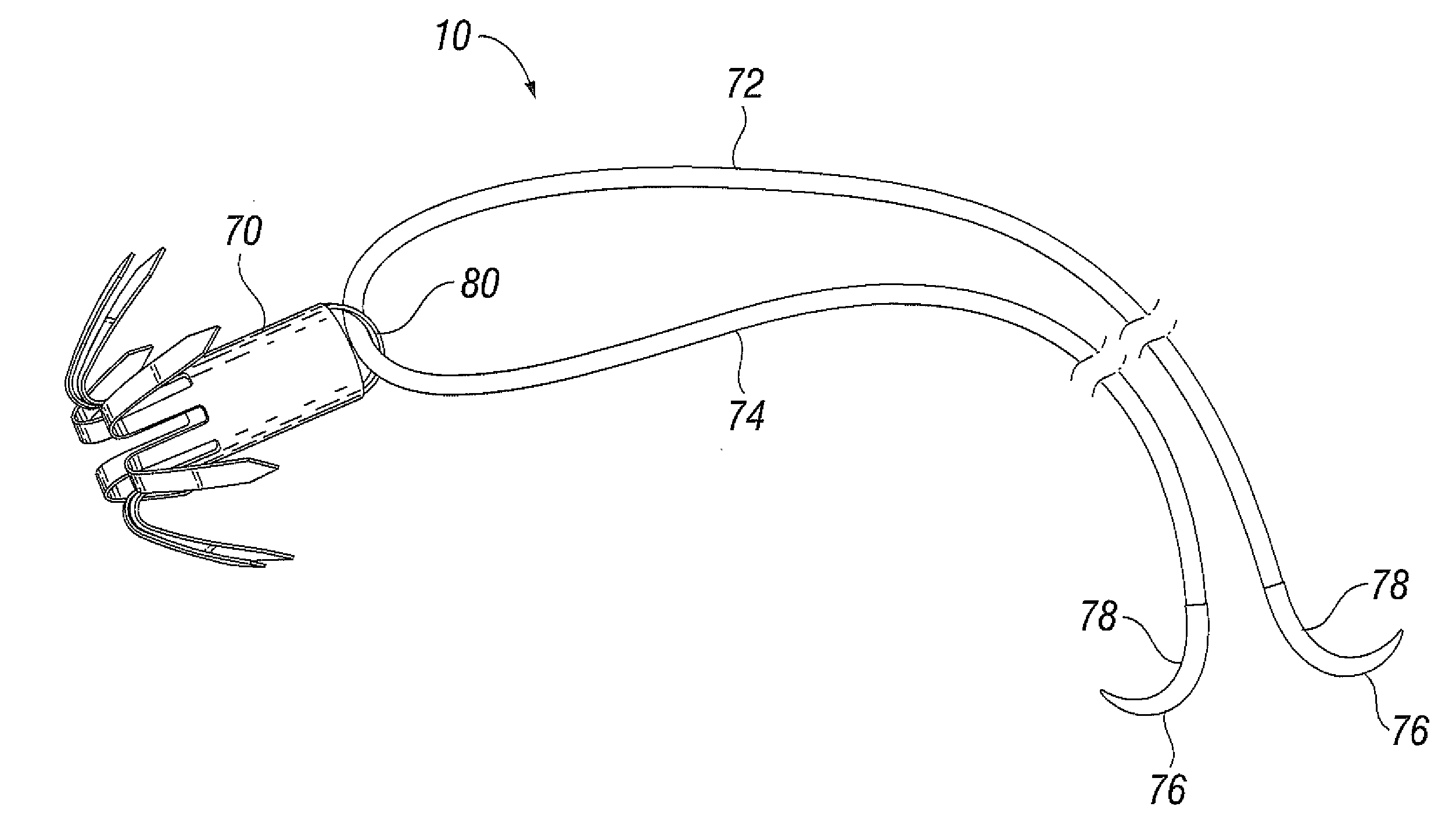
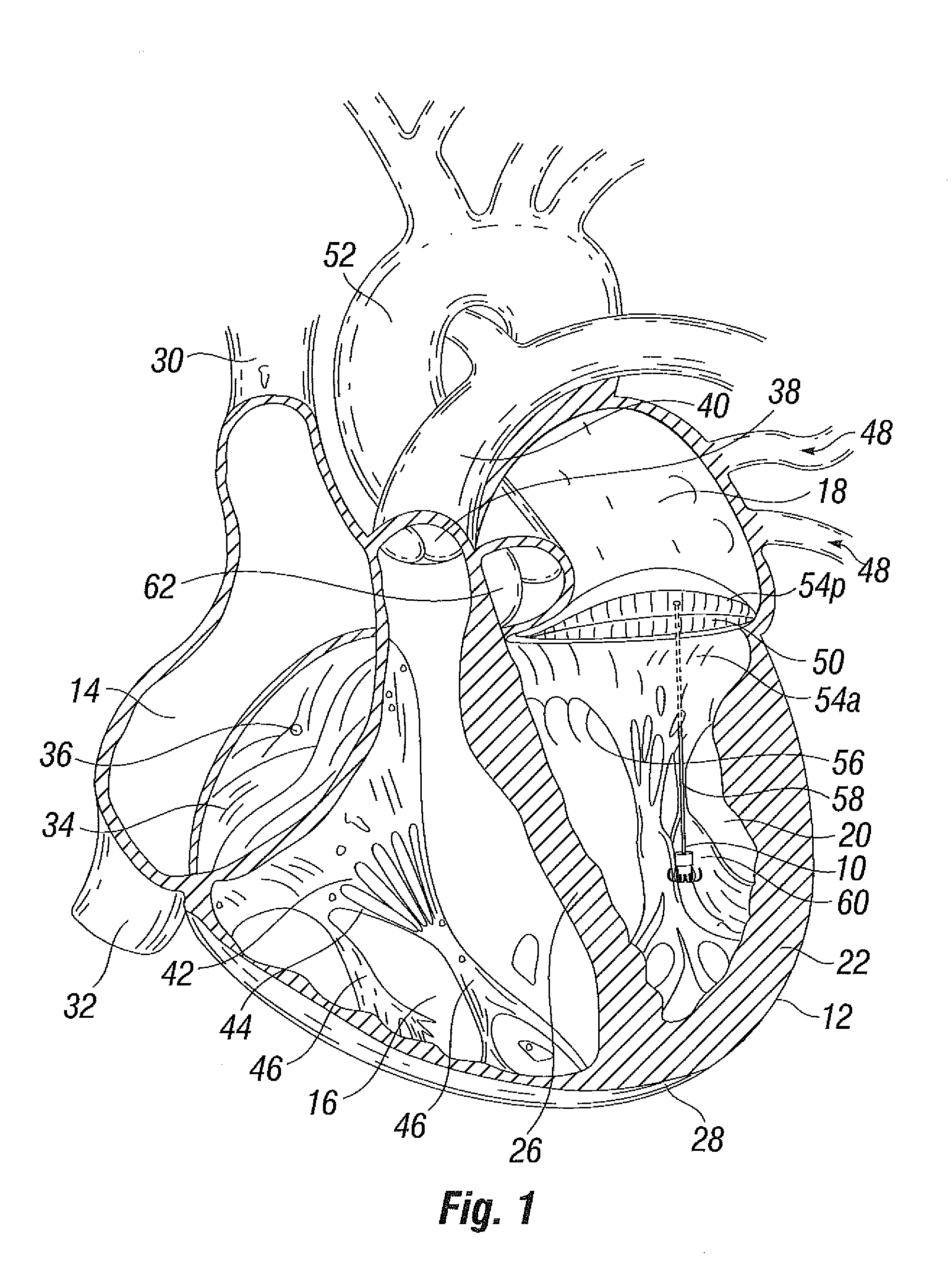
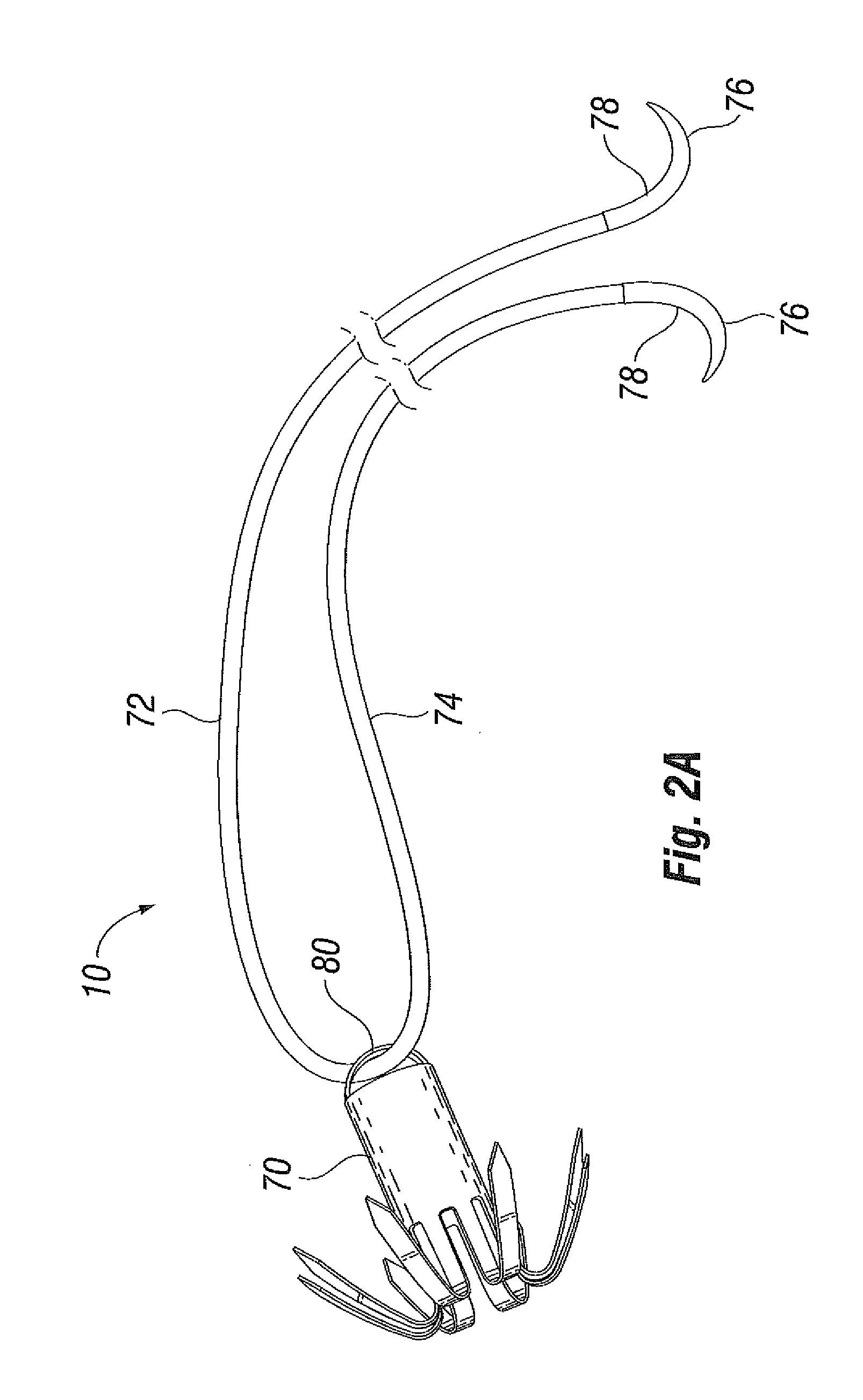
[0045]With reference to FIG. 1, a device 10 according to the invention is depicted deployed within a heart 12. The heart 12 has four chambers, known as the right atrium 14, right ventricle 16, left atrium 18, and left ventricle 20. In the particular embodiment depicted, the device 10 is deployed in the left ventricle 20. The heart 12 has a muscular outer wall 22, with an interatrial septum 24 (not visible in FIG. 1) dividing the right atrium 14 and left atrium 18, and a muscular interventricular septum 26 dividing the right ventricle 16 and left ventricle 20. At the base of the heart 12 is the apex 28.

[0046]Blood flows through the superior vena cava 30 and the inferior vena cava 32 into the right atrium 14 of the heart 12. The tricuspid valve 34, which has three leaflets 36, controls blood flow between the right atrium 14 and the right ventricle 16. The tricuspid valve 34 is closed when blood is pumped out from the right ventricle 16 to the lungs. Thereafter, the tricuspid valve 34 ...


PUM

PropertyMeasurementUnit
flexibleaaaaaaaaaa
tensionaaaaaaaaaa
volumeaaaaaaaaaa

Abstract

Devices and methods for treating or repairing a heart are disclosed. The device includes at least one anchor configured to engage tissue of a heart and a thread or other elongate member adapted to be coupled to the anchor and secured to heart tissue. The anchor may be attached to papillary muscle tissue and an elongate member may be attached to a valve leaflet for creating artificial chordae tendinae, thereby treating mitral valve prolapse. In another application, multiple anchors may be deployed within a ventricle and the threads pulled together for reducing dilation of a ventricle. A locking mechanism is provided for capturing and locking the threads together, thereby maintaining the ventricle in the reshaped condition.

Description

RELATED APPLICATIONS[0001]The present application is a division of co-pending U.S. utility patent application Ser. No. 12 / 031,490, filed Feb. 14, 2008 and entitled “Suture and Method for Repairing a Heart,” which claims priority from U.S. provisional patent application Ser. No. 60 / 889,921, filed on Feb. 14, 2007 and entitled “Suture and Method for Repairing a Heart,” the entire contents of each of which are expressly incorporated herein in their entirety.FIELD OF THE INVENTION[0002]The present invention relates generally to medical devices, and more particularly, to implantable medical devices for treating diseases, disorders, and malformations of a heartBACKGROUND OF THE INVENTION[0003]The function of the heart may be seriously impaired if any of the heart valves is not functioning properly. The heart valves may lose their ability to close properly due to disease or defects, such as (for example) by a mitral or tricuspid valve leaflet becoming partially or completely detached from ...

Claims


Application Information

Patent Timeline
Patent Type & Authority Applications(United States)
IPC IPC(8): A61F2/24
CPCA61B17/0467A61B17/0487A61B2017/00243A61B2017/003A61B2017/0409A61B2017/0417A61F2/24A61B2017/06057A61B2017/306A61F2/2457A61F2/2487A61F2002/0086A61F2210/009A61B2017/0464A61F2210/0004A61B17/0401A61B2017/0437A61B2017/0427A61F2/2466A61B17/00234A61B17/06166A61B2017/00004A61B2017/00876A61F2/0077
Inventor SOLEM, JAN OTTO
Owner EDWARDS LIFESCIENCES CORP
Why Eureka
Try Eureka