System for Cardiac MR & MR Cine Imaging Using Parallel Image Processing

a technology of image processing and parallel image processing, applied in the field of system for cardiac mr imaging, can solve the problems of relatively low signal noise ratio (snr), difficult to perform bh 3d psir lge imaging, and insufficient nullification of normal myocardium. , to achieve the effect of improving temporal and spatial image resolution

Active Publication Date: 2013-05-09
SIEMENS HEALTHCARE GMBH
View PDF5 Cites 9 Cited by
  • Summary
  • Abstract
  • Description
  • Claims
  • Application Information

AI Technical Summary

Benefits of technology

[0004]A system improves temporal and spatial image resolution compared to known 3D (three dimensional) LGE methods by whole heart dynamic LGE imaging utilizing different TIs at different phases throughout a cardiac cycle within a single breath hold. A system for cardiac MR imaging uses parallel image processing. An input processor receives a heart rate signal representing heart electrical activity. Multiple MR imaging RF coils individually receive patient heart image representative data sets. An MR image acquisition system, over multiple successive heart cycles, uses multiple MR imaging RF coils in gradient echo imaging a patient heart, synchronized with the heart rate sign

Problems solved by technology

The TI that is chosen and subsequently applied may not adequately null normal myocardium as 2D multi BH imaging proceeds over several minutes, due to gadolinium washout kinetics.
Phase-sensitive inversion recovery (PSIR) imaging minimizes the need to precisely null normal myocardium, but

Method used


Image

  • System for Cardiac MR & MR Cine Imaging Using Parallel Image Processing
  • System for Cardiac MR & MR Cine Imaging Using Parallel Image Processing
  • System for Cardiac MR & MR Cine Imaging Using Parallel Image Processing

Examples


Embodiment Construction

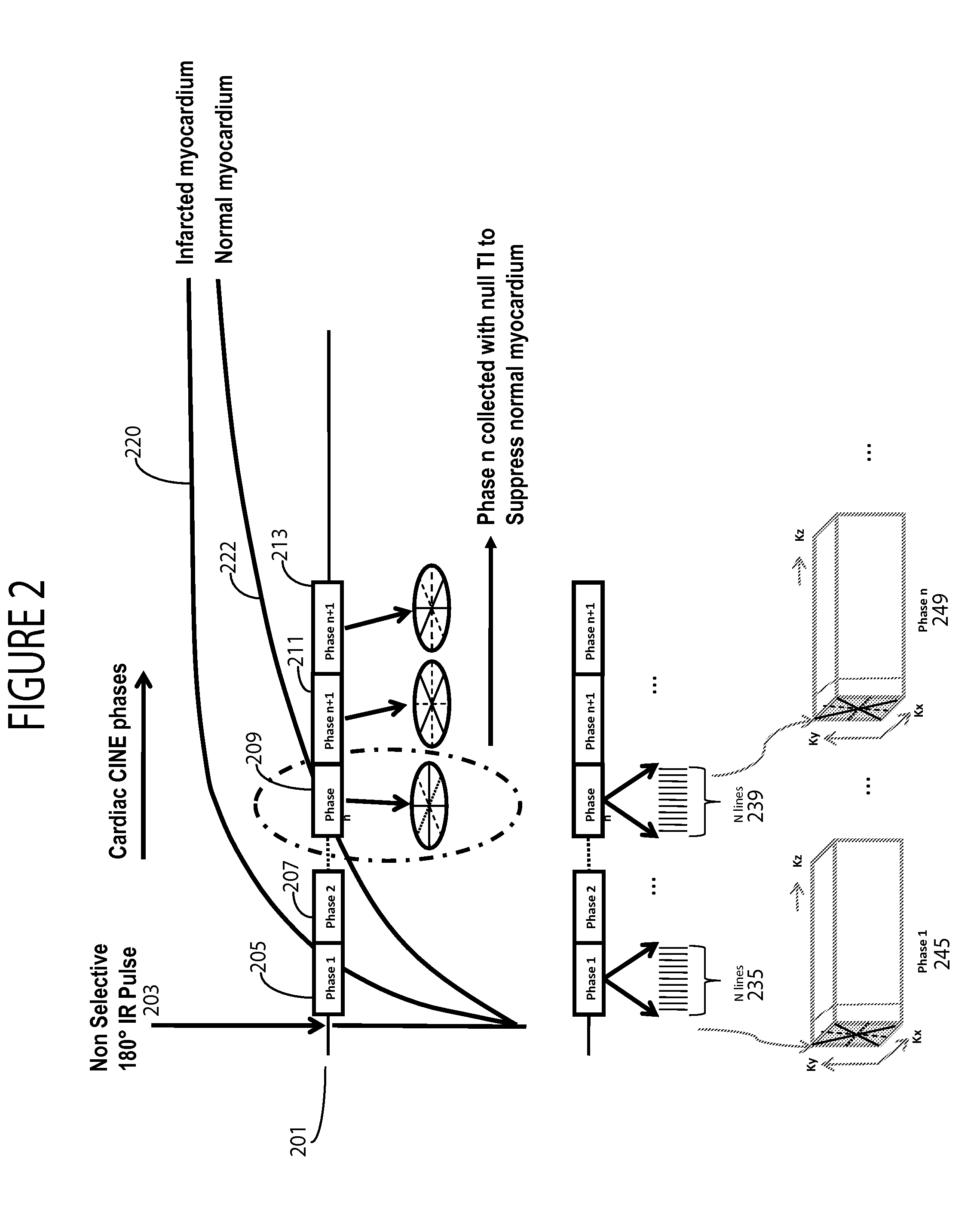
[0010]A system according to invention principles in one embodiment employs radial acquisition and a k-t SPARSE sensitivity encoding (SENSE) reconstruction (k-t RASPS) method that results in improved temporal and spatial resolution compared to known 3D LGE methods. The system performs whole heart dynamic 4D LGE imaging utilizing different TIs at different phases throughout an entire cardiac cycle within a single breath hold using 4D stack-of-star radial acquisition and k-t RASPS. This is performed without the use of a TI scout acquisition to acquire image data for determining a time at which an MR signal from normal myocardium is nulled.

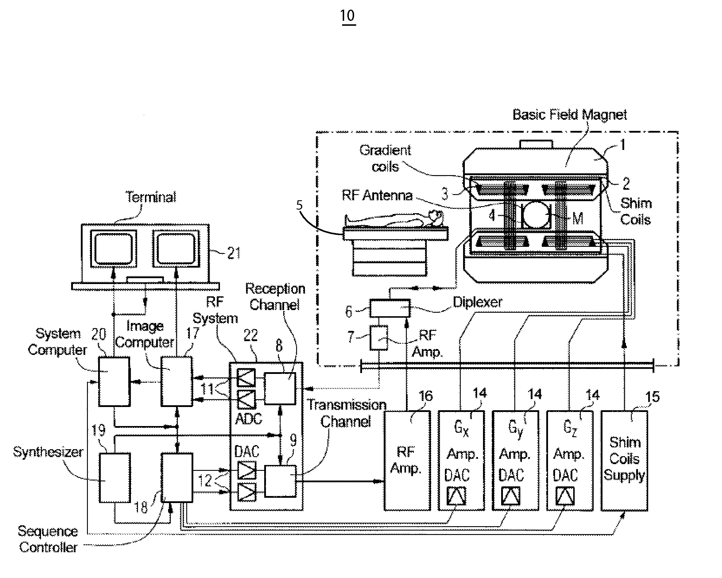
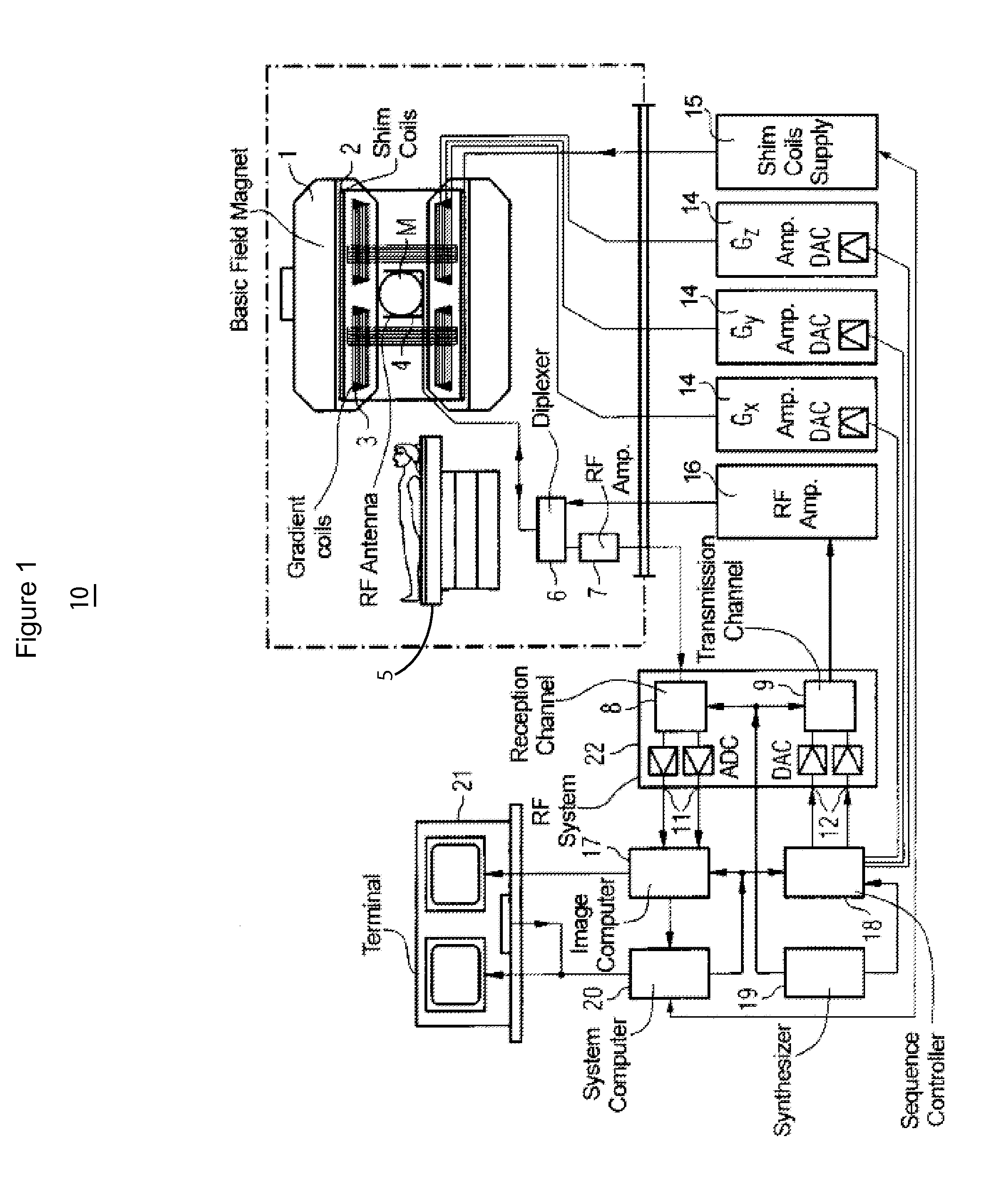
[0011]FIG. 1 shows a schematic block diagram of MR imaging system 10 for cardiac MR imaging using parallel image processing. A basic field magnet 1 generates a strong magnetic field, which is constant in time, for the polarization or alignment of the nuclear spins in the examination region of an object, such as, for example, a part of a human body to ...


PUM


Abstract

A system for cardiac MR imaging receives a heart rate signal representing heart electrical activity. The system, over multiple successive heart cycles, uses multiple MR imaging RF coils in gradient echo imaging a patient heart, synchronized with the heart rate signal and uses an inversion recovery pulse for inverting myocardium tissue MR signal for an individual heart cycle, to acquire, within multiple individual successive portions of an individual heart cycle, corresponding successive multiple patient heart images. An individual image of an individual heart cycle portion is derived from multiple heart image representative data sets comprising a reduced set of k-space data elements acquired using corresponding multiple coils of the RF imaging coils. An image generator generates an MR image of an individual heart cycle portion using the multiple heart image representative data sets comprising the reduced set of k-space data elements.

Description

[0001]This is a non-provisional application of provisional application Ser. No. 61 / 557,645 filed 9 Nov., 2011, by J. Xu.FIELD OF THE INVENTION[0002]This invention concerns a system for cardiac MR imaging using parallel image processing for generating MR images of individual heart cycle portions using multiple heart image representative data sets comprising a reduced set of k-space data elements in response to an RF inversion recovery pulse.BACKGROUND OF THE INVENTION[0003]Infarcted myocardium exhibits late gadolinium hyper-enhancement (LGE) and is usually imaged with an inversion recovery (IR) sequence. The inversion time (TI) is typically set to null normal myocardial signal in order to maximize luminance contrast between normal and infarcted myocardium. A multi-slice multi-planar 2D (two dimensional) approach requiring multiple breath-holds (BH) is commonly used in routine clinical practice. An additional BH TI scout is utilized to determine the precise null time of normal myocard...

Claims


Application Information

Patent Timeline
IPC IPC(8): A61B5/055A61B5/0402
CPCA61B5/0037A61B5/0044A61B5/7289A61B5/0402A61B5/055G01R33/5673G01R33/5602G01R33/5611G01R33/5616G01R33/56325G01R33/4822A61B5/318
Inventor XU, JIAN
Owner SIEMENS HEALTHCARE GMBH
Why Eureka
Try Eureka