Optogenetic probes for measuring membrane potential

a technology of optogenetic probes and membrane potentials, applied in the field of optogenetic probes for measuring membrane potentials, can solve the problems of slow electrophysiological experiment setup, inability to access deep buried tissues, and difficult to measure membrane potentials, so as to reduce the ion pumping activity of such proteins and reduce the ion pumping activity

Active Publication Date: 2013-08-29
PRESIDENT & FELLOWS OF HARVARD COLLEGE
View PDF0 Cites 44 Cited by
  • Summary
  • Abstract
  • Description
  • Claims
  • Application Information

AI Technical Summary

Benefits of technology

[0089]Exemplary nucleic acids and nucleic acid constructs are provided throughout this specification and their nucleic acid sequences are provided in the accompanying Sequence Listing. Typically, the modified microbial rhodopsins are modified to at least reduce the ion pumping activity of such proteins by mutating the proton acceptor proximal to the Schiff Base. However, the invention is not intended to be limited to these examples, as similar optical measurement is possible for any number of the existing microbial rhodopsin proteins. Any such protein may be used in the methods of the invention and any such protein may also be modified to reduce its ion pumping activity.

Problems solved by technology

In spite of its fundamental biological role, membrane potential is very difficult to measure.
Electrophysiological experiments are slow to set up, can only be performed on one or a few cells at a time, cannot access deeply buried tissues (e.g., in vivo), do not work for cells that are too small (e.g. bacteria) or are enclosed in a hard cell wall (e.g. yeast), or are motile (e.g., sperm) cannot be applied to long-term measurements, and usually damage or kill the cell under study.

Method used


Image

  • Optogenetic probes for measuring membrane potential
  • Optogenetic probes for measuring membrane potential
  • Optogenetic probes for measuring membrane potential

Examples


example 1

Proteorhodopsin as a Fluorescent Sensor of Protons

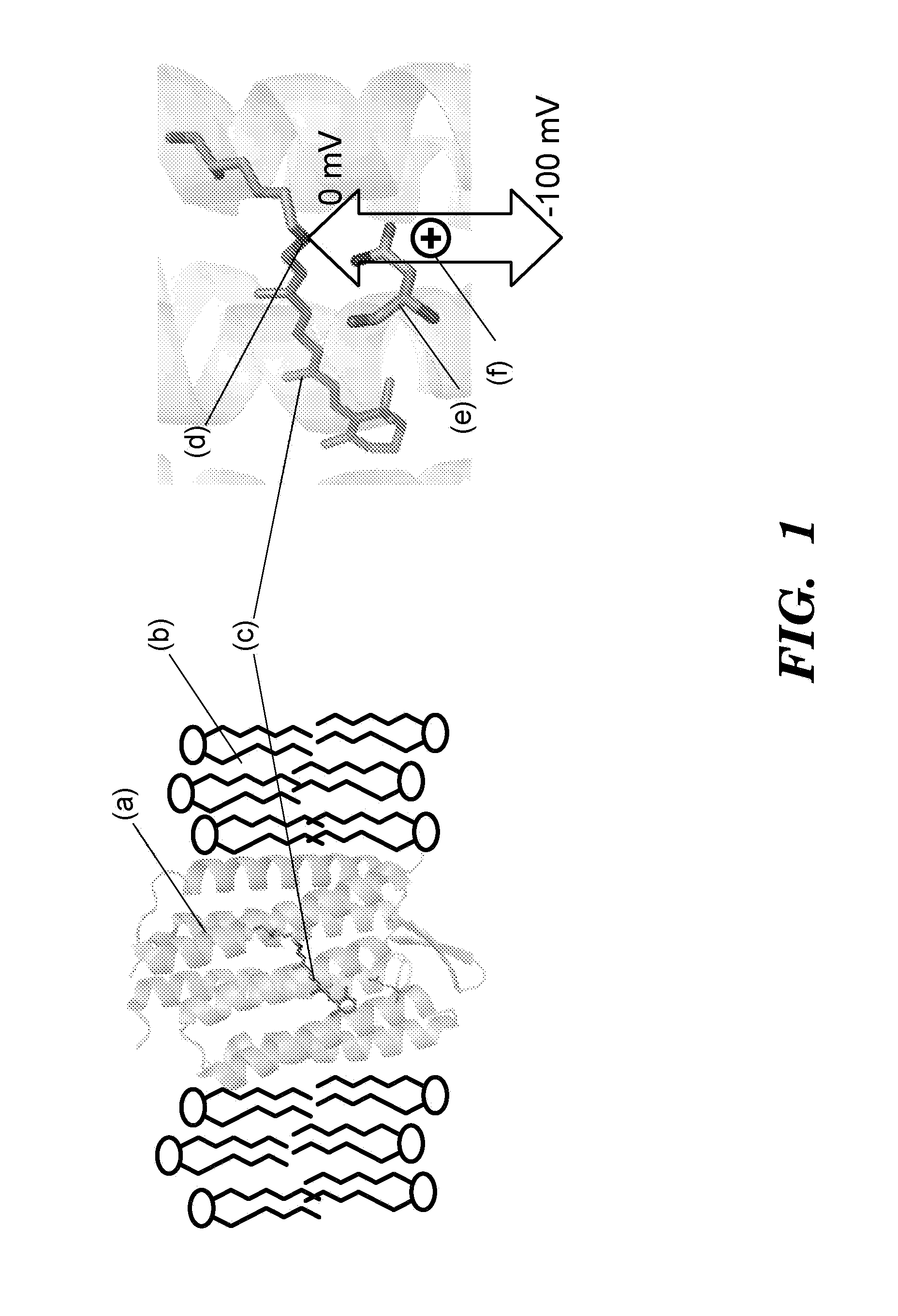
[0512]Proteorhodopsins are a large family of photoactive transmembrane proteins recently discovered in marine bacteria. Green-absorbing proteorhodopsin (GPR) is a light-driven proton pump that converts solar energy into a proton-motive force used by its host to power cellular machinery. The directional motion of a proton through GPR is accompanied by a series of dramatic color shifts in the protein. The approach of the study was to determine whether GPR could be run backward: could a transmembrane potential drive a proton through the protein and thereby alter its color? Described herein is a protein-based colorimetric indicator of membrane potential using such an approach.

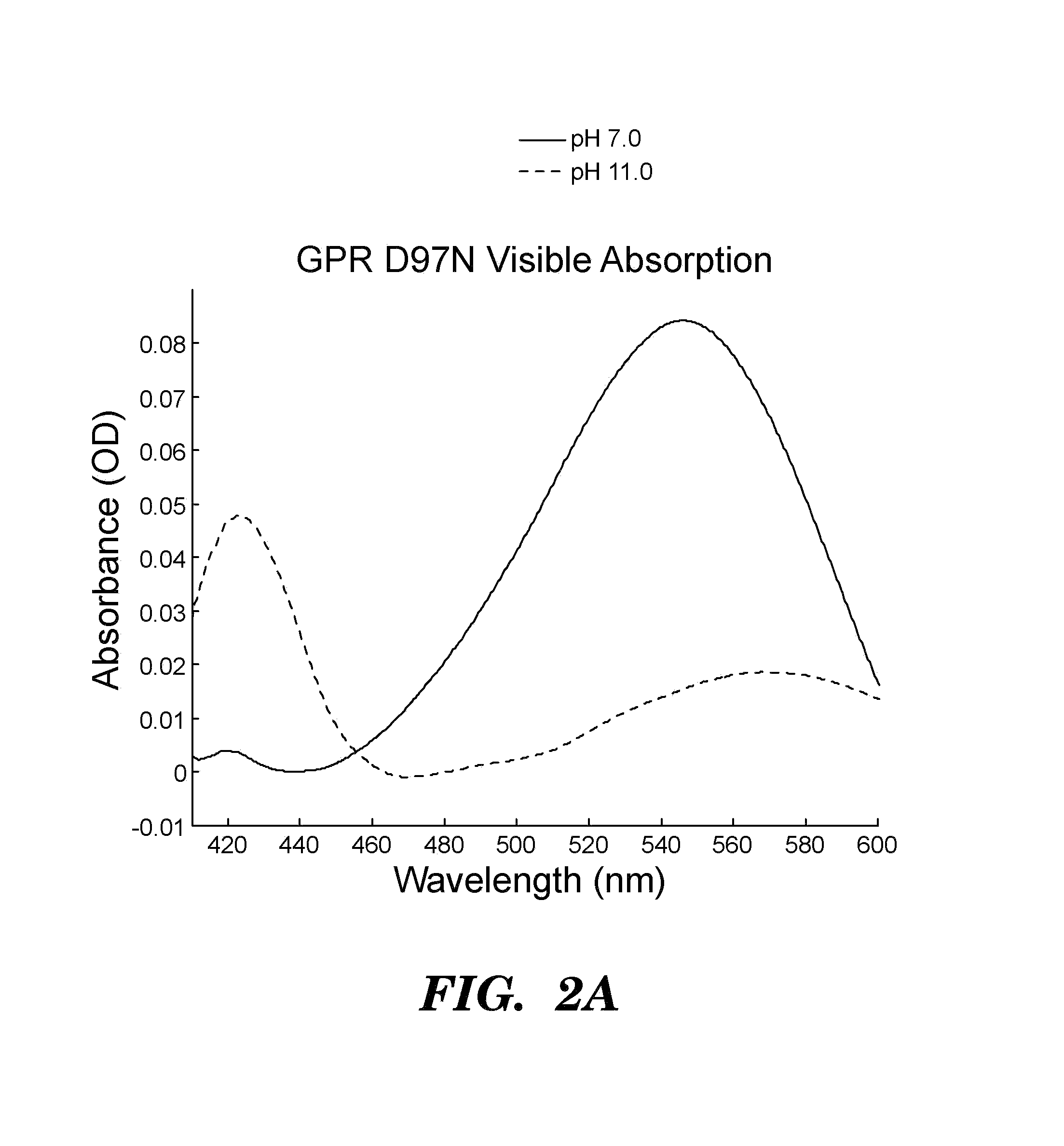
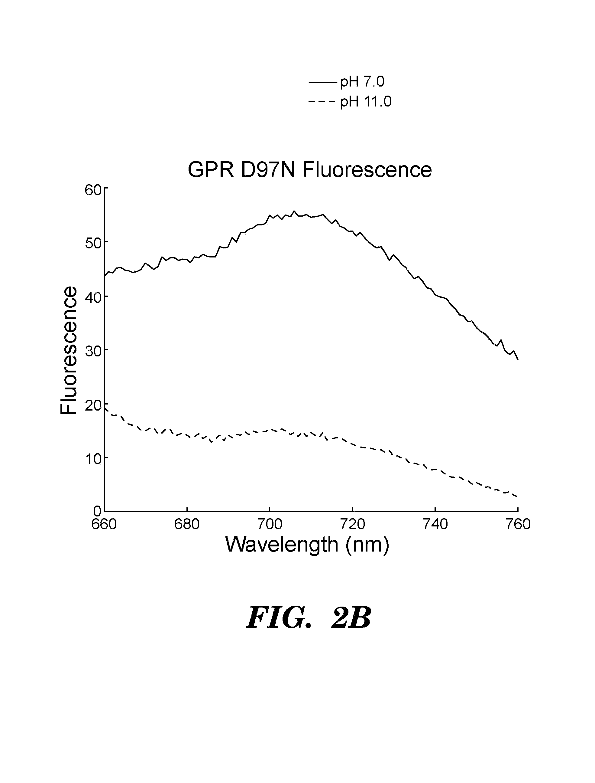
[0513]A covalently bound retinylidene is the visible chromophore of GPR. A Schiff base (SB) links the retinal to the ε-amino group of lysine 231. Early in the wild-type photocycle, a proton leaves the SB, causing a shift in the peak absorption from λmax=535 nm to λm...

example 2

Arch 3 can be Run Backward and Thereby Provide a Voltage Sensor

[0533]Archaerhodopsin 3 (Arch 3) from Halorubrum sodomense is a light-driven outward proton pump, capturing solar energy for its host (Ihara, K. et al. Evolution of the archaeal rhodopsins: evolution rate changes by gene duplication and functional differentiation. J. Mol. Biol. 285, 163-174 (1999)). Recently Arch 3 was expressed in mammalian neurons, wherein it enabled optical silencing of neural activity, and was shown to be minimally perturbative to endogenous function in the dark (Chow, B. Y. et al. High-performance genetically targetable optical neural silencing by light-driven proton pumps. Nature 463, 98-102 (2010)). We have now demonstrated that Arch 3 can be run backward: that a membrane potential can alter the optical properties of the protein, and thereby provide a voltage sensor that function through a mechanism similar to PROPS.

[0534]At neutral pH Arch 3 was pink, but at high pH the protein turned yellow (FIG...


PUM

PropertyMeasurementUnit
wavelengthaaaaaaaaaa
pHaaaaaaaaaa

Abstract

The invention provides methods, cells and constructs for optical measurement of membrane potential. These methods can be used in cells that are not accessible to presently available methods using electrodes. The methods can be directed to, for example, high-throughput drug screening assays to determine agents that can affect membrane potential of a target cell.

Description

CROSS-REFERENCE TO RELATED APPLICATIONS[0001]The present application claims benefit under 35 U.S.C. 119(e) of provisional applications No. 61 / 412,972, filed on Nov. 12, 2010, and 61 / 376,049, filed on Aug. 23, 2010, the contents of which are herein incorporated into this application by reference in their entirety.FIELD OF THE INVENTION[0002]The field of the invention relates to methods, constructs, and compositions for optically measuring electrical potential across a phospholipid bilayer.BACKGROUND[0003]Membrane-enclosed biological structures can support a voltage difference between the inside and the outside of the membrane. This voltage, also called membrane potential, serves a variety of biological functions, including carrying information (e.g., in neurons), acting as an intermediate in production of ATP (e.g., in bacteria and mitochondria), powering the flagellar motor (e.g., in bacteria), and controlling transport of nutrients, toxins, and signaling molecules across the cell m...

Claims


Application Information

Patent Timeline
Patent Type & Authority Applications(United States)
IPC IPC(8): G01N33/68C12N15/85C12N15/62
CPCC07K14/195G01N33/566G01N2333/726G01N33/6872C12N15/625C12N15/85C12N15/62C07K14/00C07K14/43595C07K2319/60C12N7/00C12N15/86C12N2740/15043C12N2750/14143C12N2810/00G01N21/6486
Inventor COHEN, ADAM E.KRALJ, JOEL M.DOUGLASS, ADAM D.
Owner PRESIDENT & FELLOWS OF HARVARD COLLEGE
Why Eureka
Try Eureka