Skeletal muscle regeneration using mesenchymal system cells

a technology of mesenchymal system cells and skeletal muscle, which is applied in the direction of skeletal/connective tissue cells, drug compositions, biocide, etc., can solve the problems of severe functional deficits, no improvement in muscle contraction force, and limited clinical application of mscs in skeletal muscle regeneration in the prior art, so as to accelerate the rehabilitation time, enhance skeletal muscle regeneration, and reduce pain in the subject

Active Publication Date: 2013-10-03
PLURI BIOTECH LTD
View PDF2 Cites 24 Cited by
  • Summary
  • Abstract
  • Description
  • Claims
  • Application Information

AI Technical Summary

Benefits of technology

[0018]The invention relates in a preferred embodiment to mesenchymal stem cells for use as a medicament for enhancing the functional recovery and / or structural regeneration of skeletal muscle damage or injury, preferably in addition to reducing pain associated with said muscle injury in a subject, whereby treatment with said MSCs occurs within 72 hours, preferably within 48 hours, more preferably within 24 hours or more preferably within 12 hours, after muscle damage or injury.
[0074]It is therefore particularly advantageous, in one embodiment of the present invention, that the MSCs are administered by local application to injured muscle and / or to the healthy tissue surrounding the damaged musculature, preferably within 0.5 mm to 100 mm, preferably between 0.5 mm to 50 mm or more preferably within 0.5 mm to 20 mm of the muscle damage. Also encompassed within the invention is the administration of cells 0.5, 1, 2, 3, 4, 5, 6, 7, 8, 9, 10, 12, 15, 20, 25, 30, 35, 40, 45, 50, 60, 70, 80, and / or 90 mm from the muscle injury. Administration of MSCs within these specific regions surrounding the muscle injury functions in synergistic combination with the surprising effect of enhanced cell migration, which is present immediately after injury, to facilitate the regenerative effect of the transplanted MSCs. Administration of cells within these particular ranges at the time points as described within the present application assists with the surprising effect that wound healing and functional muscle regeneration is enhanced beyond what would have been expected. These administration sites provide significant improvements in cell migration in comparison to administration at other sites or via other methods.

Problems solved by technology

Skeletal muscle trauma often leads to severe functional deficits due to insufficient regeneration of the musculature.
The clinical application of MSCs in skeletal muscle regeneration in the prior art is limited.
No improvement in muscle contraction forces could be found and the authors stated that MSCs were in fact currently unsuitable for the treatment of muscle dystrophies (Gang et al.
The one week delay between injury to the soleus muscle and local injection of MSCs adhered to the commonly accepted paradigm that the initial inflammatory phase, with a high activity of macrophages, would be disadvantageous for the putative integration and regenerative effect of the applied MSCs (Jarvinen, M. and Sorvari, T. Acta Pathol Microbiol Scand [A] 83, 259, 1975, Li, Y. and Huard, J. Am J Pathol 161, 895, 2002).
To our knowledge, previous work on skeletal muscular regeneration has provided no recommendation regarding application time point due to the lack of studies aimed at this question.
Previous reports, describing the transplantation of human umbilical cord blood cells into crushed gastrocnemius muscles of immunosuppressed mice or the transplantation of myoblasts into dystrophin deficient muscles after toxin injury could not demonstrate any therapeutic effect leading to the assumption that the immediate transplantation of progenitor cells is prone to a worse outcome because of the initial hostile microenvironment (Gang et al.
The general view found in the prior art is that MSCs transplanted directly after muscle trauma would not be able to exert their effect on regeneration because of the hostile microenvironment.
The prior art method of transplantation one week after injury presents multiple disadvantages regarding patient discomfort and risk.

Method used


Image

  • Skeletal muscle regeneration using mesenchymal system cells
  • Skeletal muscle regeneration using mesenchymal system cells
  • Skeletal muscle regeneration using mesenchymal system cells

Examples


example 1

Results of In Vivo Functional Muscle Testing

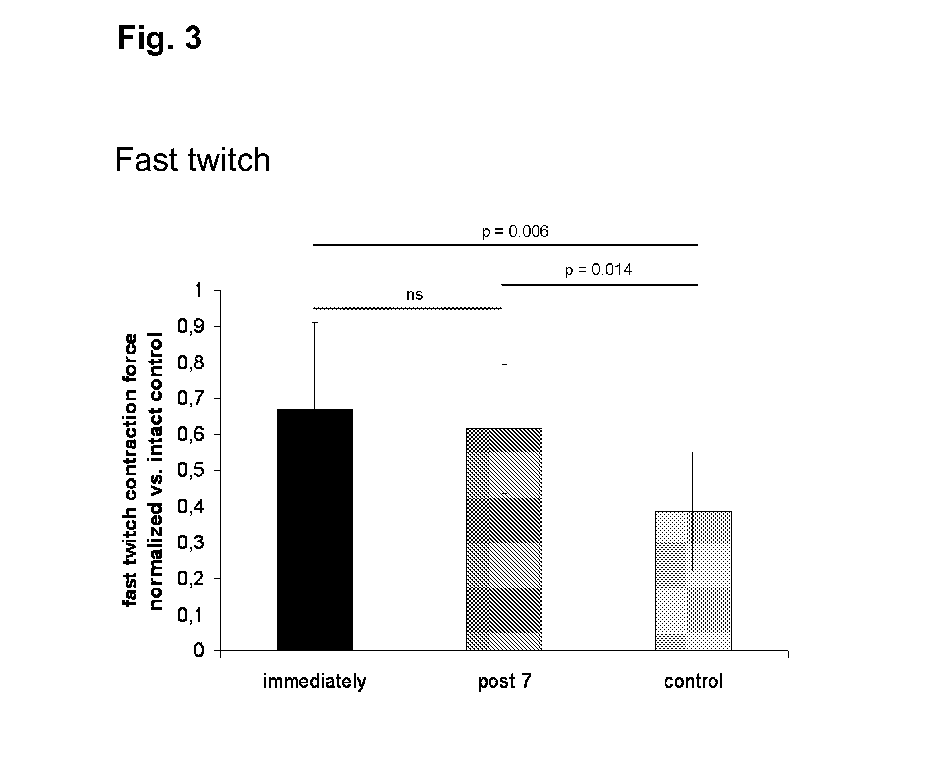
[0120]Fast twitch stimulation of the healthy right soleus muscles of all animals resulted in an average contraction force of 0.52±0.14 N. No decrease of successive contraction peaks was observed in uninjured or injured muscles irrespective of treatment. Stimulation with 9 mA and a frequency of 75 Hz at a duration of 3 seconds yielded tetanic contractions in all tested muscles reflecting their maximum contraction capacity. The latter amounted to 0.98±0.27 N in the uninjured soleus muscles. Tetanic maxima showed a continuous, linear decrease with successive stimulations in all muscles. This decrease could be described by a decline gradient, which was significantly different between the traumatized left and the non-traumatized right soleus muscles [−0.015±0.009 (left) vs. 0.031±0.014 (right), p-value<0.001]. The decline of the tetanic contractions was not influenced by cell therapy.

[0121]The ratio of fast twitch and tetanic forces represents ...

example 2

Results of Histological Analysis

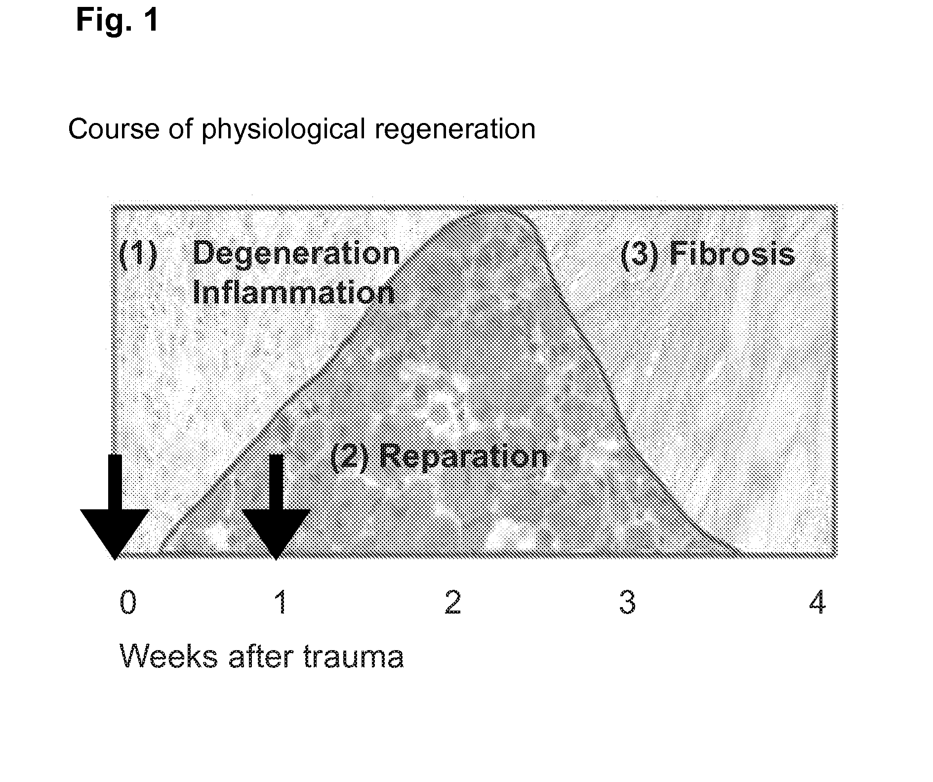
[0124]H&E stains of muscles harvested 3 days after injury still showed a pronounced inflammatory reaction throughout the soleus with the infiltration of predominantly macrophages, but also of neutrophils and lymphocytes. Most of the necrotic myofibers had already been removed and multiple groups of regenerating centronucleated fibers could be observed surrounded by loose connective tissue with the above described inflammatory infiltration. In muscles harvested 10 days after crush injury the inflammatory reaction had almost disappeared. The connective tissue showed a condensation towards a more dense type, although still with active fibroblastic nuclei.

[0125]Three days after both immediate and delayed transplantation, groups of GFP+ MSCs or single MSCs could be observed in the cryosectioned soleus muscles. Representative pictures can be seen in FIGS. 5A and B. The transplanted MSCs were found predominantly in the interstitium throughout the muscles, a ...


PUM

PropertyMeasurementUnit
timeaaaaaaaaaa
temperatureaaaaaaaaaa
tensionaaaaaaaaaa

Abstract

The present invention relates to a therapeutic substance and / or medicament and methods relating to the use of said substance and / or medicament for skeletal muscle regeneration using mesenchymal stem cells (MSCs) which can be applied directly or shortly after muscle damage or injury.

Description

[0001]The present invention relates to a therapeutic substance and / or medicament and methods relating to the use of said substance and / or medicament for skeletal muscle regeneration using mesenchymal stem cells (MSCs) which can be applied directly or shortly after muscle damage or injury.BACKGROUND OF THE INVENTION[0002]Skeletal muscle trauma often leads to severe functional deficits due to insufficient regeneration of the musculature. Approaches to reduce the collateral damage due to soft tissue trauma have until recently been predominantly conservative and based on preparing the muscular macro-structure to enable intrinsic healing mechanisms of the muscle. Such approaches include the traditional RICE approach (Rest Ice Compression Elevation) and the suturing of retracted muscle stumps, which allow the intrinsic regenerative capacities of the muscle to take effect. The central process of such intrinsic mechanisms is represented by the activation of satellite cells and their contrib...

Claims


Application Information

Patent Timeline
Patent Type & Authority Applications(United States)
IPC IPC(8): A61K35/28C12N5/0775A61K35/12
CPCA61K35/28A61K48/00C12N5/0675C12N5/0663A61K2035/124C12N5/0662C12N5/0664C12N5/0665C12N5/0666C12N5/0667C12N5/0668A61P21/00
Inventor DUDA, GEORGWINKLER, TOBIASMATZIOLIS, GEORGVON ROTH, PHILIPPPERKA, CARSTEN
Owner PLURI BIOTECH LTD
Why Eureka
Try Eureka