Method for operating an imaging diagnostic device and medical imaging system

a diagnostic device and imaging technology, applied in the field of imaging diagnostic devices and medical imaging systems, can solve the problems of affecting the comparability of measurements performed at different time points, and restrictions often apply to evaluation comparability, so as to reduce the variability of evaluation

Inactive Publication Date: 2013-10-10
SIEMENS AG
View PDF28 Cites 11 Cited by
  • Summary
  • Abstract
  • Description
  • Claims
  • Application Information

AI Technical Summary

Benefits of technology

[0015]Even though it is impossible to eliminate or compensate for all of the influences that adversely affect the comparability of quantitative measurements, it is possible to minimize the variability of the evaluation of diagnostic image data by technical measures. In the case of examinations that are performed using a contrast medium, the parameters relating to the contrast medium injection also play a role in this context. According to the application, parameter values which relate to the injection protocol governing the use of the contrast medium are automatically proposed accordingly. In this case, the proposal for the parameters relating to contrast medium use is based firstly on the current examination to be performed by the imaging diagnostic device and subsequent evaluation, and secondly on stored comparison data. A subsidiary application is understood generally to be an algorithm by which existing data can be evaluated automatically and suitable parameter values that are derived can be determined and at least proposed.

Problems solved by technology

The application takes as its point of departure the idea that the results of quantitative measurements, which are performed in the context of imaging diagnostics and can play a key role in the diagnosis, are dependent on numerous factors of influence, and this can adversely affect the comparability of measurements performed at different time points.
Even when the conditions governing the acquisition of image data remain the same, restrictions often apply to the comparability of evaluation results.

Method used


Image

  • Method for operating an imaging diagnostic device and medical imaging system
  • Method for operating an imaging diagnostic device and medical imaging system

Examples


Embodiment Construction

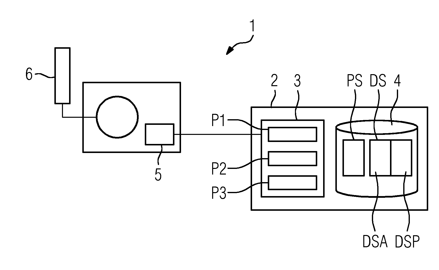
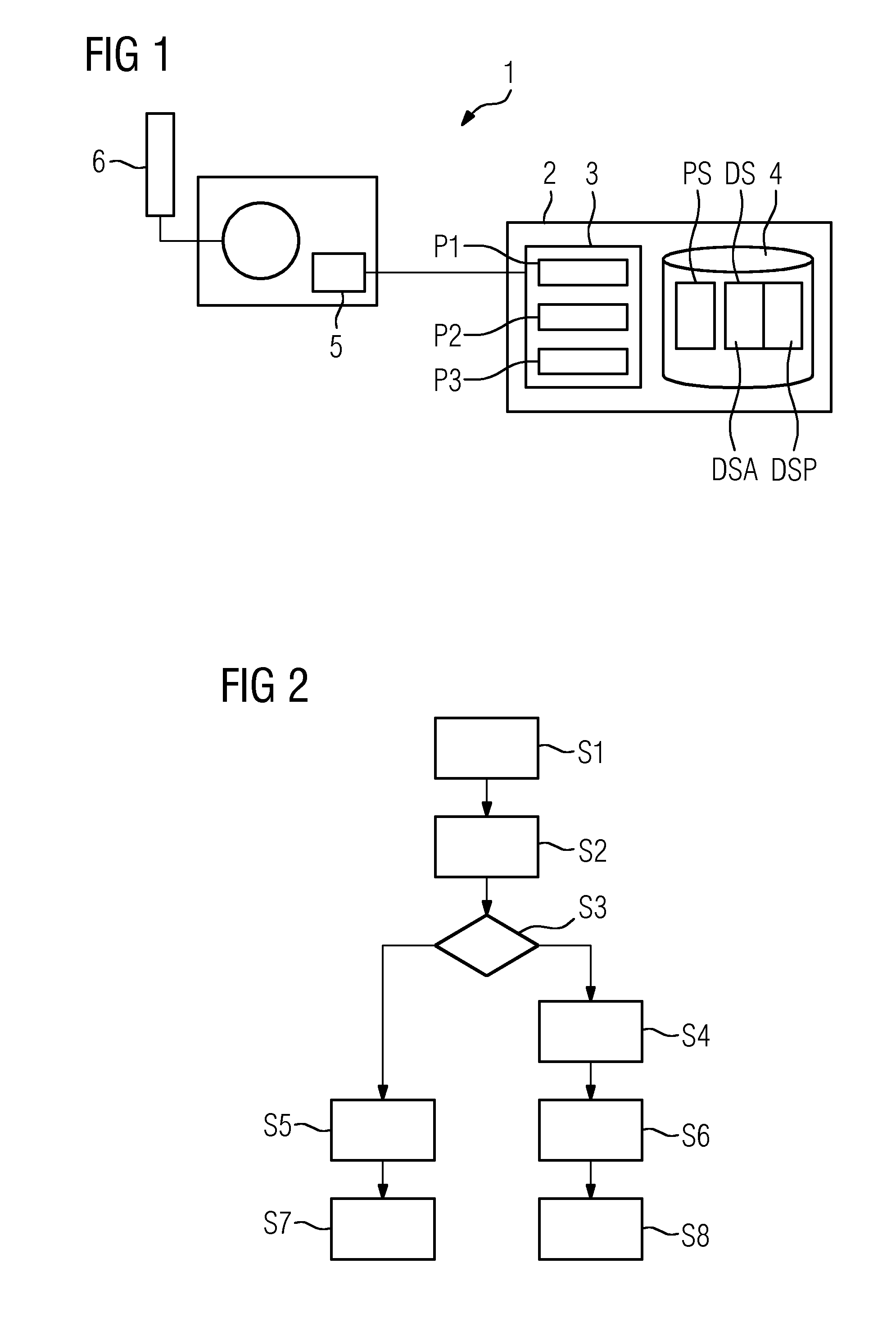
[0032]A medical diagnostic device 1, specifically a computed tomography system, which is merely outlined in FIG. 1 and whose principal function is described by the prior art cited in the introduction, has a data connection to a data processing system 2. The data processing system 2 can theoretically be realized as a single data processing device in the simplest case. However, the data processing system 2 is in fact designed as a data processing network which is connected to a radiology information system (RIS) or embedded in such a system.

[0033]The data processing system 2 comprises a computing unit 3 and a data store 4, wherein (as explained above) the schematic illustration according to FIG. 1 does not imply any hardware structures. The data store 4 has various storage areas, specifically a parameter store PS and an archive data store DS. The archive data store DS is in turn divided into a general data store DSA and a patient-specific data store DSP.

[0034]The computing unit 3 is d...


PUM


Abstract

A method for operating an imaging diagnostic device is proposed. Raw data is generated by the diagnostic device as a function of modality parameters while utilizing a contrast medium that is used in accordance with an injection protocol. Image data is generated from the raw data by image reconstruction as a function of reconstruction parameters. A quantitative measurement is performed in an image data evaluation by an analysis application. A subsidiary application automatically proposes a set of parameter values which is suited to the analysis application and relates to the injection protocol utilizing stored comparison data.

Description

CROSS REFERENCE TO RELATED APPLICATIONS[0001]This application claims priority of German application No. 10 2012 205 711.0 filed Apr. 5, 2012, which is incorporated by reference herein in its entirety.FIELD OF INVENTION[0002]The application relates to a method for operating a medical imaging diagnostic device, also referred to generally as a modality, such as a computed tomography system, and a medical imaging system for performing said method.BACKGROUND OF INVENTION[0003]DE 10 2010 043 849 B3 discloses a computed tomography system for determining and depicting the perfusion of the heart muscle on the basis of statistical CCTA recordings (coronary CT angiography). In this context, provision is made for a segmentation unit for segmenting the coronary arteries and the left-hand heart muscle in a CCTA image of the heart, a first simulation unit for simulating the blood flow through the coronary arteries, and a second simulation unit by which the local perfusion can be determined on the ...

Claims


Application Information

Patent Timeline
Patent Type & Authority Applications(United States)
IPC IPC(8): A61B6/00
CPCA61B6/481A61B6/5205A61B6/563A61B6/5217A61B6/032G16H50/30
Inventor SCHEUERING, MICHAELSOZA, GRZEGORZ
Owner SIEMENS AG
Why Eureka
Try Eureka