Methods and devices for modulating excitable tissue of the exiting spinal nerves

a technology of excitable tissue and spinal nerve, which is applied in the direction of spinal electrodes, internal electrodes, therapy, etc., can solve the problems of needing to recharge or explant the ipg, the device that utilizes a battery-powered or charge-storage component is no longer functional, and the associated wired leads, so as to reduce post-operative pain and reduce recovery time

Active Publication Date: 2013-11-21
MICRON MEDICAL LLC +1
View PDF5 Cites 95 Cited by
  • Summary
  • Abstract
  • Description
  • Claims
  • Application Information

AI Technical Summary

Benefits of technology

[0013]One of the advantages of this method is that the clinician may now provide a more permanent electrical stimulation directly to a targeted area that responds to a nerve block. Nerve blocks are often successfully used to temporarily relieve pain in areas that are not accessible with wired leads. Another advantage of the present invention is that it does not require a long row of electrodes to be implanted into the patient and then tested to determine the proper location for stimulation. Instead, the physician is able to determine this location through the application of one or more nerve blocks, and then follow-up the nerve block(s) with implantation of the wireless stimulation device in the precise location of the nerve block(s) that provide relief to the patient.
[0014]In yet another aspect of the invention, a method for treating post-operative pain by modulating excitable tissue includes performing a surgical procedure on the patient, such as a spinal fusion procedure (e.g., a TLIF), and then implanting a wireless stimulation device of the present invention in the location of the surgical procedure. An input signal is transmitted to the wireless stimulation device and converted into an electrical impulse sufficient to modulate nerves in or around the location of the surgical procedure. The application of electrical stimulation to a surgery site reduces post-operative pain and potentially decreases recovery time.
[0015]In another aspect of the present invention, a method for modulating excitable tissue includes advancing at least a distal portion of a wireless stimulation device through the intervertebral foramen into the Kambin's triangle and anchoring the distal portion of the device into the adjacent nearby exiting nerve. In a preferred embodiment, a wireless stimulation device is positioned near the intervertebral foramen opening and the wireless stimulation device is advanced through the lumen of the introducer and out of the distal opening of the lumen such that one or more electrodes pass through the intervertebral foramen opening. The lumen may be, for example, a cannula, spinal needle, endoscope or the like.
[0016]In certain embodiments, the distal portion of the wireless stimulation device may include at least one suture location. In this embodiment, the distal portion of the electrode lead is suture to tissue or bone near the Kambin's triangle, such as a facet joint, vertebral body, pedicle, annulus or a facet capsule. In other embodiments, the distal portion of the wireless stimulation device comprises one or more fixation prongs. In these embodiments, the method includes embedding the fixation prongs into tissue within Kambin's triangle. In yet another embodiment, the distal portion of the wireless stimulation device comprises screw-tips and the method includes rotating the screw-tips into the tissue within Kambin's triangle. The implantation step may further include visualizing the implanting process by fluoroscopy or through an endoscope.
[0017]In another aspect of the invention, a method for modulating excitable tissue includes advancing a distal portion of a wireless stimulation device through a sacral hiatus opening to place the device in the region of the sacral exiting nerve bundle. In a preferred embodiment, the distal portion of the wireless stimulation device is introduced through the Kambin's Triangle, and then advanced along the dorsal or ventral epidural space behind the spinal cord to a target region. Once the target region is reached, the distal portion of the wireless stimulation device is positioned and fixated in the intervertebral foraminal space so that the electrical field of conduction remains in close proximity to the exiting nerve root or nerve ganglion. An electrical impulse is applied through the electrodes sufficient to modulate one or more nerves within the exiting nerve root or nerve ganglion.
[0018]In another aspect of the invention, a device for modulating excitable tissue in a patient's body comprises a lead including one or more electrodes, circuitry and a receiving antenna, all of which are fully encapsulated in one self contained device without a connector or wires to a power source. The wireless stimulation device is sized and configured for placement adjacent to or near a target site at an exiting nerve root or a nerve ganglion. The receiving antenna is configured to receive an input signal containing electrical energy and waveform parameters through radiative coupling from a transmitter located outside of the patient's body and the circuitry is configured to generate an electrical impulse from the input signal sufficient to modulate a nerve or a nerve ganglion at the target site.

Problems solved by technology

Traditional wired leads have several disadvantages, including: a large surgical pocket to house the implantable pulse generator with a battery or charge storage component; extensions and connectors between the IPG and the proximal end of the lead that are housed under the skin, and, a need to recharge or explant the IPG.
Having the IPG tethered to the IPG within the patient's body is a disadvantage because this connection can cause lead migration and the possibility for loss of therapy from a disconnection from the IPG exists and has plagued the industry.
However, devices that utilize a battery-powered or charge-storage component are no longer functional once the battery cannot be recharged or charge cannot be stored.
Wired leads are associated with numerous failure modes, including, for example, mechanical dislodgement due to motion, acceleration and impingement of the lead electrode assembly, infection and uncomfortable irritation.
In particular, longitudinal movement of the lead can move the surface electrodes, making them bear on a different portion of the spinal cord, no longer treating the pain.
A transverse or side-to-side movement of the lead can have the same effect, and can also move the electrodes further away from the spinal cord, weakening the signal and / or requiring greater power to reach the targeted nerves.
Increased power consumption can decrease battery life, which may require more frequency surgical replacement of the implanted battery.

Method used


Image

  • Methods and devices for modulating excitable tissue of the exiting spinal nerves
  • Methods and devices for modulating excitable tissue of the exiting spinal nerves
  • Methods and devices for modulating excitable tissue of the exiting spinal nerves

Examples


Embodiment Construction

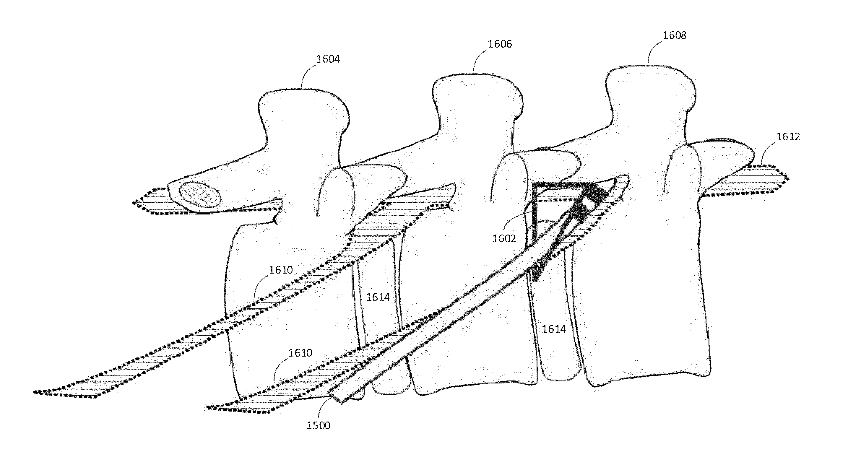
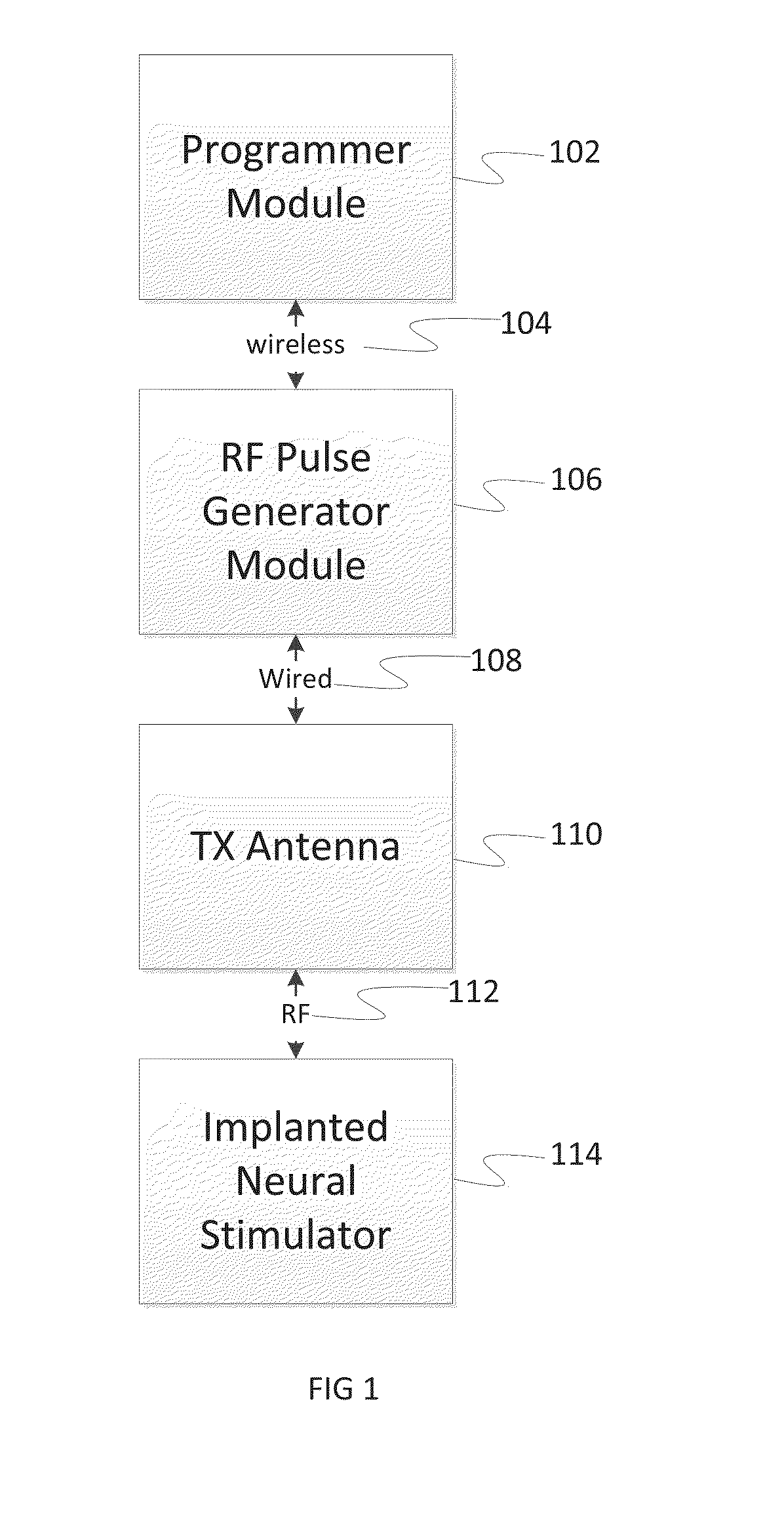
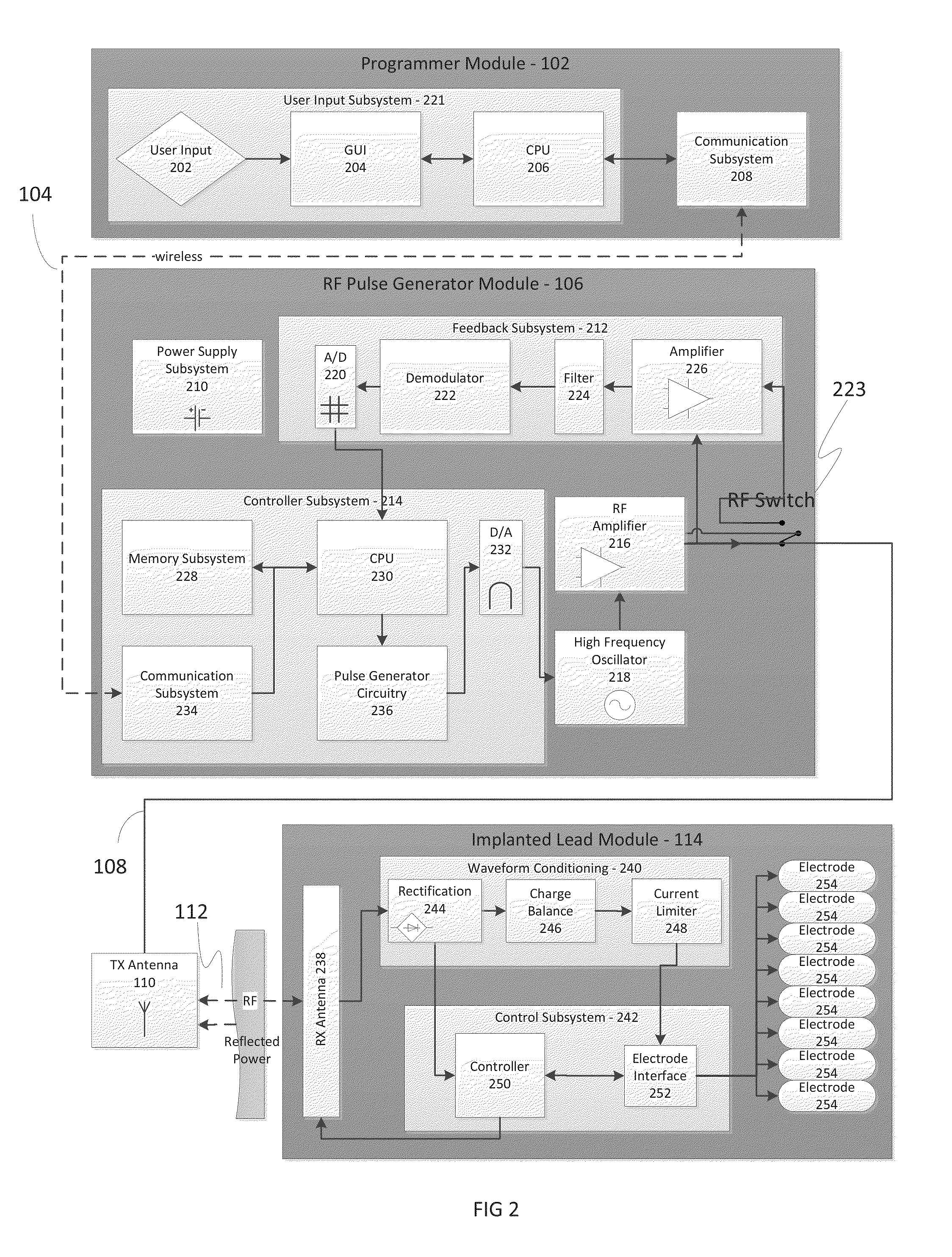
[0060]In various implementations, systems and methods are disclosed for applying one or more electrical impulses to targeted excitable tissue, such as nerves, for treating chronic pain, inflammation and / or other disorders. In certain embodiments, a wireless stimulation device may be used to send electrical energy to targeted nerve tissue by using remote radio frequency (RF) energy with neither cables nor inductive coupling to power the passive implanted wireless stimulation device. The targeted nerves can include, but are not limited to, the spinal cord and surrounding areas, including the dorsal horn, dorsal root ganglion, the exiting nerve roots, nerve ganglions, the dorsal column fibers and the peripheral nerve bundles leaving the dorsal column and brain, such as the vagus, occipital, trigeminal, hypoglossal, sacral, coccygeal nerves and the like.

[0061]The wireless stimulation device of the present invention can include an implantable lead body that includes an enclosure that hou...


PUM


Abstract

A method for modulating nerve tissue in a body of a patient includes implanting a wireless stimulation device in proximity to a dorsal root ganglion or an exiting nerve root such that an electrode, circuitry and a receiving antenna are positioned completely within the body of the patient. An input signal containing electrical energy and waveform parameters is transmitted to the receiving antenna(s) from a control device located outside of the patient's body via radiative coupling. The circuitry within the stimulation device generates one or more electrical impulses and applies the electrical impulses to the dorsal root ganglion or the exiting nerve roots through the electrode.

Description

CROSS-REFERENCE TO RELATED APPLICATIONS[0001]This application claims the benefit of priority of U.S. Provisional Patent Application No. 61 / 649,834 filed May 21, 2012 and is related to the following commonly-assigned co-pending patent applications: U.S. patent application Ser. Nos. 13 / 551,050 (filed Jul. 17, 2012), 13 / 562,221 (filed Jul. 30, 2012) and 13 / 584,618 (filed Aug. 13, 2012) and PCT Patent Application Nos. PCT / US2012 / 023029 (filed Jan. 27, 2012), PCT / US2012 / 032200 (filed Apr. 4, 2012), PCT / US2012 / 048903 filed Jul. 30, 2012), PCT / US2012 / 050633 (filed Aug. 13, 2012) and PCT / US2012 / 055746 (filed Sep. 15, 2012). The complete disclosures of all of the above patent applications are hereby incorporated by reference in their entirety for all purposes.BACKGROUND OF THE INVENTION[0002]The field of the present invention relates generally to the delivery of energy impulses (and / or fields) to bodily tissues for therapeutic purposes and more specifically to devices and methods for modulat...

Claims


Application Information

Patent Timeline
Patent Type & Authority Applications(United States)
IPC IPC(8): A61N1/05A61N1/372
CPCA61N1/0558A61N1/37229A61N1/025A61N1/36125A61N1/36171A61N1/3787
Inventor PERRYMAN, LAURA TYLERANDRESEN, CHADKING, GARYYEUNG, ANTHONY
Owner MICRON MEDICAL LLC
Why Eureka
Try Eureka