Method and apparatus for accelerated phase contrast magnetic resonance angiography and blood flow imaging

a phase contrast and magnetic resonance angiography technology, applied in the direction of nmr measurement, instruments, diagnostic recording/measuring, etc., can solve the problems of affecting the accuracy of nmr measurement, long scan time, and limited spatial and temporal resolution, so as to increase image processing speed and high accuracy in reconstructed images

Inactive Publication Date: 2014-02-27
BETH ISRAEL DEACONESS MEDICAL CENT INC
View PDF7 Cites 22 Cited by
  • Summary
  • Abstract
  • Description
  • Claims
  • Application Information

AI Technical Summary

Benefits of technology

[0016]In addition, the apparatus and method of the present invention process magnetic resonance image (MRI) data to generate and output a reconstructed image of bodily fluid flow, such as blood flow, in the living subject with increased image processing speed and having high accuracy in the reconstructed images. The apparatus includes an input device, a processor, and an output device. The input device receives the MRI data of the fluid flow in the living subject from an MRI device having a plurality of coils, and generates selected image data from received inputs. The processor operates predetermined software, receives the selected image data, and applies a compressed sensing (CS) reconstruction method to the image data to generate reconstructed images from the image data. The output device outputs the reconstructed image of the fluid flow in the living subject.

Problems solved by technology

However, despite the potential of PC CMR as an alternative to Doppler ultrasound for evaluation of these indices, its accuracy is impacted by several limitations including background offset, eddy currents, and long scan time.
One of the challenges of PC CMR is the long scan time which limits spatial and temporal resolution.
While k-t approaches could surpass the acceleration that can be achieved using acceleration only in the spatial domain, k-t approaches could also cause temporal blurring.
However, the method of King does not use sparsifying in the complex difference domain, and the method only related to use of CS in addition to parallel imaging for angiography.
The method of King uses CD images by combining all of such CD images to obtain one flow weighted image, which does not allow for extraction of flow measurements.

Method used


Image

  • Method and apparatus for accelerated phase contrast magnetic resonance angiography and blood flow imaging
  • Method and apparatus for accelerated phase contrast magnetic resonance angiography and blood flow imaging
  • Method and apparatus for accelerated phase contrast magnetic resonance angiography and blood flow imaging

Examples


Embodiment Construction

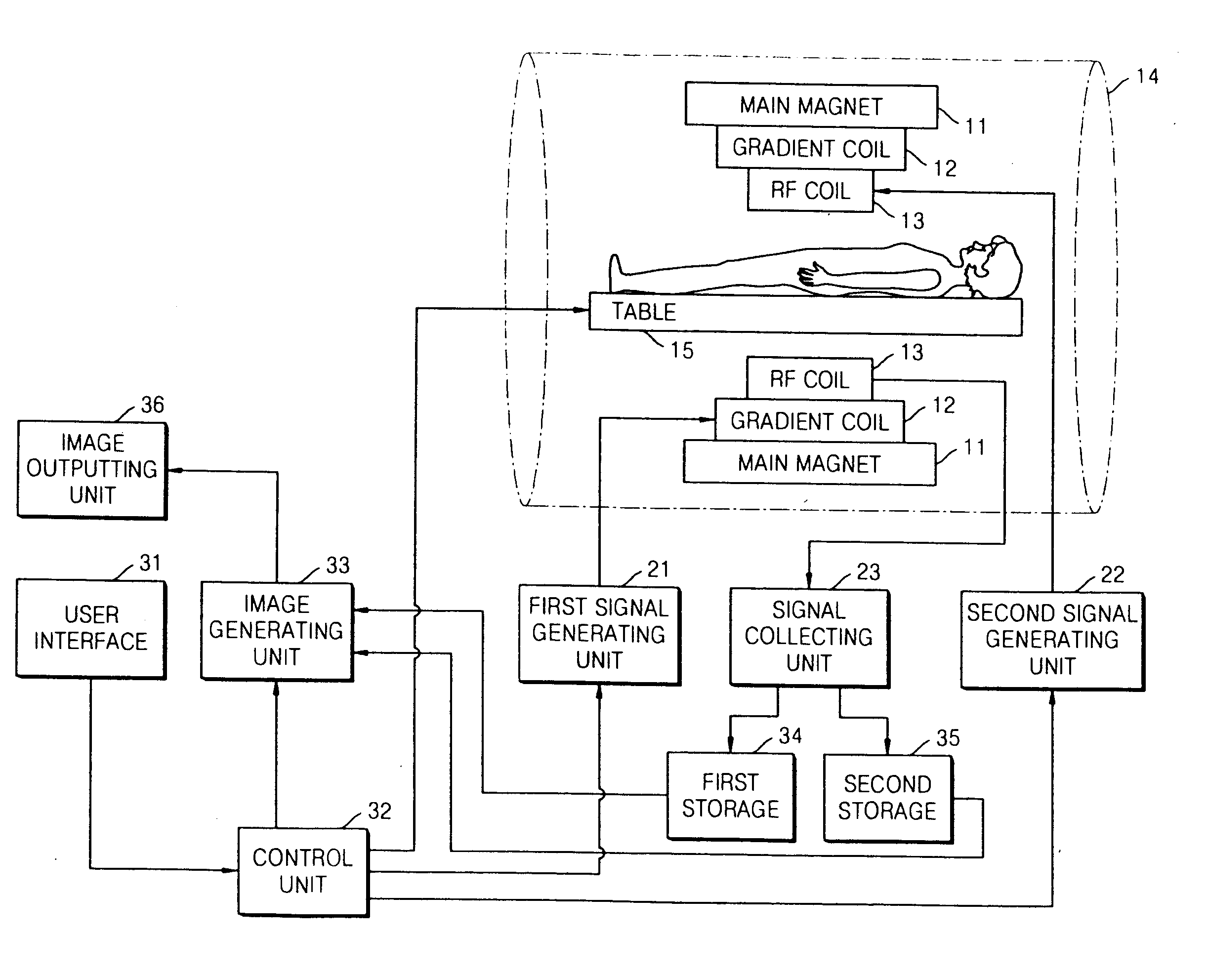
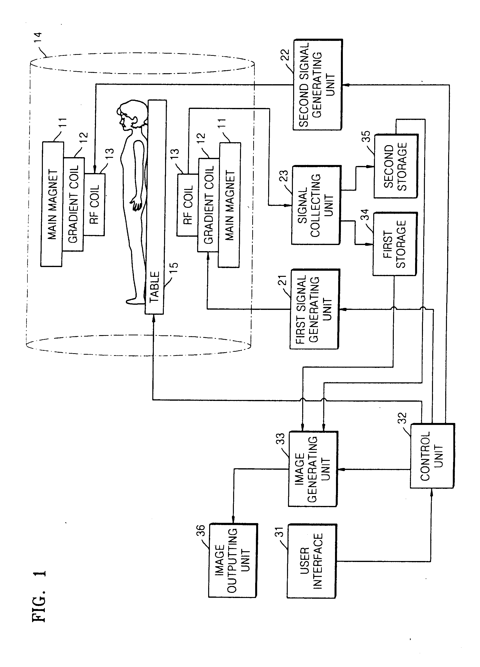
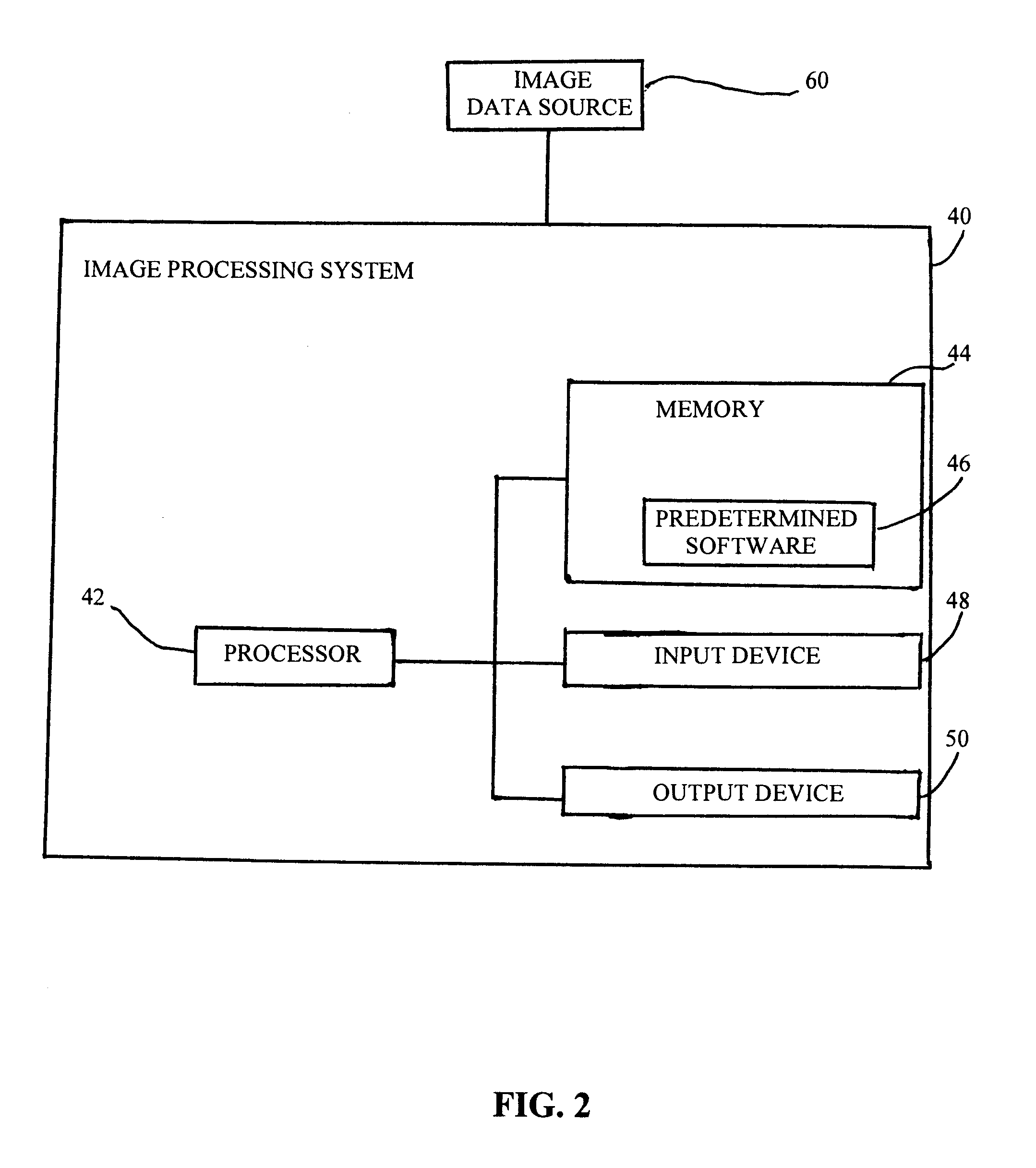
[0026]Hereinafter, preferred embodiments of the present invention will be described with reference to the accompanying drawings. In the following description, a detailed explanation of known related functions and constructions may be omitted to avoid unnecessarily obscuring the subject matter of the present invention. This invention may, however, be embodied in many different forms and should not be construed as limited to the exemplary embodiments set forth herein. The same reference numbers are used throughout the drawings to refer to the same or like parts. Also, terms described herein, which are defined considering the functions of the present invention, may be implemented differently depending on user and operator's intention and practice. Therefore, the terms should be understood on the basis of the disclosure throughout the specification. The principles and features of this invention may be employed in varied and numerous embodiments without departing from the scope of the in...


PUM


Abstract

An apparatus and method process magnetic resonance image (MRI) data and other data from a subject, including image data corresponding to at least one of angiographic data, four-dimensional blood flow, neurological blood flow, abdominal blood flow, and peripheral blood flow in the subject, and applies a compressed sensing (CS) reconstruction method utilizing a complex difference of the image data as a sparsifying transform for imaging of at least one of blood flow and magnetic resonance angiography to output a reconstructed image of the blood flow and magnetic resonance angiography, in the subject with increased processing speed and having high accuracy. The apparatus receives the MRI data of the fluid flow from an MRI device. The processor operates predetermined software, receives the MRI data, and applies the CS reconstruction method to generate the reconstructed image. An output device outputs the reconstructed image of the fluid flow.

Description

BACKGROUND OF THE INVENTION[0001]1. Field of the Invention[0002]The present invention relates to accelerated data acquisition for magnetic resonance imaging, and more particularly, to a compressed sensing reconstruction apparatus and method for imaging of the blood flow and angiography in the human body.[0003]2. Description of the Related Art[0004]Phase contrast (PC) cardiac magnetic resonance (CMR) imaging is widely used in the prior art for the clinical in-vivo assessment of blood flow in the diagnosing of various diseases and the depiction of the vessels in the body; for example, as described in Nayler et al., “Blood flow imaging by cine magnetic resonance”, J Comput Assist Tomogr 1986; 10(5):715-722; Pelc et al., “Phase contrast cine magnetic resonance imaging”, Magn Reson Q 1991; 7(4):229-254; and Rebergen et al., “Magnetic resonance measurement of velocity and flow: technique, validation, and cardiovascular applications”, Am Heart J 1993; 126(6):1439-1456.[0005]Through-plane a...

Claims


Application Information

Patent Timeline
Patent Type & Authority Applications(United States)
IPC IPC(8): G01R33/56G06K9/36
CPCG01R33/56316A61B5/055A61B5/0263G01R33/5611G06F18/2136G06T1/00
Inventor KWAK, YONGJUNNEZAFAT, REZA
Owner BETH ISRAEL DEACONESS MEDICAL CENT INC
Why Eureka
Try Eureka