

Compositions and Methods for Treating Brain Tumors
a brain tumor and composition technology, applied in the field of brain tumor compositions and methods, can solve the problems of frequent ineffective chemotherapy treatment, and achieve the effects of decreasing brain tumor deposition, reducing brain tumor cell proliferation, and reducing brain tumor vascularity
- Summary
- Abstract
- Description
- Claims
- Application Information
AI Technical Summary
Benefits of technology
Problems solved by technology
Method used
Image

Examples
example 1
Cell Cultures and Treatment Conditions
[0139]Human glioblastoma (U87MG and T98) tumor cells (Tumorbank DKFZ Heidelberg, Germany) were cultured in DMEM medium with 10% FCS at 37° C. with 5% CO2 and 95% humidity. GBM CSLCs were isolated from a human glioblastoma surgical sample. (See Galli et al. Can Res. 2004; 64: 7011-7021) Glioblastoma CSLCs were maintained in their undifferentiated state using Neurobasal Media (Life Technologies GmbH, Frankfurt, Germany), supplemented with epidermal growth factor (10 μg / 500 mL media) and fibroblastic growth factor (10 μg / 500 mL media), sodium pyruvate, glutamine, B27, non-essential amino acids and penicillin / streptomycin (Gibco, Grand Island, N.Y.). GBM CSLCs displayed typical characteristics of stem cells as they grew as neurospheres, had increased expression of CD133 and nestin, and were highly tumorigenic after implantation in SCID-beige mice.
[0140]The anti-CTGF antibody, CLN-1, (FibroGen Inc., San Francisco, Calif., USA), was reconstituted in w...
example 2
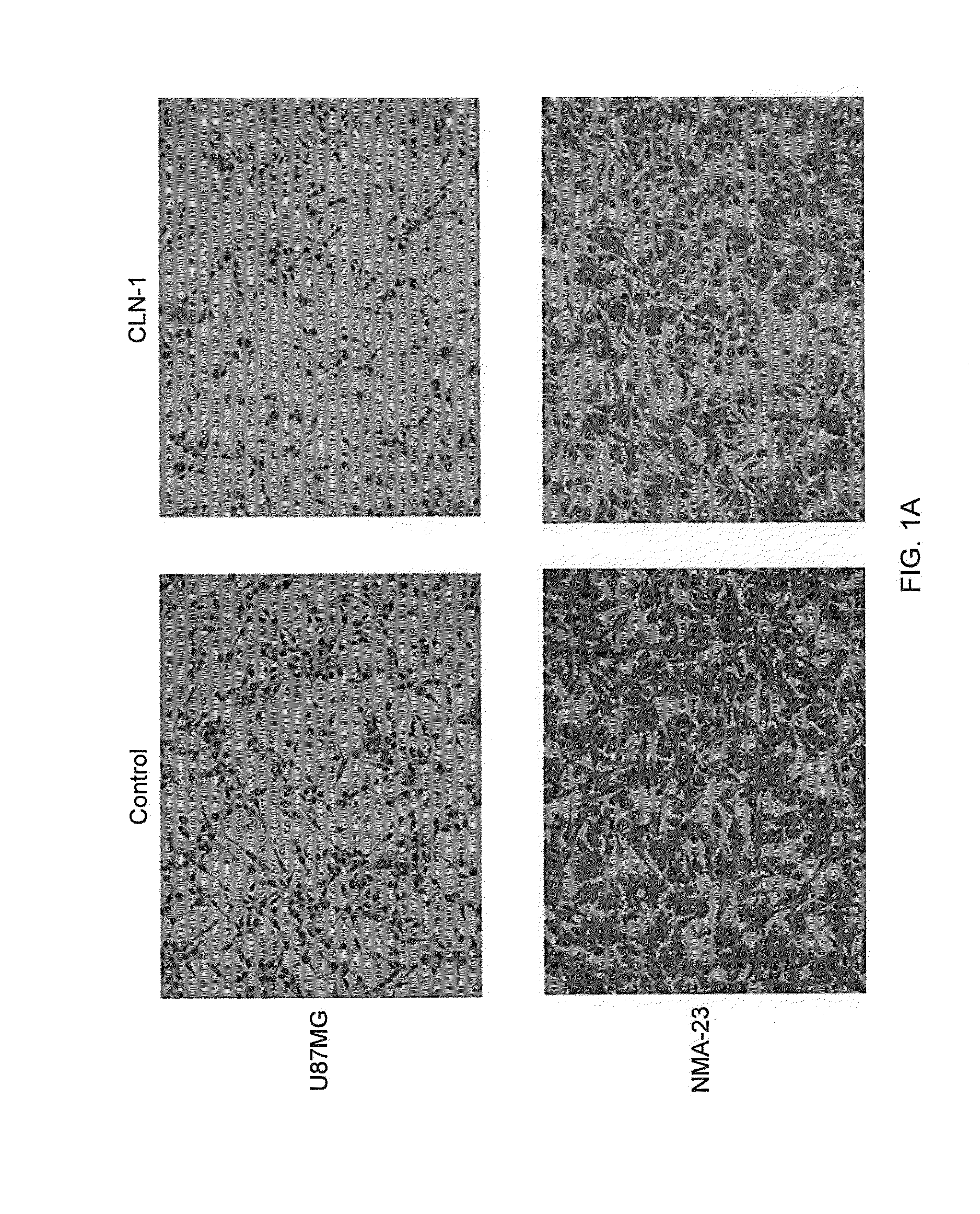
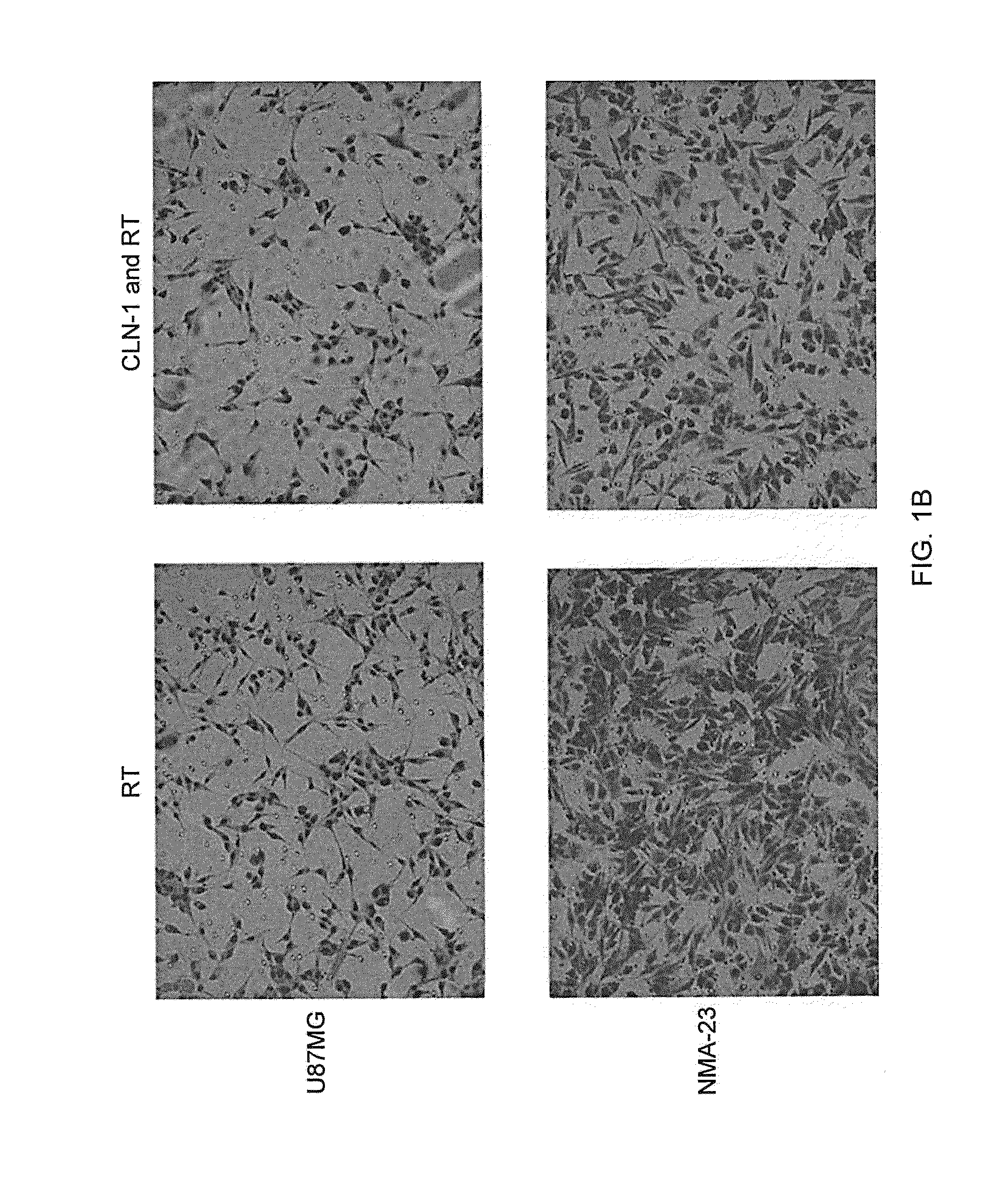
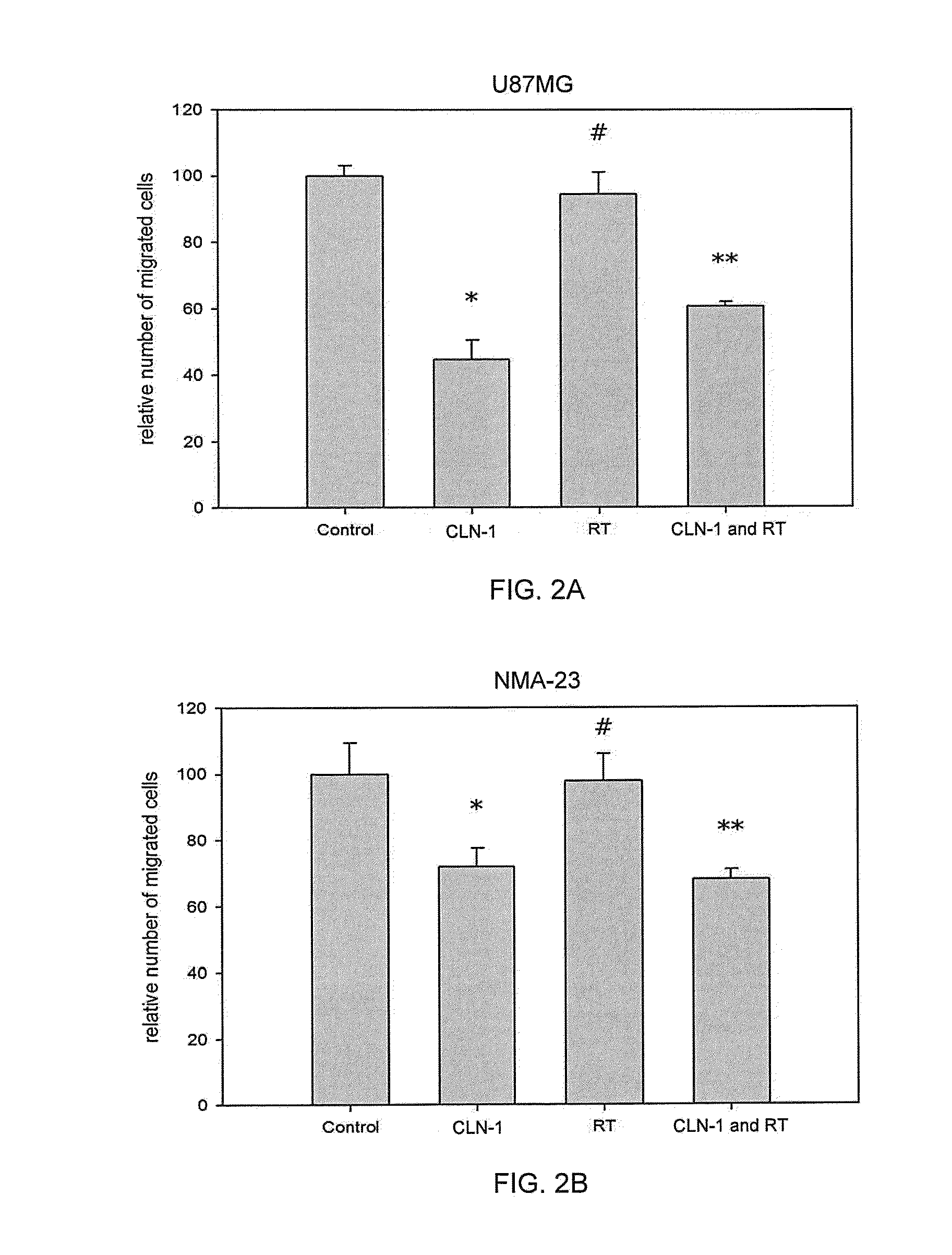
Inhibition of Glioma Cells Migratory Ability with Anti-CTGF Antibody Treatment
[0141]A transwell migration assay was used to study the effect of the anti-CTGF antibody (CLN-1, 30 μg / ml) and / or 4 Gy irradiation on the migration ability of U87MG and NMA-23 CSLCs through a collagen IV coated membrane (Life Technologies GmbH). The NMA-23 cells required subculturing in DMEM media (Life Technologies GmbH) with 10% FCS prior to the experiment in order for them adhere to the surface of the flasks. The media was removed and replaced with fresh DMEM media without any additives to starve the cells for 24 hours. This was followed by incubation with / without the anti-CTGF antibody for 2 hours. Specific flasks were then irradiated with 4 Gy. After treatment, cells were trypsinized and then plated onto the upper chamber of the transwells (10,000 cells in 50 μl / well). The lower chamber was previously filled with 280 μl of DMEM media containing 10% BSA. After 6 hours, the collagen IV coated membrane w...
example 3
Anti-CTGF Antibody Treatment Decreases Clonogenicity of Glioma Cell Lines
[0143]The effect of an anti-CTGF antibody on clonogenicity of two glioma cell lines, U87MG and T98, was tested by plating increasing numbers of cells (103 to 8×10³) in 25 cm2 flasks (Becton Dickinson, Heidelberg, Germany). Subsets were treated with an anti-CTGF antibody (CLN-1, 30 μg / ml) for 2 h. Further subsets were then irradiated (4 Gy). The cells were allowed to grow for 10-14 days before being fixed and then stained with crystal violet (Sigma, Germany). The flasks were examined using a microscope and only colonies with at least 50 cells were counted.
[0144]Treatment of U87MG cells with the anti-CTGF antibody (CLN-1, 30 μg / ml) decreased the number of clones to 66.95±3.11% (p-value<0.05) of control. FIG. 3A. Irradiation decreased the number of clones to 11.86±0.98% (p-value<0.05) of control. The combined modalities decreased the number of clones to 6.89±2.13% (p-value<0.05) of control.
[0145]Treatment of T98G ...
PUM
| Property | Measurement | Unit |
|---|---|---|
| Time | aaaaa | aaaaa |
| Time | aaaaa | aaaaa |
| Time | aaaaa | aaaaa |
Abstract
Description
Claims
Application Information