Method for determining differentiation level of pluripotent stem cells

a technology differentiation levels, which is applied in the field of determining the differentiation level of pluripotent stem cells, can solve the problems of unsuitability for regenerative medicine or the like, loss of pluripotency, and inability to subculture pluripotent stem cells, so as to increase the number of samples and determine the flatness more easily and effectively. , the effect of increasing the number of samples

Active Publication Date: 2014-03-13
HAMAMATSU PHOTONICS KK
View PDF0 Cites 6 Cited by
  • Summary
  • Abstract
  • Description
  • Claims
  • Application Information

AI Technical Summary

Benefits of technology

[0013]Using the flatness in a microscopic visual field unit, the differentiation level in the whole culture system can be determined, and the flatness can also be determined more easily and effectively and the differentiation level can be determined more effectively.
[0014]Using the flatness in a region of interest unit within a microscopic visual field allows increasing the number of samples easily.
[0015]The flatness may also be a flatness of a cell (one cell). For cells in a microscopic visual field, using the flatness of each cell, the flatness can be determined in a more accurate physiological nature-based manner, and thus the differentiation level can be determined more accurately.
[0016]The flatness may also be a standard deviation of height of the surface of the cell or the surface of the cell population. In accordance with this, the flatness can be determined easily. Note that the height of the surface of the cell is a distance from a bottom surface of the cell attaching to a culture vessel to a surface of the cell not attaching to the culture vessel, and the height of the surface of the cell population is a distance from a bottom surface of the cell population attaching to a culture vessel to a surface of the cell population not attaching to the culture vessel.
[0017]The flatness may also be a value of mean square height difference (hereinafter also referred to as “MSHD”) in a predetermined horizontal distance. In accordance with this, a value of the flatness in which a position of the object in the space is reflected can be obtained, thereby determining the differentiation level of pluripotent stem cell more accurately.

Problems solved by technology

However, an iPS cell is different from an established cultured cell in that the iPS cell tends to nonspecifically and easily differentiate into a more mature cell during a subculture.
There is a problem in that it becomes gradually impossible to subculture a differentiated iPS cell and further pluripotency is lost, making its application in the regenerative medicine or the like unsuitable.
Conventionally, a method for determining the differentiation level by visual observation based on a phase contrast microscopic image is known; however, a phase contrast microscope lacks quantitativity and thus does not accurately reflect the differentiation level of cells.
However, these methods require destroying the cells.
However, a pluripotent stem cell such as an iPS cell is sensitive to environmental change, and an external reagent such as a fluorescence reagent may affect the differentiation and nature of the pluripotent stem cell.

Method used


Image

  • Method for determining differentiation level of pluripotent stem cells
  • Method for determining differentiation level of pluripotent stem cells
  • Method for determining differentiation level of pluripotent stem cells

Examples


example 1

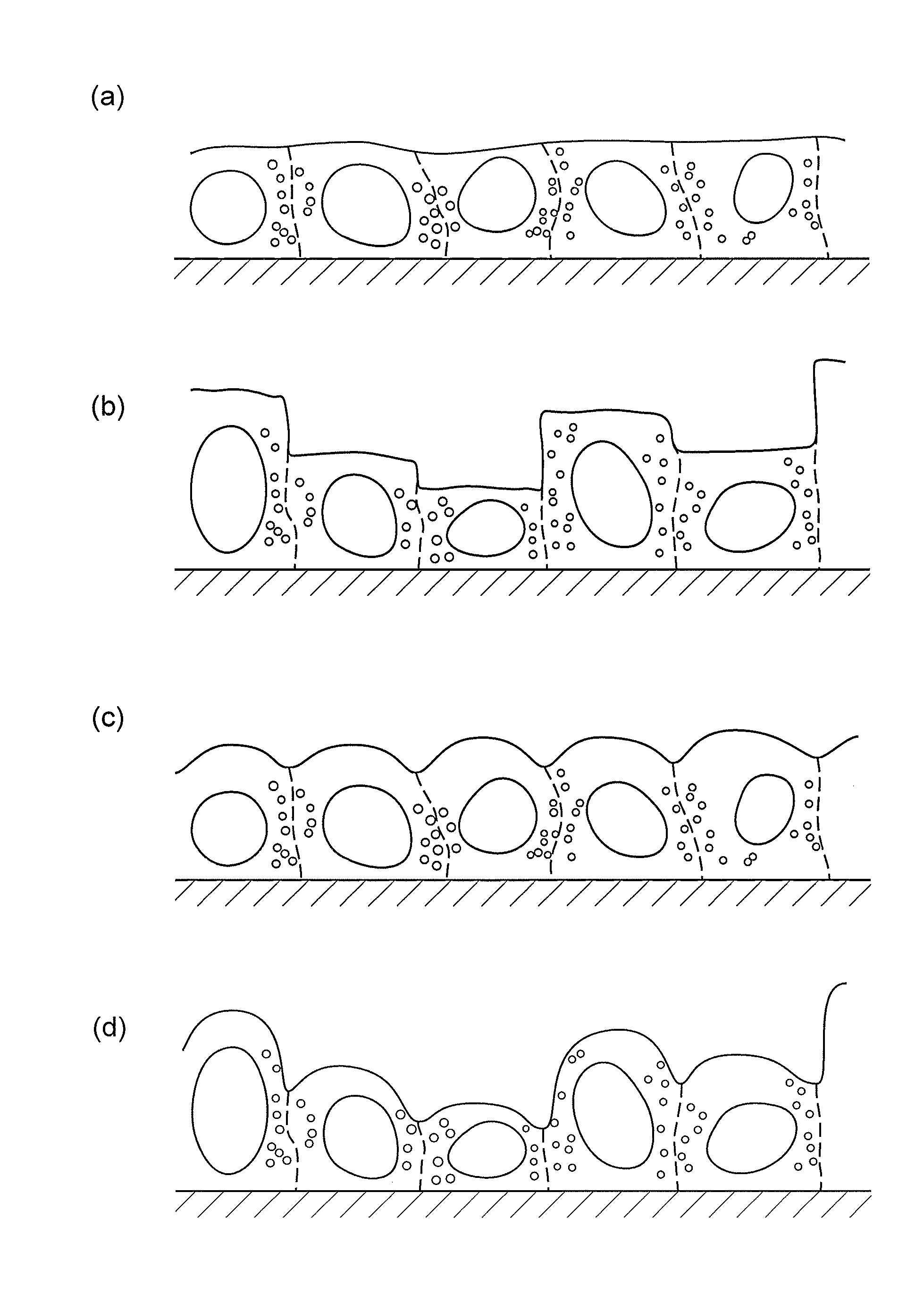
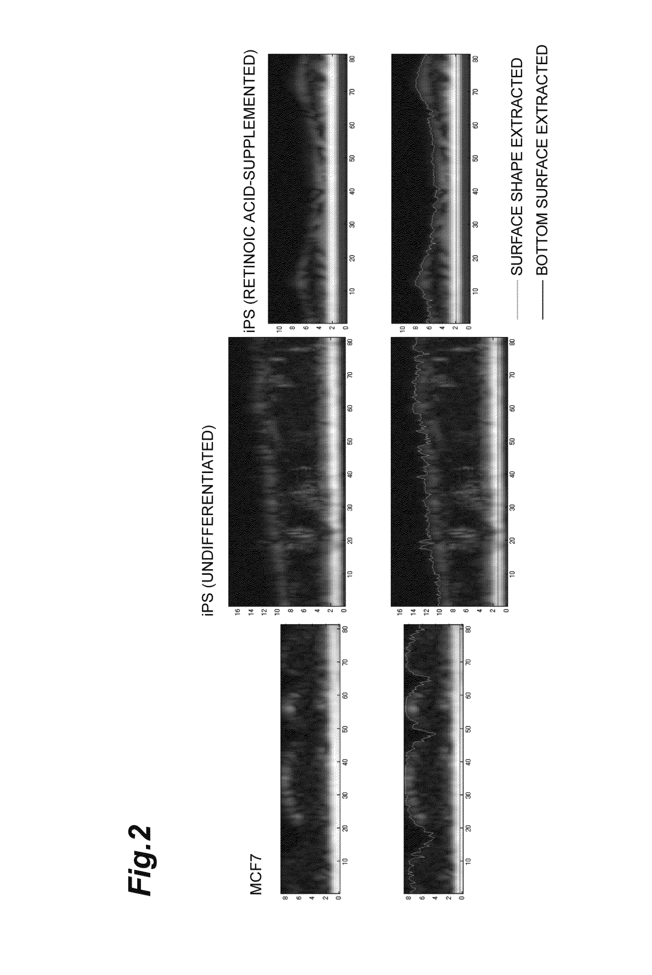
[0072]Onto an antireflectively coated glass bottom dish with 35 mm diameter, a 253G1 iPS cell line derived from a human adult fibroblast was seeded at 10 to 20 colonies / dish and cultured on feeder using a Primate ES Medium (manufactured by ReproCELL Incorporated, a commercial name: RCHEMD001) containing 4 ng / mL human basic FGF in the final concentration. The size of one colony was around 200 μn. As the feeder cells, mouse lung fibroblasts were used. The cells were cultured until the third day after the seeding. The medium was replaced once a day.

[0073]A sample of the iPS cells for differentiation, the medium of which was replaced with a Primate ES Medium containing 4 ng / mL human basic FGF and 12.5 μM retinoic acid in the final concentration on the second day after the seeding, was cultured for another 4 days. Retinoic acid is generally used as a reagent for inducing differentiation. In addition, for a control of differentiated cells, MCF7 cells were used which were human breast canc...

example 2

[0077]As to 3 colonies of the undifferentiated iPS cells and 3 colonies of the iPS cells cultured by supplementing retinoic acid, 7 to 15 visual fields around the center of each colony were analyzed. Each visual field was partitioned into six ROIs, and the bottom surfaces and surface shapes of the cells were extracted for each ROI unit to measure the height of the surface of the cells under the same condition as that of Example 1. The size of the ROI was 23 μM×23 μm. The flatness was calculated as a standard deviation of height of the surface of the cells which was standardized with a mean value of height of the surface of the cells, and a statistical processing was done to perform Student's t-test. The result was shown in Table 2 and FIG. 4. The flatness of the iPS cells cultured by supplementing retinoic acid was higher than the flatness of the undifferentiated iPS cells, in which the difference was significant.

TABLE 2UndifferentiatedRetinoic acid-addediPS cellsiPS cellsCont-1Cont...

example 3

[0078]Under the same condition as that of Example 2, three-dimensional data for the undifferentiated iPS cells and the iPS cells cultured by supplementing retinoic acid were obtained every 0.125 μm in the horizontal direction and every 0.32 μm in the vertical direction, and from the obtained data, tomogram images were created. From the created tomogram images, the bottom surfaces and surface shapes of the cells were extracted and a variogram analysis was performed. Specifically, a value of MSHD when the Δr was within a range of 0.25 μm to 15 μm was calculated from formula (I) as described above, and standardized with a mean value of height of the surface of the cells. As shown in FIG. 5, the flatness of the iPS cells cultured by supplementing retinoic acid was higher than the flatness of the undifferentiated iPS cells.

[0079]Next, a value of MSHD when the Δr was 15 μl was calculated and standardized with a mean value of height of the surface of the cells to calculate the flatness. Th...


PUM

PropertyMeasurementUnit
heightaaaaaaaaaa
heightaaaaaaaaaa
flatnessaaaaaaaaaa

Abstract

The present invention provides a method for determining differentiation level of pluripotent stem cell, comprising a step of determining a flatness of cultured pluripotent stem cell, wherein the flatness is an indication.

Description

CROSS-REFERENCE TO RELATED APPLICATION[0001]This application claims priority to Provisional Application (61 / 700,622) filed on Sep. 13, 2012, which is hereby incorporated by reference in its entirety.BACKGROUND OF THE INVENTION[0002]1. Technical Field[0003]The present invention relates to a method for determining differentiation level of pluripotent stem cells.[0004]2. Background Art[0005]An embryonic stem cell has a pluripotency to differentiate into various cells, so that its application to repair or regeneration of various tissues is expected in the field of regenerative medicine, for example. Since cells derived from a fetus are used, however, its practical application in the medicine is presumably difficult because of limited cell source, controversial ethical questions, and the like. In order to solve such problems, an induced Pluripotent Stem cell (hereinafter also referred to as “iPS cell”) has been developed. An iPS cell is a pluripotent stem cell artificially induced from a...

Claims


Application Information

Patent Timeline
Patent Type & Authority Applications(United States)
IPC IPC(8): G01N33/483
CPCG01N33/4833G01N15/1456G01N2015/1006G01N2015/1497G06T7/50C12N5/0696C12N15/09C12Q1/04G06T7/0012G06T2207/10056G06T2207/30004
Inventor YAMAUCHI, TOYOHIKOSUGIYAMA, NORIKAZUFUKAMI, TADASHIIWAI, HIDENAO
Owner HAMAMATSU PHOTONICS KK
Why Eureka
Try Eureka