Tumour necrosis factor receptor 1 antagonists
a tumor necrosis factor and receptor technology, applied in the field of tumor necrosis factor receptor 1 antagonists, can solve the problems of affecting the development of lymphoma, affecting the survival of patients, and blocking the tnf-mediated host defence, so as to prevent the multimerisation of tnf-tnfr1 trimeric complexes, and preventing the amplification of tnfr1 signal transduction
- Summary
- Abstract
- Description
- Claims
- Application Information
AI Technical Summary
Benefits of technology
Problems solved by technology
Method used
Image
Examples
example 1
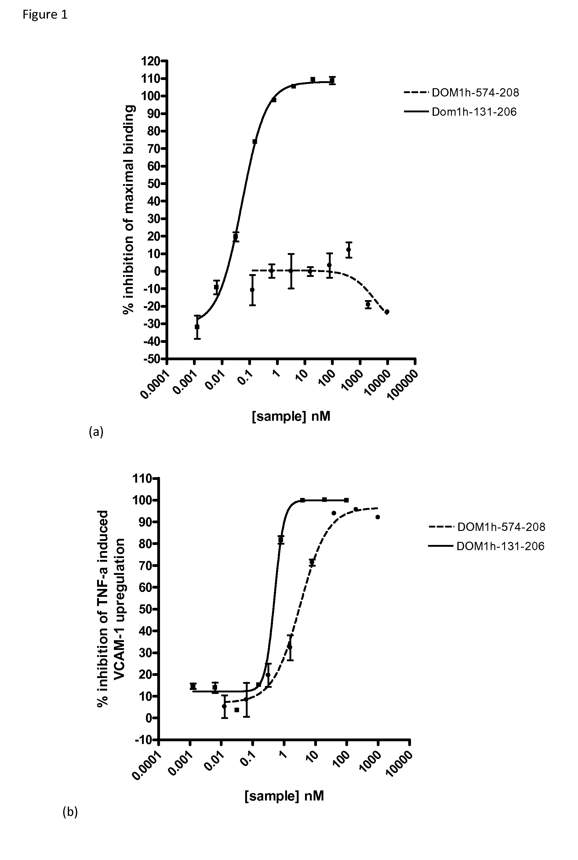
Functional Characterisation of DOM1h-574-208
[0174]Signalling through TNF receptor 1 (TNFR1, p55) can be inhibited either directly through competitive inhibition of TNFα binding to its receptor or indirectly by a non-competitive mechanism in which the binding of TNFα to its receptor is not affected by the presence of the inhibitor.
[0175]To discriminate between these two classes of TNFR1-signaling inhibitors, the combined information from a receptor-binding assay and a cell-based, TNFα-induced, functional assay can be used. Suitable assays are described in WO2011051217.
[0176]Briefly, in the standard receptor binding assay TNFR1-Fc fusion (R&D Systems (Cat #372-RI), sequence is human TNFR1 (Leu30-Thr211 & Asp41-Thr211)-IEGRMD-Human IgG1 (Pro100-Lys330)-6 His-tag) is coated on anti-IgG beads and incubated with a concentration range (e.g. 0.01 nM-10 μM) of a domain antibody directed against TNFR1. Subsequently, TNFα is added followed by addition of a biotinylated anti-TNFα antibody and f...
example 2
Summary of Epitope Mapping with TNFR1 Binding Protein DMS5541
[0181]DMS5541 comprises, as a TNFR1 binding protein, the TNFR1 dAb DOM1h-574-208 (SEQ ID NO:2), coupled to a human serum albumin (HSA) binding dAb by a short linker (Ala-Ser-Thr). It is described further in WO2011051217. The epitope of this molecule (referred to as DMS5541) on TNFR1 was determined using hydrogen deuterium exchange mass spectrometry.
[0182]Methods and principles on using H / D exchange perturbation for epitope mapping are discussed in a review by Hamuro et al J. Biomol. Tech. (2003) 14:171-182; and Coales et al, Rapid Comm. In Mass Spec. (2009) 23(5):639-647.
[0183]For the epitope mapping of TNFR1, H / D exchange analysis of the antigen in the presence and absence of DMS5541 was carried out. The regions of TNFR1 which exchange slower in the presence of DMS5541 compared to speed of exchange when the binding protein is absent is considered to define the epitope on TNFR1. To identify the epitope one requires firstly...
example 3
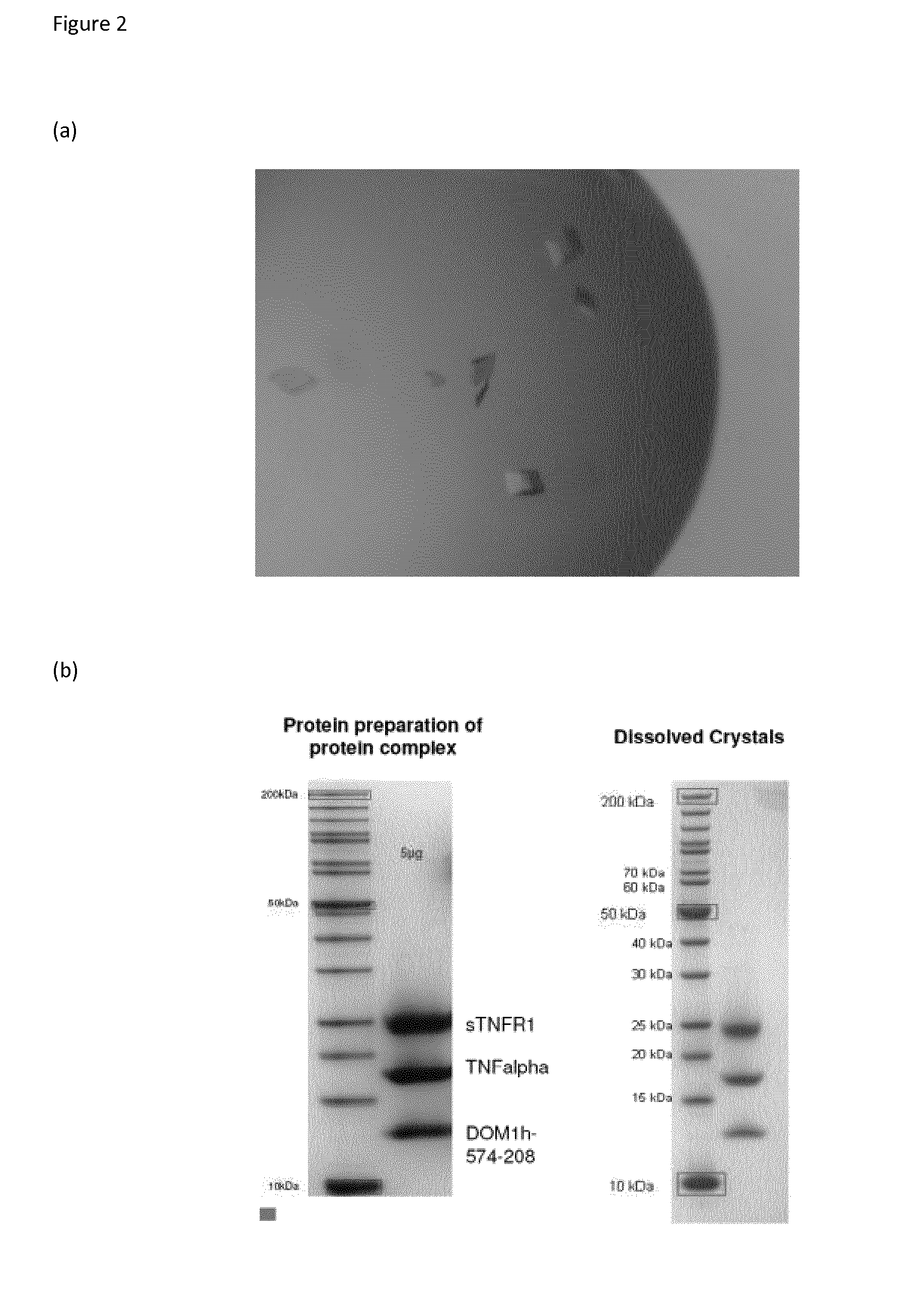
Elucidation of the TNFR1 / TNFα / DOM1h-574-208 Complex
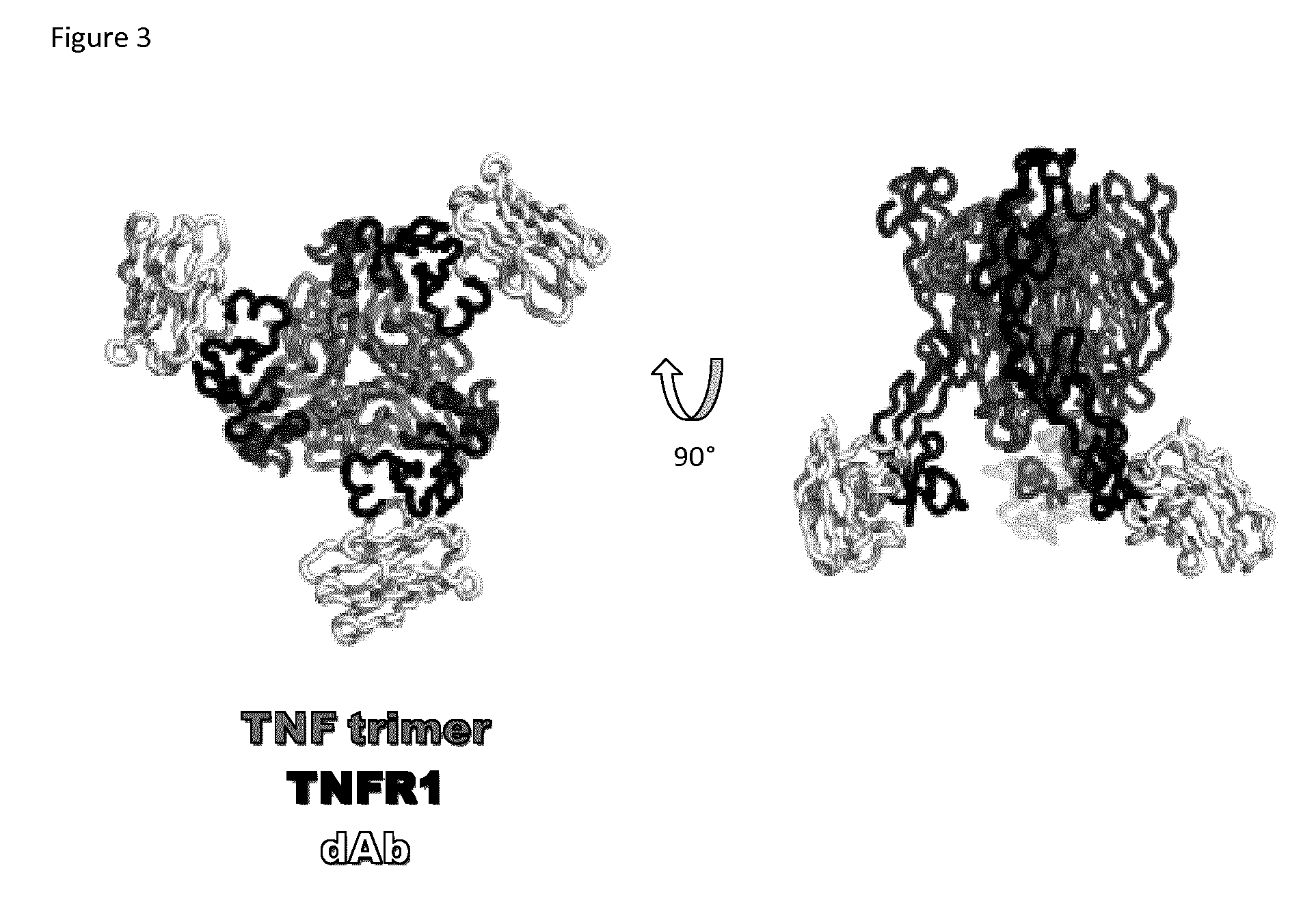
[0185]Co-crystallography of DOM1h-574-208 (SEQ ID NO:2) with TNFR1 and TNFα, refined at a resolution of 2.9 Å, now confirms that DOM1h-574-208 binds predominantly to residues within Domain 4 of TNFR1 (crystallographic data collection and model refinement statistics are given below).
[0186]Moreover, the crystallography also reveals that the TNFα ligand is indeed trimeric, and that the TNFα-TNFR1-DOM1h-574-208 complex is also trimeric, forming around, and driven by, the trimeric ligand molecule. The structure is shown graphically in FIG. 3. This is the believed to be the first time the TNFα-TNFR1 structure has been fully described and experimentally isolated.
[0187]The structure reveals DOM1h-574-208 as binding to an epitope on the opposite side of the TNFαbinding site on TNFR1. Accordingly, DOM1h-574-208, and other TNFR1 binding molecules which bind in the same area as DOM1h-574-208, cannot disrupt the formation of the TNFα-TNFR1 trime...
PUM
| Property | Measurement | Unit |
|---|---|---|
| assay time | aaaaa | aaaaa |
| concentration | aaaaa | aaaaa |
| concentration | aaaaa | aaaaa |
Abstract
Description
Claims
Application Information
 - R&D
- Intellectual Property
- Life Sciences
- Materials
- Tech Scout
- Unparalleled Data Quality
- Higher Quality Content
- 60% Fewer Hallucinations
Browse by: Latest US Patents, China's latest patents, Technical Efficacy Thesaurus, Application Domain, Technology Topic, Popular Technical Reports.
© 2025 PatSnap. All rights reserved.Legal|Privacy policy|Modern Slavery Act Transparency Statement|Sitemap|About US| Contact US: help@patsnap.com


