Intracavitary Brachytherapy Device for Insertion in a Body Cavity and Methods of Use Thereof

a brachytherapy device and intracavitary technology, applied in the field of brachytherapy devices and methods of cervical brachytherapy treatment, can solve the problems of affecting patient outcomes, uterine perforation risk, and difficulty in maintaining in situ position, so as to achieve reproducible insertion, more safety, and efficient

Inactive Publication Date: 2014-05-01
PF BIOMEDICAL SOLUTIONS
View PDF5 Cites 159 Cited by
  • Summary
  • Abstract
  • Description
  • Claims
  • Application Information

AI Technical Summary

Benefits of technology

[0019]The objects described above and other advantages and features of the invention are incorporated in the application as set forth herein, and the associated drawings, related to systems for more safely, efficiently, and reproducibly inserting an applicator assembly into a body cavity of a patient using an image guidance system.

Problems solved by technology

The risk of uterine perforation using this technique has been described at rates of 2-14%, and has been postulated to adversely affect patient outcomes [Kim, R. Y., et al., Radiology, Vol. 147, pp.
Although the Fletcher-Suit applicator has been widely used, maintaining its position in situ can be difficult due to their weight and the difficulty of ensuring a secure connection between the colpostats and tandem.
However, they can be uncomfortable and / or difficult to insert into the appropriate region of the patient due to their rigidity and incapability of accommodating variations in anatomy, e.g., variations in the size, shape, and orientation of the uterus among patients, or postoperative distortions in anatomy.
Such techniques have limited the use of shielded ovoids used in ICBT applicators because the shields can interfere with these various methods of planning by distorting images of the implant localization and causing streak artifacts, making a determination of the optimal position of the applicator within the body cavity very difficult to determine.
For example, the Weeks applicator is not is adaptable to remote after-loading (loading the radioactive source into the applicator post-insertion and positioning within the body cavity) thereby increasing the radiation exposure from LDR brachytherapy; and, it cannot be used at all for HDR or pulsed dose rate (PDR) applications.
In addition, in order to accommodate the after-loading shields, the arms connected to the ovoids are much more bulky than those of a standard FSD applicator.
The increased size of the arms makes it more difficult to insert the vaginal packing needed to distance the bladder and rectum from the radiation sources.
This added bulk also has a potentially negative impact on the comfort of the patient undergoing treatment.
This applicator was not designed for use with any shielding, however, and thus its use results in exposure of the rectum and bladder or other surrounding tissue to high doses of radiation which may lead to clinical complications.
Although all of these devices improve three dimensional localization of the applicator within the patient, none decrease the inherent difficulty of placing the tandem optimally within an orifice of the patient to be treated.
Improper tandem placement may increase the risk of tissue perforation or damage during intra-uterine insertion and placement.
Compounded by, and in addition to, a lack of reproducible positioning when considering multiple procedures for a single patient, these uncertainties may lead to unpredictable treatment results.
However, given the poor image quality and resolution issues inherent with the is use of ultrasound, such guidance still results in it being difficult for the physician to place the instrument accurately within the patient.
Additionally, ultrasonic visualization of the tandem is only possible once the tandem is within the uterus.
As such, the use of ultrasonics will not assist with localizing and entering the uterus via the cervical os.

Method used


Image

  • Intracavitary Brachytherapy Device for Insertion in a Body Cavity and Methods of Use Thereof
  • Intracavitary Brachytherapy Device for Insertion in a Body Cavity and Methods of Use Thereof
  • Intracavitary Brachytherapy Device for Insertion in a Body Cavity and Methods of Use Thereof

Examples


Embodiment Construction

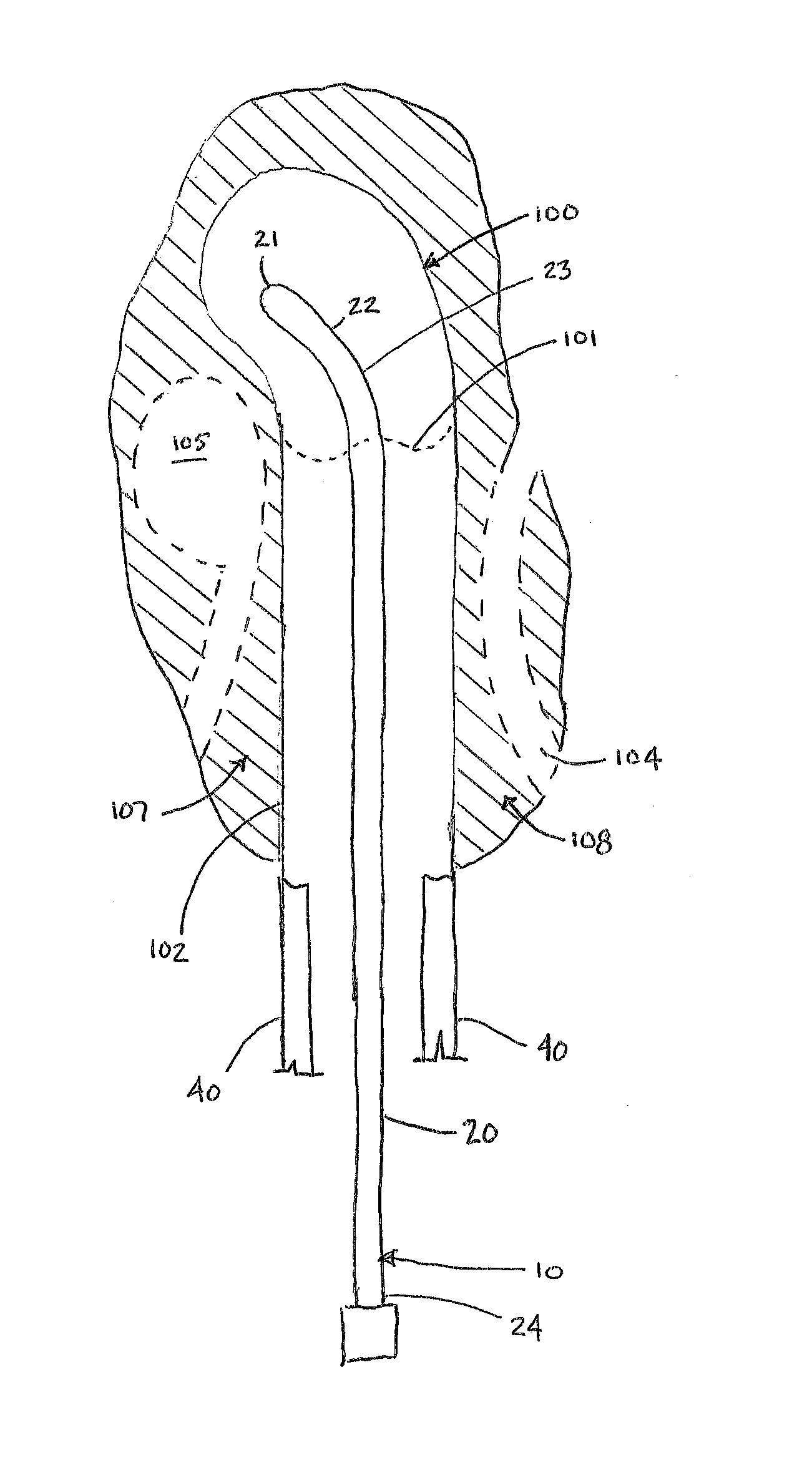
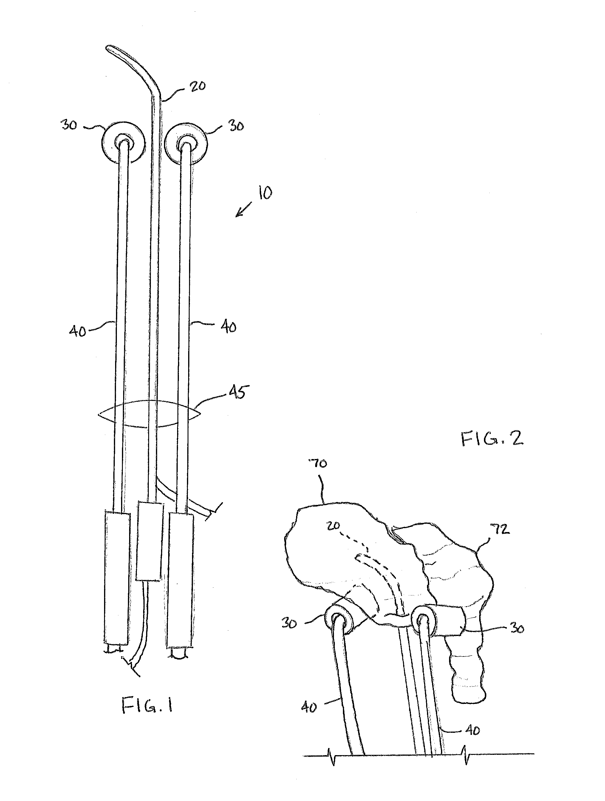
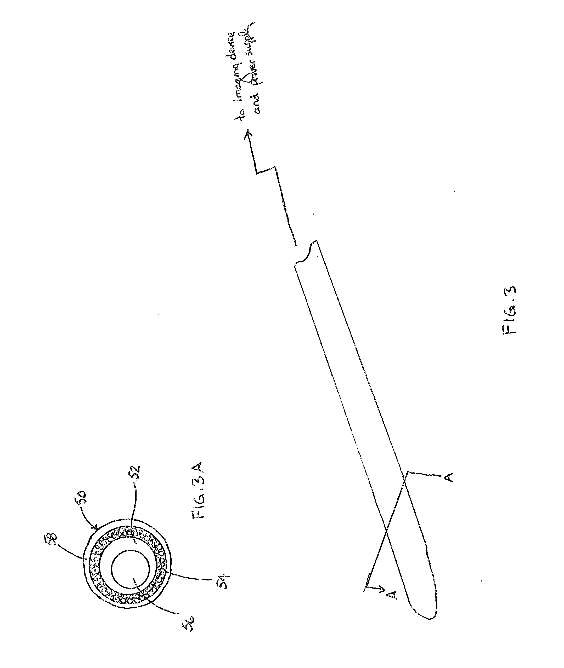
[0030]The Figures described above and the written description of specific structures and functions below are not presented to limit the scope of what Applicants have invented or the scope of the appended claims. Rather, the Figures and written description are provided to teach any person skilled in the art to make and use the inventions for which patent protection is sought. Those skilled in the art will appreciate that not all features of a commercial embodiment of the inventions are described or shown for the sake of clarity and understanding. Persons of skill in this art will also appreciate that the development of an actual commercial embodiment incorporating aspects of the present inventions will require numerous implementation-specific decisions to achieve the developer's ultimate goal for the commercial embodiment. Such implementation-specific decisions may include, and likely are not limited to, compliance with system-related, business-related, government-related and other c...


PUM


Abstract

A brachytherapy application device is described, which includes a tandem having a transparent region at its front end, and which is coupled with a fiber-optic illumination means and endoscope. This improved tandem assembly allows the user to guide the tandem into the uterus of a patient in a safer, more reproducible manner with the reduction in occurrence of uterine perforation during tandem advancement and placement.

Description

CROSS REFERENCE TO RELATED APPLICATIONS[0001]This application claims priority to U.S. Provisional Patent Application Ser. No. 61 / 719,431, filed Oct. 28, 2012, the contents of which are incorporated herein by reference in its entirety.STATEMENT REGARDING FEDERALLY SPONSORED RESEARCH OR DEVELOPMENT[0002]Not applicable.REFERENCE TO APPENDIX[0003]Not applicable.BACKGROUND OF THE INVENTION[0004]1. Field of the Invention[0005]The inventions disclosed and taught herein relate generally to brachytherapy devices and to methods for cervical brachytherapy treatment using such brachytherapy devices. More particularly, the inventions disclosed herein relate to image-guided brachytherapy devices and image-guided interuterine tandem placement systems having illuminated imaging devices integral with the tandem, for more reproducible and safer placement of the device into the uterus of a subject. Using this device, radioactive therapeutics may be delivered to a region of the uterus, for example, the...

Claims


Application Information

Patent Timeline
Patent Type & Authority Applications(United States)
IPC IPC(8): A61N5/10A61B1/303A61B1/00A61B1/05A61B1/07
CPCA61N5/1016A61B1/00087A61B1/05A61B1/07A61B1/303
Inventor FONTENOT, JONAS D.PRICE, MICHAEL J.
Owner PF BIOMEDICAL SOLUTIONS
Why Eureka
Try Eureka