

Biopsy and sonography method and apparatus for assessing bodily cavities
- Summary
- Abstract
- Description
- Claims
- Application Information
AI Technical Summary
Benefits of technology
Problems solved by technology
Method used
Image

Examples
Embodiment Construction
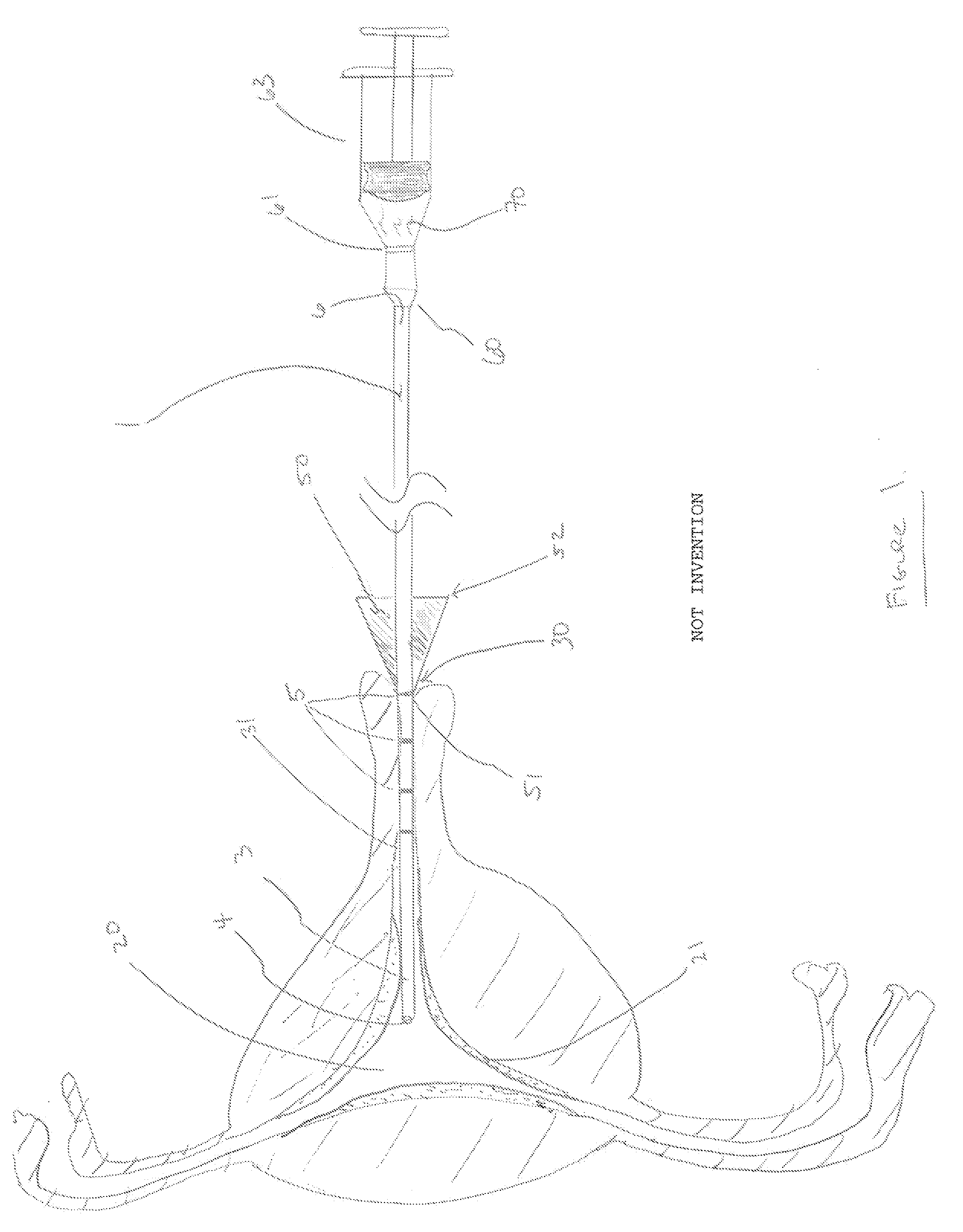
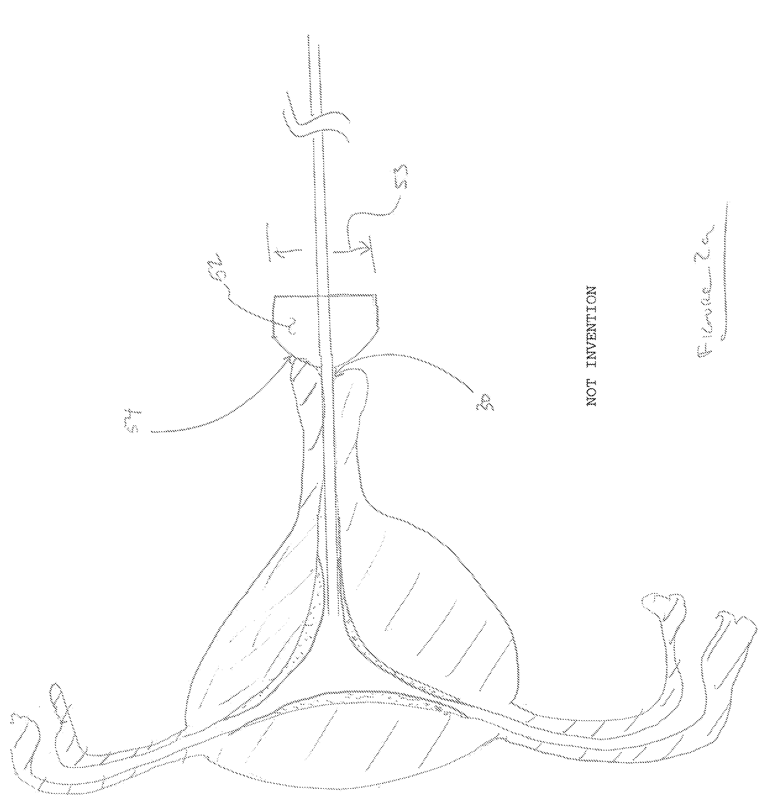
[0024]In FIG. 1 illustrates a device 1 within a cross-sectional view of the uterine cavity 20. The distal end of the device 3 is shown with a distal end opening 4, inner lumen 6, and indicia 5 for determining the depth of insertion within the uterine cavity using the exocervix 30 of the cervix or endocervical canal 31 as a frame of reference. Located at the exocervical opening 30, an acorn tip 50 is placed and pressed into the exocervical opening 30 to provide a seal for media within the uterine cavity 20. A connector or coupling 60 can be located on the proximal end of the device 1. This coupling 60 can be configured as a stopcock, T-connector or Y-connector to allow for the injection of fluids or media into connector opening 61. This connector opening 61 can be configured as a luer connector to allow for the connection of a syringe 63 or other fluid supplying source. In operation the syringe 63 or other fluid supplying source 64 can be used to distend the uterine cavity 20 when pe...
PUM
 Abstract
Description
Claims
Application Information