Nanosplasmonic imaging technique for the spatio-temporal mapping of single cell secretions in real time

Active Publication Date: 2014-09-18
THE UNITED STATES OF AMERICA AS REPRESENTED BY THE SECRETARY OF THE NAVY
View PDF3 Cites 11 Cited by
  • Summary
  • Abstract
  • Description
  • Claims
  • Application Information

AI Technical Summary

Benefits of technology

This patent describes the use of nanoplasmonic imaging to study extracellular signaling. This technology has several advantages, including measuring protein secretion in real-time, using standard imaging techniques like fluorescence and bright field imagery. The nanostructures are calibrated for accurate protein concentration measurements. The technique can be used with both adherent and non-adherent cell lines and is not affected by blinking or photobleaching, which are problems with other methods. Overall, nanoplasmonic imaging provides a better understanding of cell-cell communication and can help identify potential therapeutic targets.

Problems solved by technology

There is currently no alternative method for the label-free and real time imaging of protein secretions from individual cells that integrates the standard glass coverslips widely used for cell imaging and culture.

Method used


Image

  • Nanosplasmonic imaging technique for the spatio-temporal mapping of single cell secretions in real time
  • Nanosplasmonic imaging technique for the spatio-temporal mapping of single cell secretions in real time
  • Nanosplasmonic imaging technique for the spatio-temporal mapping of single cell secretions in real time

Examples


Embodiment Construction

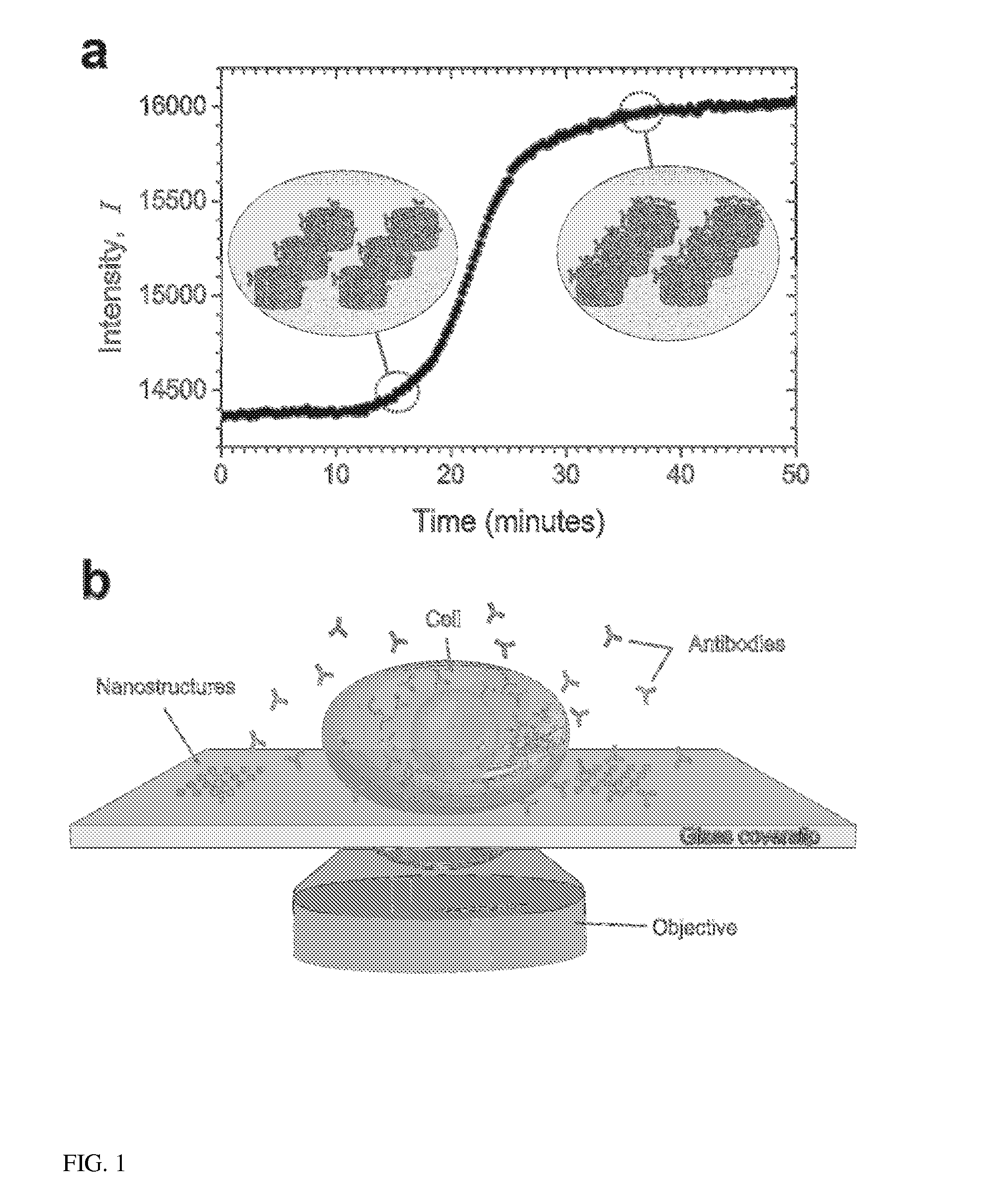
[0016]The present invention provides a label-free approach based upon localized surface plasmon resonance (LSPR) imaging for the real-time measurement of protein secretions from individual cells. LSPR biosensing is founded upon the fact that the plasmonic resonance of a metallic nanostructure exhibits both a red shift and an increase in scattering intensity when analyte binding creates small perturbations in the local index of refraction. When imaged on a CCD camera these spectroscopic signatures are manifested as an increase in the brightness of the nanostructures (FIG. 1(a)) and can be quantified in terms of the fractional occupancy of surface bound receptors. In contrast to thin-film based surface plasmon resonance (SPR) approaches that require total internally reflected light for the excitation of the surface plasmons, nanoplasmonic resonances can be excited with visible light using the same optical configurations used in traditional wide-field microscopy setups (FIG. 1(b)).

[001...


PUM


Abstract

A label-free method for the spatio-temporal mapping of protein secretions from individual cells in real time by using a chip for localized surface plasmon resonance (LSPR) imaging. The chip is a glass coverslip compatible for use in a standard microscope having at least one array of functionalized plasmonic nanostructures patterned onto it. After placing a cell on the chip, the secretions from the cell are spatially and temporally mapped using LSPR imaging. Transmitted light imaging and / or fluorescence imaging may be done simultaneously with the LSPR imaging.

Description

PRIORITY CLAIM[0001]The present application is a non-provisional application claiming the benefit of U.S. Provisional Application No. 61 / 778,652, filed on Mar. 13, 2013 by Marc P. Raphael et al., entitled “Nanoplasmonic Imaging Technique for the Spatio-Temporal Mapping of Single Cell Secretions in Real Time,” the entire contents of which is incorporated herein by reference.BACKGROUND OF THE INVENTION[0002]1. Field of the Invention[0003]The present invention relates generally to paracrine signal measuring and, more specifically, to label-free mapping of single cell secretions in real time.[0004]2. Description of the Prior Art[0005]Paracrine signaling is a form of close-range communication between cells, typically mediated by the secretion of proteins. The types of proteins secreted as well as their spatial and temporal distributions give rise to a broad range of possible responses amongst the receiving cells, including cell migration and proliferation. Not surprisingly then, paracrin...

Claims


Application Information

Patent Timeline
IPC IPC(8): G01N33/569
CPCG01N33/56966G01N21/274G01N21/554G01N21/59G01N21/6458G01N21/648G01N33/54373G01N2021/6491G01N33/551
Inventor RAPHAEL, MARC P.CHRISTODOULIDES, JOSEPH A.BYERS, JEFF M.DELEHANTY, JAMES B.
Owner THE UNITED STATES OF AMERICA AS REPRESENTED BY THE SECRETARY OF THE NAVY
Why Eureka
Try Eureka