Methods for suppressing cancer by inhibition of tmcc3

a technology of tmcc3 and cancer, applied in the field of cancer treatment, can solve the problems of recurrent disease, recurrence disease remains a challenge, and no reliable and validated biomarkers specific for early detection of cancer

Active Publication Date: 2014-12-11
DEV CENT FOR BIOTECHNOLOGY +2
View PDF4 Cites 8 Cited by
  • Summary
  • Abstract
  • Description
  • Claims
  • Application Information

AI Technical Summary

Benefits of technology

[0047]FIG. 28 shows that anti-TMCC3 intercoil domain antibodies can enrich stem cell-like cancer cells. In this experiment, breast cancer cell line BC0145 was used. As shown, the anti-TMCC3 Ab significantly enriched the stem cell-like cancer cell population.

Problems solved by technology

One possibility is that most cancer cells lack the ability to form a new tumor such, that only the dissemination of rare cancer stem cells can lead to metastatic disease.
However, because most cells within a cancer have limited proliferative potential, an ability to shrink a tumor mainly reflects an ability to kill these cells.
Although many chemotherapeutic drugs and targeted therapeutic agents have improved the cure rate of cancer, recurrent disease remains to be a challenge due to the development of drug resistance.
So far, there is no reliable and validated biomarkers specific for early detection of cancer.

Method used


Image

  • Methods for suppressing cancer by inhibition of tmcc3
  • Methods for suppressing cancer by inhibition of tmcc3
  • Methods for suppressing cancer by inhibition of tmcc3

Examples


example 1

Western Blot Analysis

[0129]Cells were lysed in RIPA buffer supplemented with phosphatase and protease inhibitors and boiled after mixing with protein loading buffer. Aliquots of cell lysates were subjected to SDS-PAGE, and the proteins were transferred to PVDF membranes. The membrane lots were blocked with TBST buffer containing 3% BSA for 1 hour at room temperature and then incubated with the respective primary antibodies diluted in TBST (containing 0.05% Tween20 and 3% BSA) overnight at 4° C. Subsequently, blots were washed and incubated with appropriate secondary antibodies in TBST. Anti-phospho-mTOR (Ser 2448), mTOR, phospho-Akt (Ser 473), Akt, p-FAK (Tyr 397), FAK, p-ERK1 / 2 (p44 / 42) and ERK1 / 2 antibodies were obtained from Cell signaling Technology. Anti-GAPDH antibody was from GeneTex Inc.

example 2

Mammosphere Formation Assay

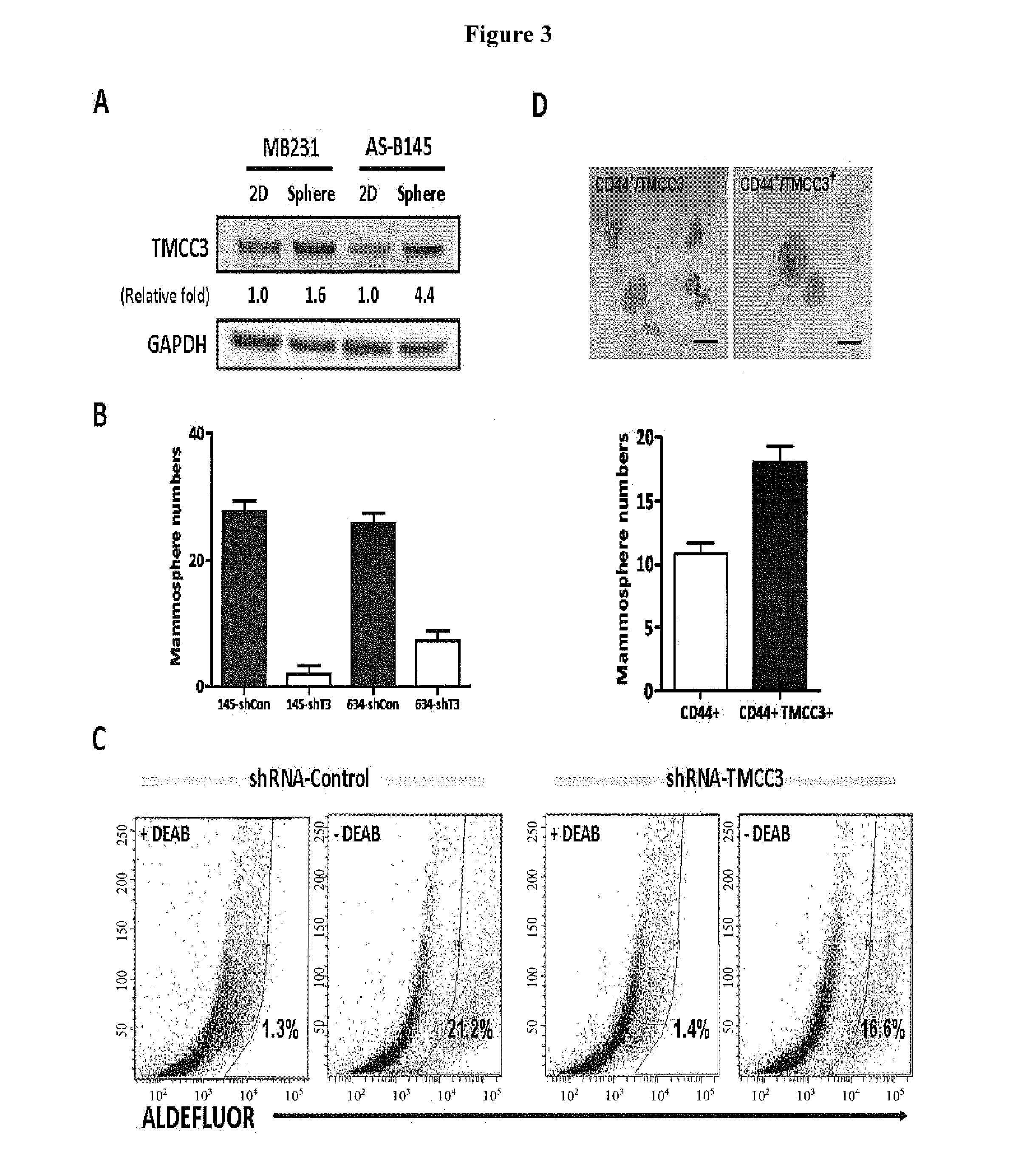
[0130]Single cells were plated at a density of 1,000 cells per well in ultralow attachment plates (35 mm; Corning). Cells were grown in serum-free DMEM / F12, supplemented with B27 (1:50, Invitrogen), 20 ng / mL EGF and 20 ng / mL bFGF (BD Biosciences), and 10 ng / mL insulin (Sigma). The mammospheres were cultured for 7-10 d. Then the mammospheres with diameter >100 μm were counted.

example 3

Aldefluor Assay and Flow Cytometry

[0131]To measure the ALDH activity of lentiviral knocked down cells, the Aldefluor assay was performed according to manufacturer's (Stemcell Technologies) guidelines. sh-TMCC3 and sh-control transfected cells were suspended in Aldefluor assay buffer containing ALDH substrate, Bodipyaminoacetaldehyde (BAAA) and incubated for 30 minutes at 37° C. To distinguish between ALDH-positive and -negative cells, a fraction of cells was incubated under identical condition in the presence of the ALDH inhibitor, diethylamino benzaldehyde (DEAB). This results in a significant decrease in the fluorescence intensity in ALDH-positive cells and was used to compensate in the flow cytometric analysis.


PUM

PropertyMeasurementUnit
dissociation constantaaaaaaaaaa
pHaaaaaaaaaa

Abstract

A method for treating cancer includes administering to a subject in need thereof an antibody against a transmembrane and coiled-coil domains protein 3 (TMCC3), wherein the antibody binds to an epitope in an extracellular domain of TMCC3. The antibody binds to an epitope in an intercoil domain of TMCC3. A method of diagnosing or assessing a cancer condition includes assessing a level of expression or activity of a transmembrane and coiled-coil domains protein 3 (TMCC3) in a sample, wherein an increase in the level of expression of activity of TMCC3 as compared to a standard indicates the presence of cancer stem cells in the sample.

Description

CROSS REFERENCE TO RELATED APPLICATIONS[0001]This application claims the benefits of Provisional Application No. 61 / 804,751, filed on Mar. 24, 2013. The disclosure of this provisional application is incorporated by reference in its entirety.TECHNICAL FIELD OF THE INVENTION[0002]This invention relates to the field of cancer treatment. In particular, the application relates to biosignatures of cancer cells for early detection, prognostication and prediction of therapeutic responses for cancer. More particularly, the invention relates to methods for diagnostics and treatment of cancer via a cancer stem cell biomarker, transmembrane-coiled coil domain family 3 (TMCC3).BACKGROUND OF THE INVENTION[0003]The majority of cancer deaths occur as a result of recurrent or metastatic disease rather than from the effects of the primary tumor. A tumor can be viewed as an aberrant organ initiated by a tumorigenic cancer cell that acquired the capacity for indefinite proliferation through accumulated...

Claims


Application Information

Patent Timeline
Patent Type & Authority Applications(United States)
IPC IPC(8): C07K16/30G01N33/574C12Q1/68A61K51/10
CPCC07K16/3015C07K16/30A61K51/1051C07K2317/732G01N33/57496C12Q1/6816C07K2317/734A61K51/1045A61P15/00A61P35/00C07K2317/55C07K2317/622C07K2317/92C12Q1/6886C12Q2600/158A61K39/39558
Inventor YU, ALICE L.YU, JOHNWANG, YA-HUIHSU, CHUAN-LUNGCHEN, YI-CHANGLOK, YING-YUNG
Owner DEV CENT FOR BIOTECHNOLOGY
Why Eureka
Try Eureka