Dental imaging with photon-counting detector

a detector and dental imaging technology, applied in the field of dental imaging with photon-counting detectors, can solve the problems of compounding problems, unable to carry expensive detectors capable of carrying out both imaging functions, so as to reduce exposure levels

Active Publication Date: 2015-01-01
CARESTREAM DENTAL TECH TOPCO LTD
View PDF16 Cites 31 Cited by
  • Summary
  • Abstract
  • Description
  • Claims
  • Application Information

AI Technical Summary

Benefits of technology

[0011]Embodiments of the present invention address the need for advancing the CT imaging art, particularly for imaging of the head. Embodiments of the present invention adapt photon-counting and related imaging solutions to the problem of CT imaging for dental, ENT, and related applications. Using embodiments of the present invention, a medical practitioner can obtain useful images for patient treatment, taking advantage of reduced exposure levels and other advantages that photon-counting solutions provide.

Problems solved by technology

However, the proposed imaging apparatus performs both CT and panoramic imaging using only one detector.
This requires an expensive detector capable of carrying out both imaging functions in a satisfactory manner.
Conventional digital radiography detectors have some limitations related to how attenuation of radiation energy at a single exposure is interpreted.
For example, it can be very difficult, from a single exposure, to distinguish whether an imaged object has a given thickness or a given attenuation coefficient.
This problem can be compounded for CT imaging, in which multiple images are obtained, one from each of a number of angles of revolution about the patient.
Conventional CT imaging provides useful information that aids in diagnosis and treatment, but is constrained by limitations of the imaging sensor apparatus itself, and there are concerns over exposure levels needed for obtaining the desired image quality.

Method used


Image

  • Dental imaging with photon-counting detector
  • Dental imaging with photon-counting detector
  • Dental imaging with photon-counting detector

Examples


Embodiment Construction

[0053]The following is a detailed description of the exemplary embodiments of the invention, reference being made to the drawings in which the same reference numerals identify the same elements of structure in each of the several figures.

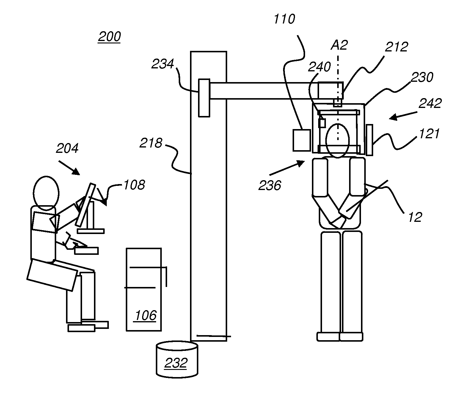
[0054]According to certain exemplary embodiments, there is provided a radiographic imaging apparatus and / or methods (e.g., a dental, extra-oral dental) for obtaining an image based on received radiation (e.g., from a patient) that can provide the capability to obtain the image using at least one imaging modality (e.g., panoramic imaging, cephalometric imaging and CT imaging) and configured with at least one detector (e.g., sensor) of the photon counting type. When multiple imaging modalities are present in a single radiographic imaging apparatus, either additional detectors of the photon counting type or other types (e.g., CMOS-based, CCD-based, flat panel area detectors, sensors that use amorphous or polycrystalline semiconductor materials such as ...


PUM


Abstract

An extra-oral dental imaging apparatus for obtaining an image from a patient has a radiation source; a first digital imaging sensor that provides, for each of a plurality of image pixels, at least a first digital value according to a count of received photons that exceed at least a first energy threshold; a mount that supports the radiation source and the first digital imaging sensor on opposite sides of the patient's head; a computer in signal communication with the digital imaging sensor for acquiring a first two-dimensional image from the first digital imaging sensor; and a second digital imaging sensor that is alternately switched into place by the mount and that provides image data according to received radiation.

Description

FIELD OF THE INVENTION[0001]The invention relates generally to the field of extraoral dental imaging and more particularly to apparatus and methods for obtaining volumetric images from the head of a patient.BACKGROUND OF THE INVENTION[0002]Radiological imaging is recognized to have significant value for the dental practitioner, helping to identify various problems and to validate other measurements and observations related to the patient's teeth and supporting structures. Among x-ray systems with particular promise for improving dental care is the extra-oral imaging apparatus that is capable of obtaining one or more radiographic images in a series and, where multiple images of the patient are acquired at different angles, combining these images to obtain a contiguous panoramic radiograph of the patient showing the entire dentition of the jaw, a tomographic image that contains more depth detail, or a computerized tomography (CT) volume image. To obtain images of any of these types, a...

Claims


Application Information

Patent Timeline
Patent Type & Authority Applications(United States)
IPC IPC(8): A61B6/04A61B6/03A61B6/14
CPCA61B6/14A61B6/04A61B6/032A61B6/501A61B6/4441A61B6/4241A61B6/4458A61B6/4085A61B6/5235A61B6/4452A61B6/588A61B6/025A61B6/54A61B6/4233A61B6/0421A61B6/4266
Inventor INGLESE, JEAN-MARCBOTHOREL, SYLVIELOUSTAUNEAU, VINCENTRANKIN-PAROBEK, DONNA
Owner CARESTREAM DENTAL TECH TOPCO LTD
Why Eureka
Try Eureka