Geometry enhancement of nanoscale energy deposition by x-rays
- Summary
- Abstract
- Description
- Claims
- Application Information
AI Technical Summary
Benefits of technology
Problems solved by technology
Method used
Image

Examples
example 1
Enhanced Nanoscale Energy Deposition Via Geometry Enhancement
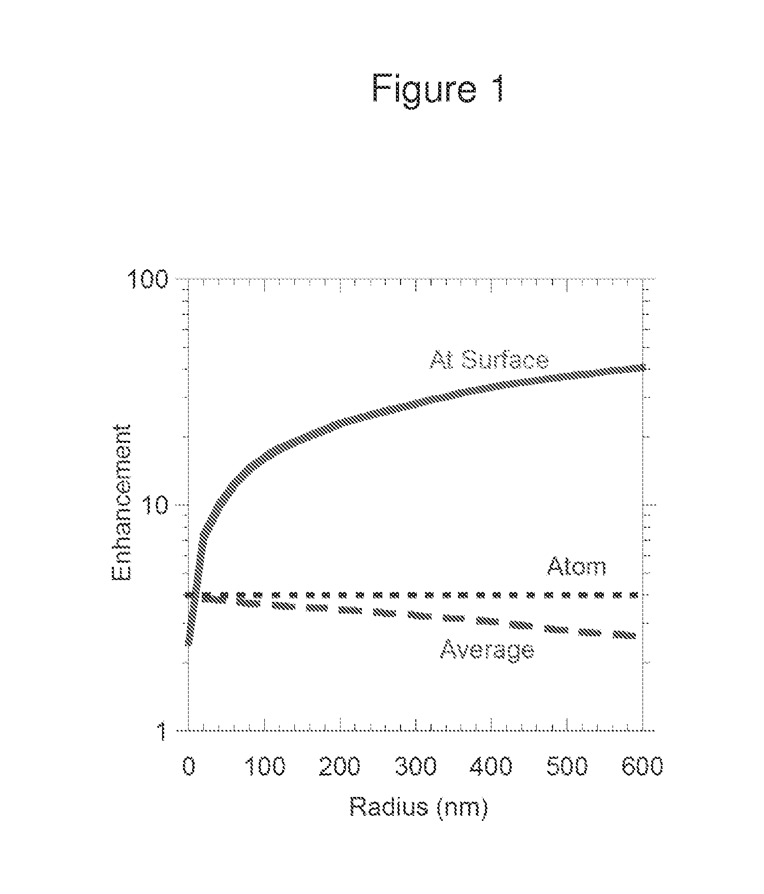
[0042]This example demonstrates that nanoscale energy deposition in water by X-rays can be greatly enhanced via the geometry of nanostructures. We have investigated for the first time using X-ray absorbing nanostructures for optimized nanoscale energy deposition. It is found that nanoshells can greatly enhance the energy deposition density on the nanometer scale. The highest enhancement from a single nanoshell with 8 mass units over the background water is approximately 60 times. Other concepts include satellite, matrix, and composition effects are introduced and studied here. Although even higher enhancement may be possible, in practice enhancement between 10 and 50 times may be more readily achievable. This work clearly shows that it is possible to use X-rays to generate localized energy deposition to activate and direct chemical and biological reactions. When combined with other effects such as chemical and biological e...
example 2
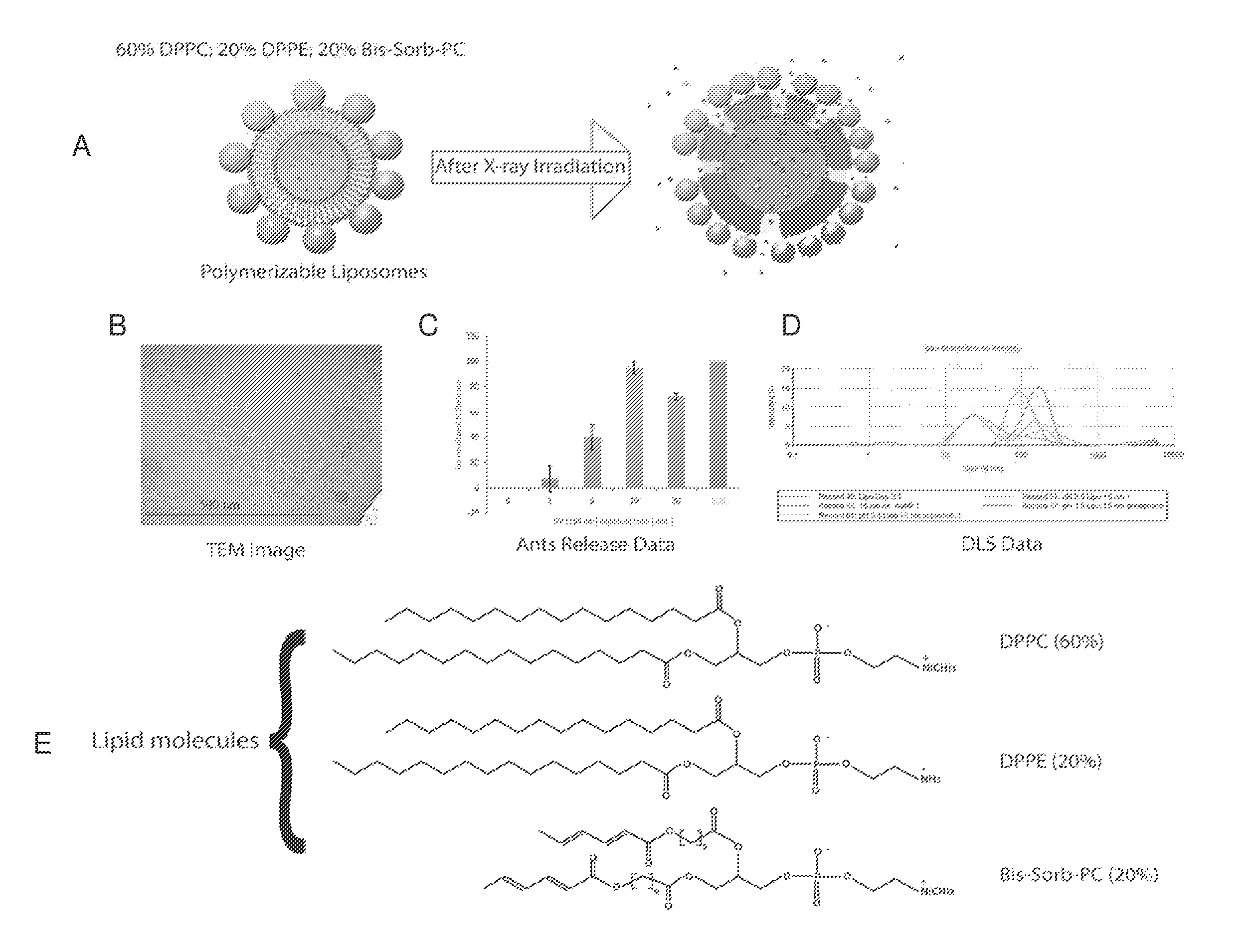
Liposomes Coated with Gold Nanoparticles
[0086]This Example demonstrates the formation of polymerizable liposomes and their cargo-carrying capacity. This Example further demonstrates that polymerizable liposomes can be coated in gold nanoparticles for use in geometry enhancement of cargo release from the liposomes.
Materials and Methods
[0087]Liposome Preparation.
[0088]Liposomes were produced through the standard extrusion method of making liposomes. A combination of membrane filters from 100 to 600 nm was used in the extrusion method. Three kinds of lipid molecules were used to construct the gold nanoparticle-coated X-ray polymerizable liposomes, as illustrated in FIG. 15E. Three types of lipids form the liposomes: DPPC, DPPE, and Bis-SorbPC. DPPC are regular lipid molecules. DPPE has amine groups that can attract citrate-covered gold nanoparticles. Bis-Sorb-PC polymerize under X-ray irradiation, which renders the liposome open to release cargos inside.
Results
[0089]Liposomes coated wi...
example 3
Polymersomes Made with Gold Nanoparticles
[0093]This Example demonstrates the construction of polymersomes formed with gold nanoparticles. The polymersomes formed with gold nanoparticles exhibit enhanced release of cargo from the polymersome upon X-ray irradiation.
Materials and Methods
[0094]Construction of Polymersomes Coated with Gold Nanoparticles.
[0095]Polymersomes were formed by mixing amphiphillic nanoparticles in water. A shell of gold nanoparticles embedded in a shell of polymers was formed.
Results
[0096]As shown in FIG. 16A and FIG. 16B, polymersomes formed from 15-nm gold nanoparticles coated with the amphiphillic ligands of polyethylene glycol (PEG) and poly methyl methacrylate and vinylpyridine (PMMAVP). Polymersomes formed with gold nanoparticles were incubated with the dye molecule sulforhodamine B (SRB). These polymersome illustrate an example of a shell of gold nanoparticles embedded in a shell of polymers. This is shown in FIG. 16A. FIG. 16B shows a transmission electr...
PUM
 Abstract
Description
Claims
Application Information
 - R&D Engineer
- R&D Manager
- IP Professional
- Industry Leading Data Capabilities
- Powerful AI technology
- Patent DNA Extraction
Browse by: Latest US Patents, China's latest patents, Technical Efficacy Thesaurus, Application Domain, Technology Topic.
© 2024 PatSnap. All rights reserved.Legal|Privacy policy|Modern Slavery Act Transparency Statement|Sitemap