
Spinal Cord Devices and Methods for Promoting Axonal Regeneration

a technology of axonal regeneration and spinal cord, which is applied in the field of spinal cord injury treatment, can solve the problems of no therapy which restores or even significantly improves the function of the spinal cord, incontinence, and partial or complete paralysis, and achieve the effects of promoting axonal regeneration, promoting axonal regeneration, and promoting axonal regeneration

Inactive Publication Date: 2015-03-26
BIOARCTIC NEUROSCI AB
View PDF0 Cites 5 Cited by
  • Summary
  • Abstract
  • Description
  • Claims
  • Application Information

AI Technical Summary

Benefits of technology

The present invention provides improved devices, kits, and methods for treatment of Spinal Cord Injury (SCI) by bridging an injured spinal cord and promoting axonal regeneration. The spinal cord device has a body with through channels for connection of motor and sensory pathways, and is designed to match the shape, size, and dimensions of the injury. The device can be selected based on the cross-section surface area and anteroposterior diameter / transverse diameter ratio of the injury. The device can also be soaked in a solution containing pharmaceutically active substances and can be implanted to bridge the gap in the injured spinal cord or nerve. The devices, kits, and methods provide for optimal connection between spinal cord tracts and promote axonal regeneration, leading to improved outcomes for patients with SCI.

Problems solved by technology

A spinal cord injury occurs when trauma or disease damages the spinal cord and results in partial or complete paralysis.
Besides paralysis there are usually signs of sensory loss, incontinence, intractable pain and pressure sores.
As today, there is no therapy which restores or even significantly improves the spinal cord function in those severe cases.
General designs are disclosed, but there is no teaching of devices adopted for the level, size and shape of the spinal cord injury.
However, in spite of considerable time from early disclosures of the basic principle of connecting the routing of tracts in white to gray matter, the translation into a clinically applicable method has been absent.
Still, no product is available on the market.
The spinal cord is an extremely important part of the central nervous system, where a patient with a complete injury faces a permanent loss of function below the site of injury, with devastating consequences for the patient's quality of life, as well as long-term costs to finance the assistance to the patient.
In many cases the patient is a fairly young person who has been involved in a serious accident and will have to spend the rest of life with no chance for functional improvements.

Method used


Image

  • Spinal Cord Devices and Methods for Promoting Axonal Regeneration
  • Spinal Cord Devices and Methods for Promoting Axonal Regeneration
  • Spinal Cord Devices and Methods for Promoting Axonal Regeneration

Examples


example 1

[0050]This example evaluates a biodegradable calcium sulphate device with heparin-activated rhFGF1 for treatment of spinal cord injury in rat.

[0051]SCI devices fabricated from α-calcium sulfate hemihydrate with 12 channels with similar geometry as shown in FIG. 5 were loaded with heparin-activated rhFGF1. The test animals, Sprague Dawley rats, were allocated into 5 study groups. Laminectomy was carried out on all animals independent of study group. The spinal cord of the control groups were either transected (group 1, negative control) or left intact (group 2, positive control). The spinal cords of the rats of groups 3-5 were transected and the removed spinal cord tissue was replaced by devices containing nerve grafts. The SCI-devices employed for study group 4 were soaked in 500 μg / ml heparin-activated rhFGF1 (rhFGF1:heparin, 1:1, w / w). For study group 5 the SCI-devices were soaked in 50 μg / ml heparin-activated rhFGF1. Due to autophagy, a few animals had to be sacrificed prior to t...

example 2

[0053]This example evaluates dose-finding of heparin-activated rhFGF1 administered in a biodegradable calcium sulphate device for treatment of spinal cord injury in rat.

[0054]The same type of devices as in Example 1 were used. Each device was soaked in heparin-activated rhFGF1 (FGF1:heparin, 1:100 molar ratio) solution for 1 h at room temperature. Heparin solution without preservatives (10000 IE / ml H2O, Leo Pharma Denmark) was employed. The devices were soaked in 50 μg / ml, 0.5 μg / ml, 0.005 μg / ml and 0 μg / ml concentrations of heparin-activated rhFGF1 corresponding to a dose of 45, 0.9, 0.01 and 0 ng / mg device (based on solution uptake and adsorption).

[0055]FGF1-dependent recovery of bilateral MEPs in the hindlimbs of treated animals is illustrated in FIG. 6. All animals showed undetectable MEPs in the hindlimbs 1 week post-surgery, while already after 2 weeks positive MEPs were recorded. The results indicate that an effective dose of heparin-activated rhFGF1 in the disclosed device i...

example 3

Implantation of Heparin-Activated rhFGF1 Loaded SCI-Devices with Peripheral Nerve Grafts in Pig—Development of Operation Technique and Limited Safety Study

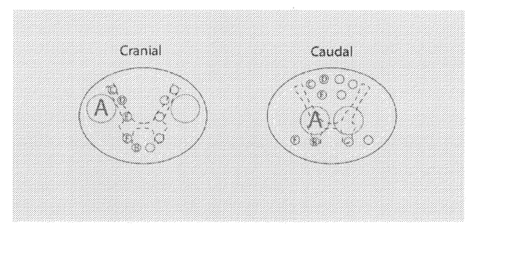
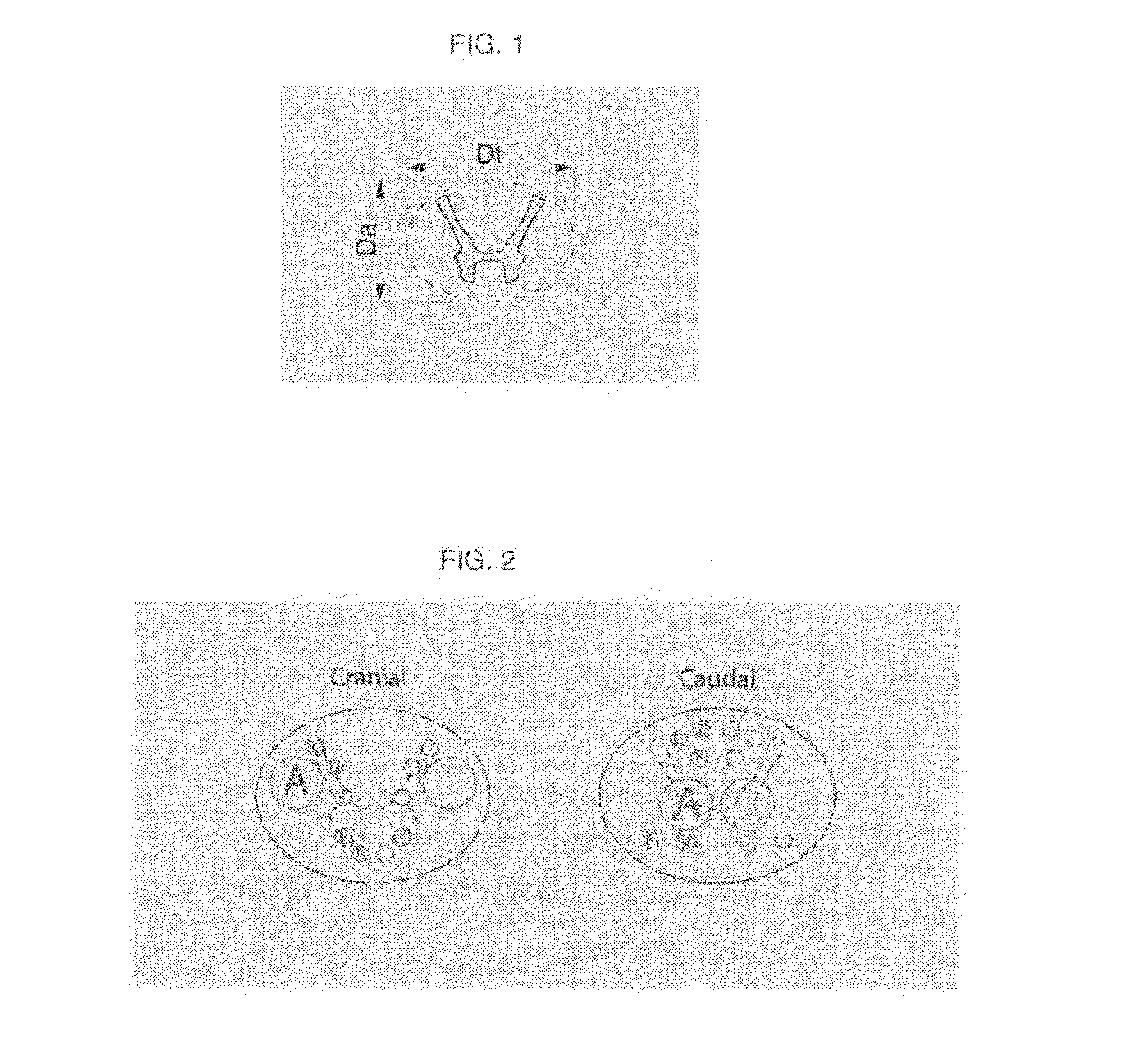
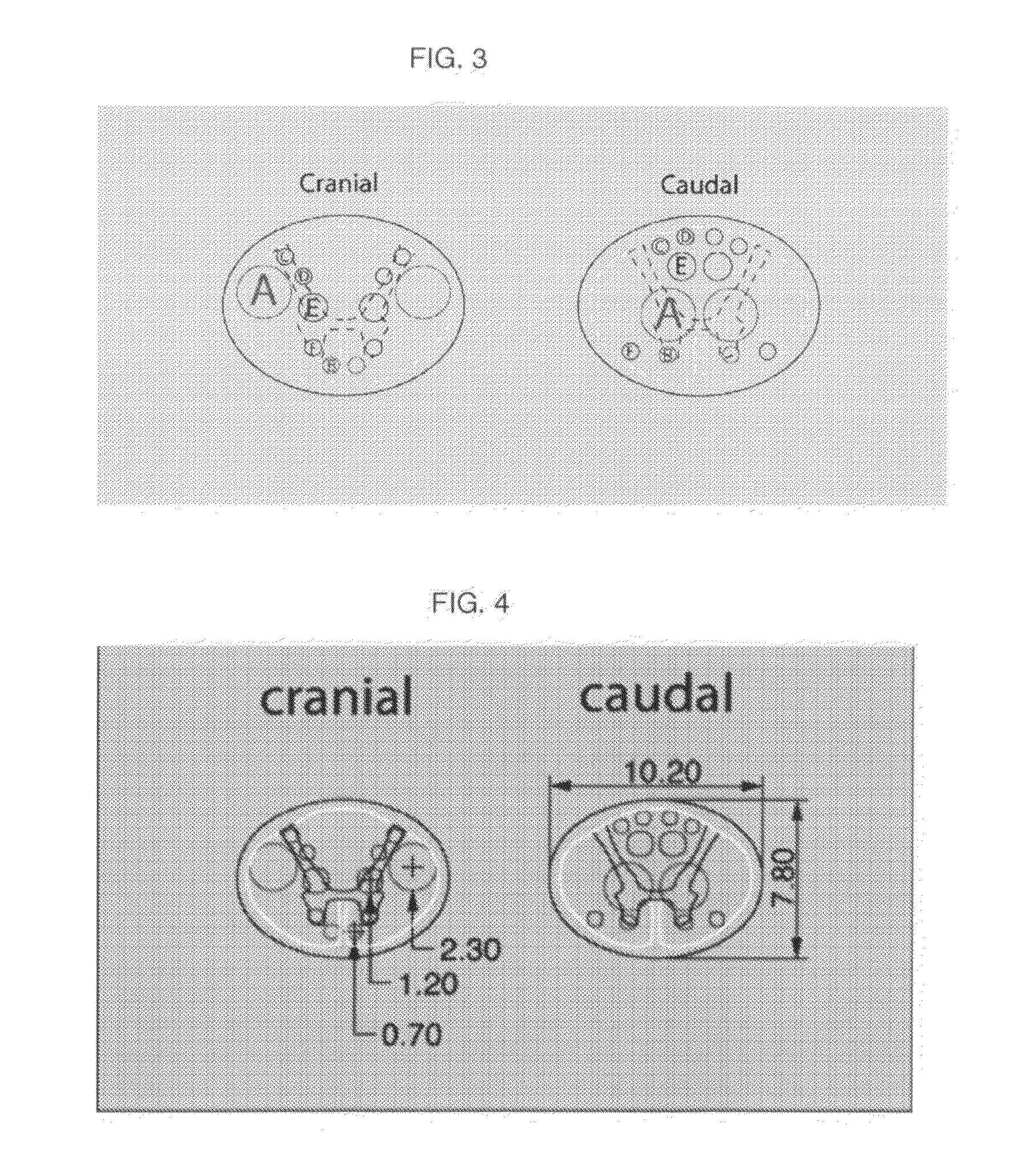
[0056]Clinical SCI-devices as illustrated in FIG. 5, 15 mm high and having an oval cross section of 9.0-6.9 mm, comprising 12 channels for nerve graft positioning are made from α-calcium sulphate hemihydrate with subsequent sterilization.

[0057]Each device is placed in 30 ml of a soaking solution consisting of 5 μg / ml rhFGF1, 80 μg / ml Gentamicin, 10 mM NaPO4, 150 mM NaCl, 0.3 mM EDTA at pH 7 for 1 hour to allow the solution to be adsorbed into the device. The heparin concentration is 430 μg / ml and the ratio rhFGF1:heparin 1:100 (molar).

[0058]The animals are female Landrace pig. After 10 days of acclimatization, the pig is anaesthetized with a combination of fentanyl, midazolam and propofol and prepared for surgery. During the surgical procedure, the intravenous anaesthesia is maintained with fentanyl 0.004 mg / kg / h, midazolam 0.5 mg...


PUM

PropertyMeasurementUnit
anteroposterior diameter/transverse diameteraaaaaaaaaa
diameteraaaaaaaaaa
diameteraaaaaaaaaa

Abstract

A spinal cord device comprises a body formed of a biocompatible, biodegradable matrix. The body includes proximal, cranial and distal, caudal surfaces for connection to two ends of an injured spinal cord after removal of an injured section and has through channels with openings in the cranial and caudal surfaces for connection of descending motor pathways and ascending sensory pathways. The device has a transversal diameter (Dt), an anteroposterior diameter (Da) and a length (L), wherein Dt is from 9 to 13 mm and the ratio anteroposterior diameter / transverse diameter (RAPT) is from 0.5 to 1.0 and wherein the position and dimension of the channels, RAPT value, and cranial surface area and / or caudal surface area of the device are adopted to the shape, level, dimension of white and gray matter, and size of the injured spinal cord for optimal connection between spinal cord tracts. Kits and methods employ such devices.

Description

FIELD OF THE INVENTION[0001]The present invention relates to treatment of Spinal Cord Injury (SCI) and is directed to biodegradable devices having dimensions adopted to the shape, level, size and dimensions of white and gray matter of the injured spinal cord. These devices are to be surgically inserted at the site of injury for promotion of axon regeneration and outgrowth for bridging a gap in the spinal cord. The devices are designed to provide motor as well as sensory connections from white to gray matter between two spinal cord ends. The invention is also directed to kits comprising a range of devices, methods for selecting an optimal device for a specific patient, and surgical methods for implantation of a device according to the invention.BACKGROUND OF THE INVENTION[0002]A spinal cord injury occurs when trauma or disease damages the spinal cord and results in partial or complete paralysis. The level of paralysis is determined by where the damage occurs, i.e. in the neck or in t...

Claims


Application Information

Patent Timeline
Patent Type & Authority Applications(United States)
IPC IPC(8): A61F2/44A61F2/46A61F2/30
CPCA61F2/4455A61F2/30771A61F2/4657A61F2002/4659A61F2310/00976A61F2310/0097A61F2002/30616A61F2002/30772A61B17/1128A61L27/50A61L27/54A61L27/58A61B2017/00004A61L2430/38A61L2430/32
Inventor FROSTELL, ARVIDMATTSSON, PERSVENSSON, MIKAEL
Owner BIOARCTIC NEUROSCI AB
Learn More