Dental devices for extraction site reconstruction

a technology for extraction site and dental implants, applied in dental surgery, bone implants, medical science, etc., can solve the problems of affecting the healing effect of the extraction socket, and the current membrane is flat, so as to improve the bone and tissue, enhance general healing, and maintain the integrity of the alveolar ridge.

Inactive Publication Date: 2015-06-11
ZADEH HOMAYOUN H
View PDF25 Cites 6 Cited by
  • Summary
  • Abstract
  • Description
  • Claims
  • Application Information

AI Technical Summary

Benefits of technology

[0007]The present invention is surgical devices, systems, and methods for maintaining the integrity of an the alveolar ridge following tooth or implant extraction or other dental or maxillofacial surgery. Specifically, the devices are dental extraction socket caps and socket cages that seal an extraction site and provide support for repair, regrowth, or surgical intervention at the site, including the surrounding bone and soft tissue, especially where part of the socket structure is damaged or destroyed. These devices are suitable for any surgical procedure that creates an extraction site where the healing of bone and soft tissue is contemplated such that the integrity of the bone and soft tissue, the quality and geometry at the revascularization of the site, and the microbiological environment is desired to be controlled. The devices and methods of the invention apply to enhance general healing at the site and improve the bone and tissue to prepare for future placement of a prosthetic device.
[0008]The devices are pre-formed, although a set of devices with specific sizes may be offered to accommodate the individual clinical situations presented by different patients. In some embodiments, the size and configuration of the devices is modified by design or by the clinician using available materials, e.g., either through construction of the devices from thermoplastic or by manipulating elements at the device formed from a material that is moldable. In other embodiments, a series of size-customized devices are constructed for selection according to each patent site using three-dimensional printing, rapid prototyping or other manufacturing processes. Because the devices have structural integrity that extends into the interior of extraction sockets, they offer a simpler method for sealing the socket and maintaining the space to support proper healing than existing membranes that require customized surgical intervention to merely cover the extraction site. Because the devices are solid and pre-formed, placement is simpler, less expensive, and preserves time and materials while creating a more uniform final result at the healed extraction site. One of the most common complications of current methods of ridge preservation with membranes is exposure of graft material because of loss of integrity of membranes. Due to their structural integrity and design features that are complementary to the shape of common extraction sockets, the socket devices are more likely to protect the graft material during their the healing period.

Problems solved by technology

One of the most common complications of current methods of ridge preservation with membranes is exposure of graft material because of loss of integrity of membranes.
Moreover, current membranes are flat and do not conform well to the complex anatomy of extraction sockets.

Method used


Image

  • Dental devices for extraction site reconstruction
  • Dental devices for extraction site reconstruction
  • Dental devices for extraction site reconstruction

Examples


first embodiment

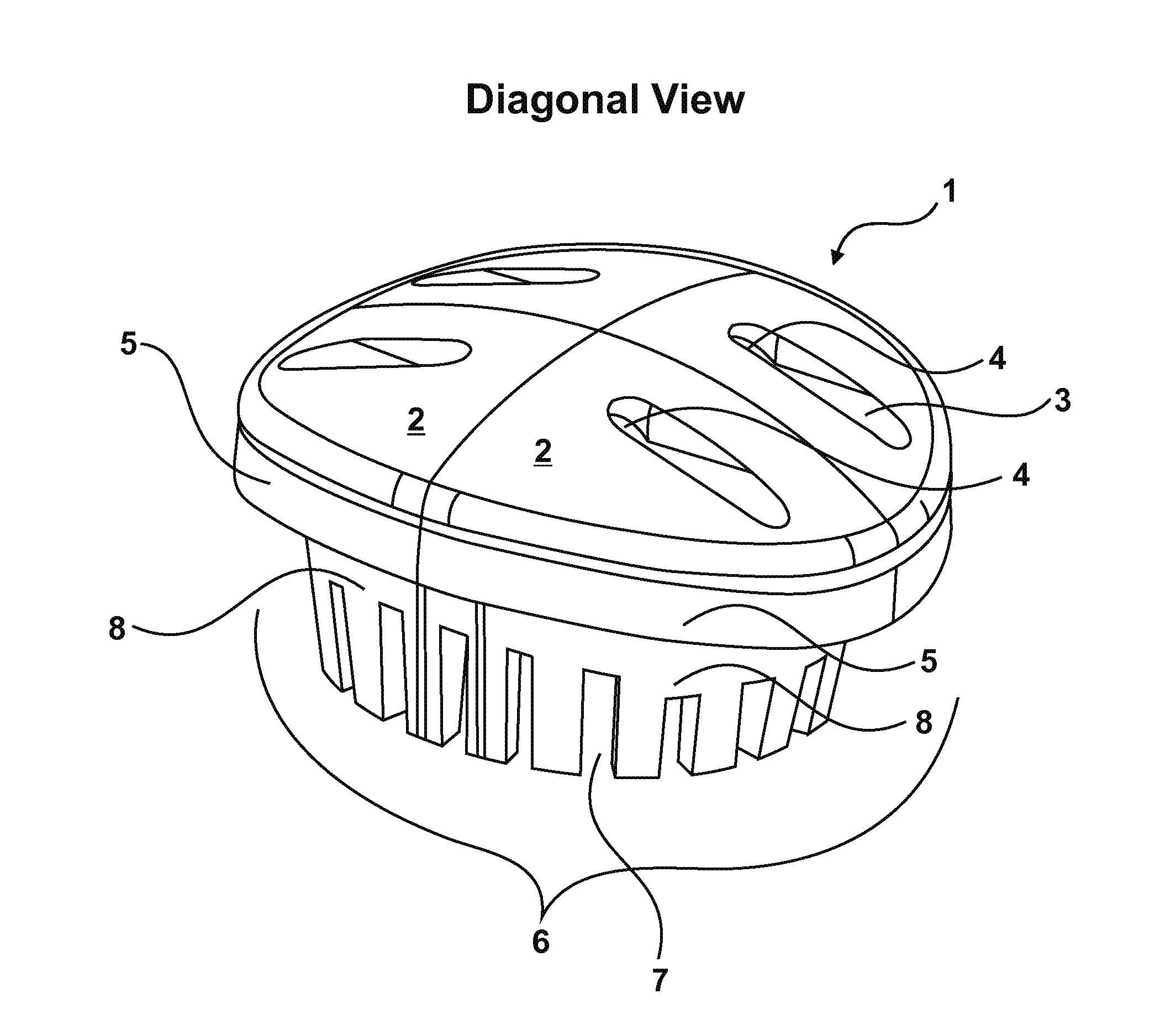
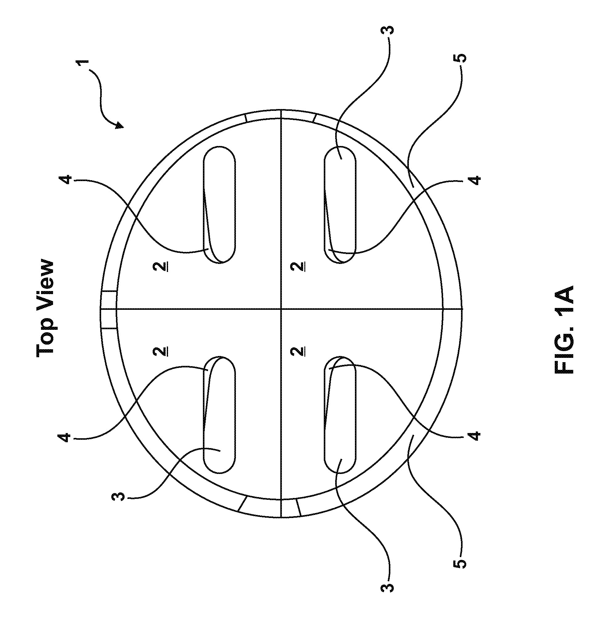
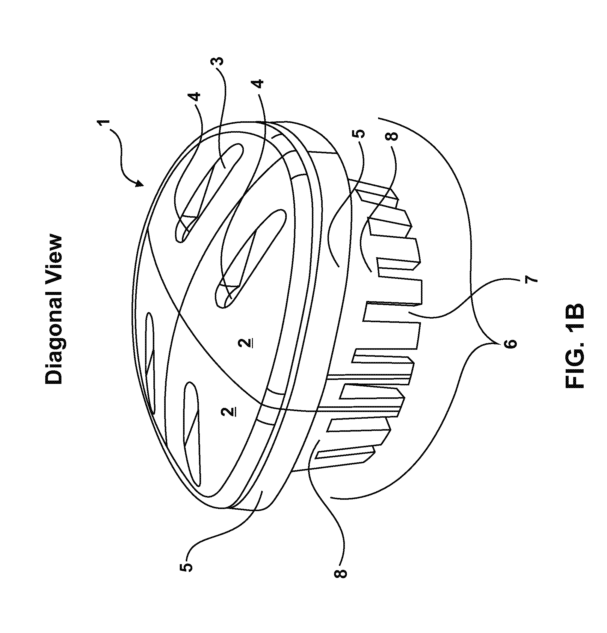
[0013]the invention is an extraction socket cap that is shaped and sized to seal an extraction site about the periphery thereof during a healing period. The shape of the socket cap can be rounded square, rounded triangle, circular, oval, or any desired shape depending on the anatomic characteristics of the extraction sites. The design incorporates features to allow the surgeon to advantageously place and position the socket cap on the extraction site while insuring that proper positioning and effective sealing about the gingival margin is secure prior to affixing the cap by suture attachment, adhesives, or fixation screws to the surrounding soft tissue.

[0014]The general dimensions and orientation of the structures shown in FIGS. 1A through F are examples of different embodiments of the invention. The socket cap features a top or cap portion designed to seal about the periphery of an extraction site. The top portion is mated to a bottom portion that extends downward and away from the...

second embodiment

[0027]Referring to FIGS. 2A-E, the present invention is an extraction site socket cage that provides support for reconstruction of the extraction socket site, including both bone and soft tissue, particularly where portions of the structural aspect of the socket walls have been destroyed by infection, disease, trauma or damaged as a result of the surgical process to extract the tooth. The complete socket cage 10 assembly is comprised of a series of substantially horizontal beams with curved semi-circular structure, which are arranged in a manner analogous to ribs. The horizontal ribs are position with pre-defined space between them, which are connected with the aid of two vertical struts. The substantial horizontal orientation provides that the individual beams are roughly parallel and an individual beam does not contact an adjacent beam along the length thereof.

[0028]Referring to FIGS. 2A-2B, the vertical strut 11 provides the vertical length and points of attachment for the horizo...


PUM


Abstract

The extraction socket devices are designed to protect the extraction socket immediately after tooth extraction. These devices protect the socket from oral bacteria and food, support a space to prevent collapse of the alveolar ridge during healing, contain any bone graft material inside of the socket, and provide comfort to patient during the healing process. Procedures are also disclosed for placing the devices in an extraction site to obtain the benefits of the disclosed devices.

Description

FIELD OF THE ART[0001]The present invention is dental devices for preservation and / or reconstruction of extraction sockets. Specifically, caps protect and seal the extraction socket and bone graft material placed within the sockets. Cages provide structural support following tooth extraction when portions of the extraction socket wall are pathologically destroyed.BACKGROUND[0002]Dental extraction of teeth or dental implants is necessitated by pathological diseases or trauma that damages the bone and tissue surrounding the socket. When the tissue or bone structure of the socket is compromised, the prognosis for successful repair is diminished. Extraction of teeth or implants initiates a cascade of healing responses that typically accompany growth of tissues to quickly fill and cover the resulting socket defects. The sequence of healing initially entails filling of the socket with a blood clot, the blood clot is replaced in turn by granulation tissue, followed by deposition of woven b...

Claims


Application Information

Patent Timeline
Patent Type & Authority Applications(United States)
IPC IPC(8): A61C8/00A61C8/02
CPCA61C8/0006A61C8/008A61C19/00A61C19/06A61C2201/00A61F2/2846
Inventor ZADEH, HOMAYOUN H.
Owner ZADEH HOMAYOUN H
Why Eureka
Try Eureka