Method for the analysis of progression of heterotopic ossification by Raman spectroscopy
a technology of raman spectroscopy and heterotopic ossification, which is applied in the direction of diagnostics using spectroscopy, instruments, catheters, etc., can solve the problems of prohibitively undesirable, side effects of steroidal anti-inflammatory drugs (nsaids), and cannot be recommended for widespread us
- Summary
- Abstract
- Description
- Claims
- Application Information
AI Technical Summary
Benefits of technology
Problems solved by technology
Method used
Image
Examples
example 1
Development of HO and determination of HO composition
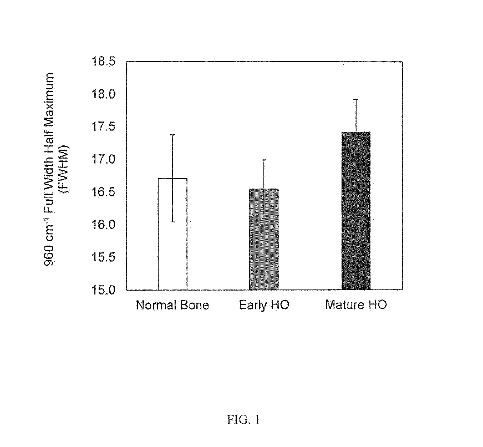
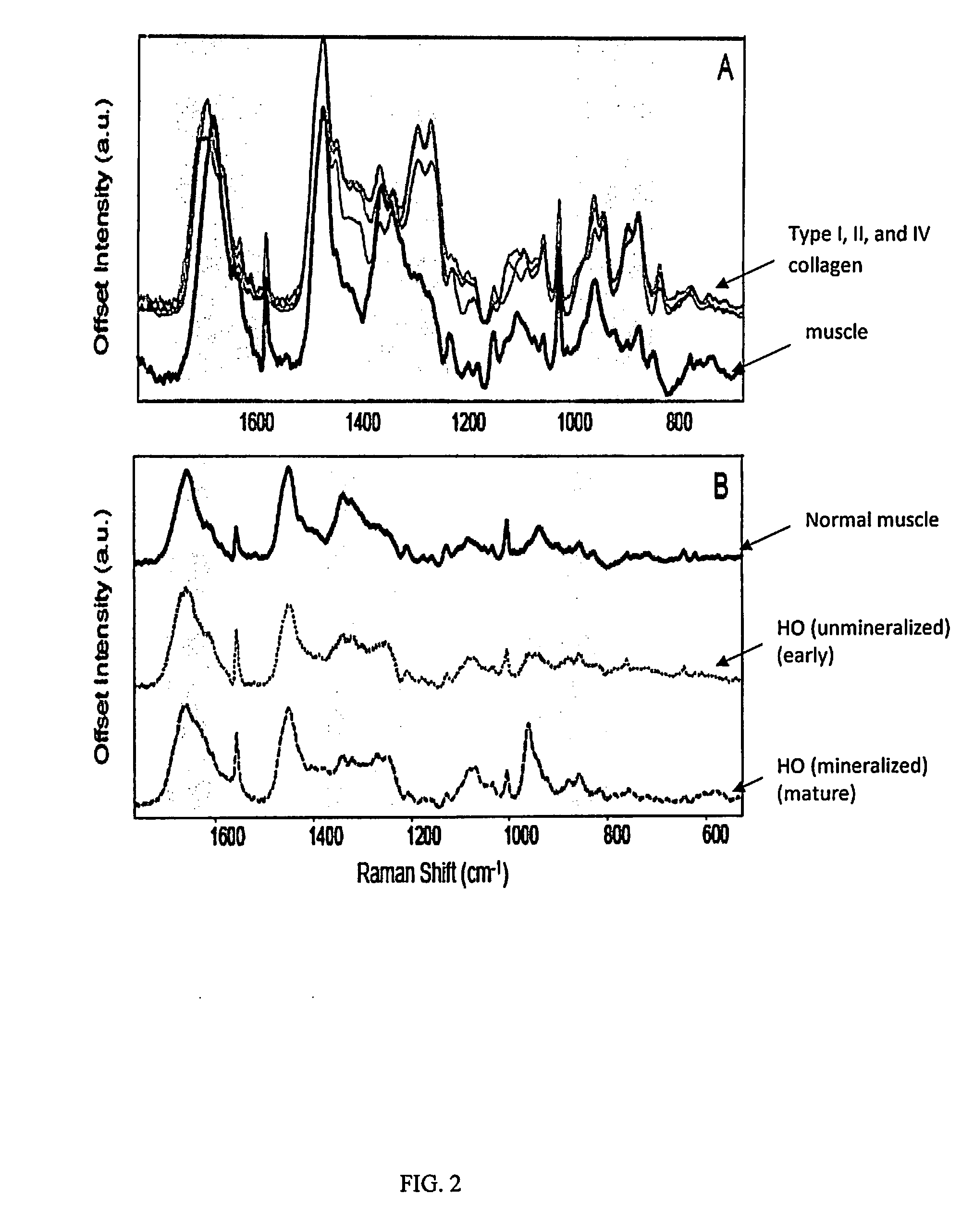
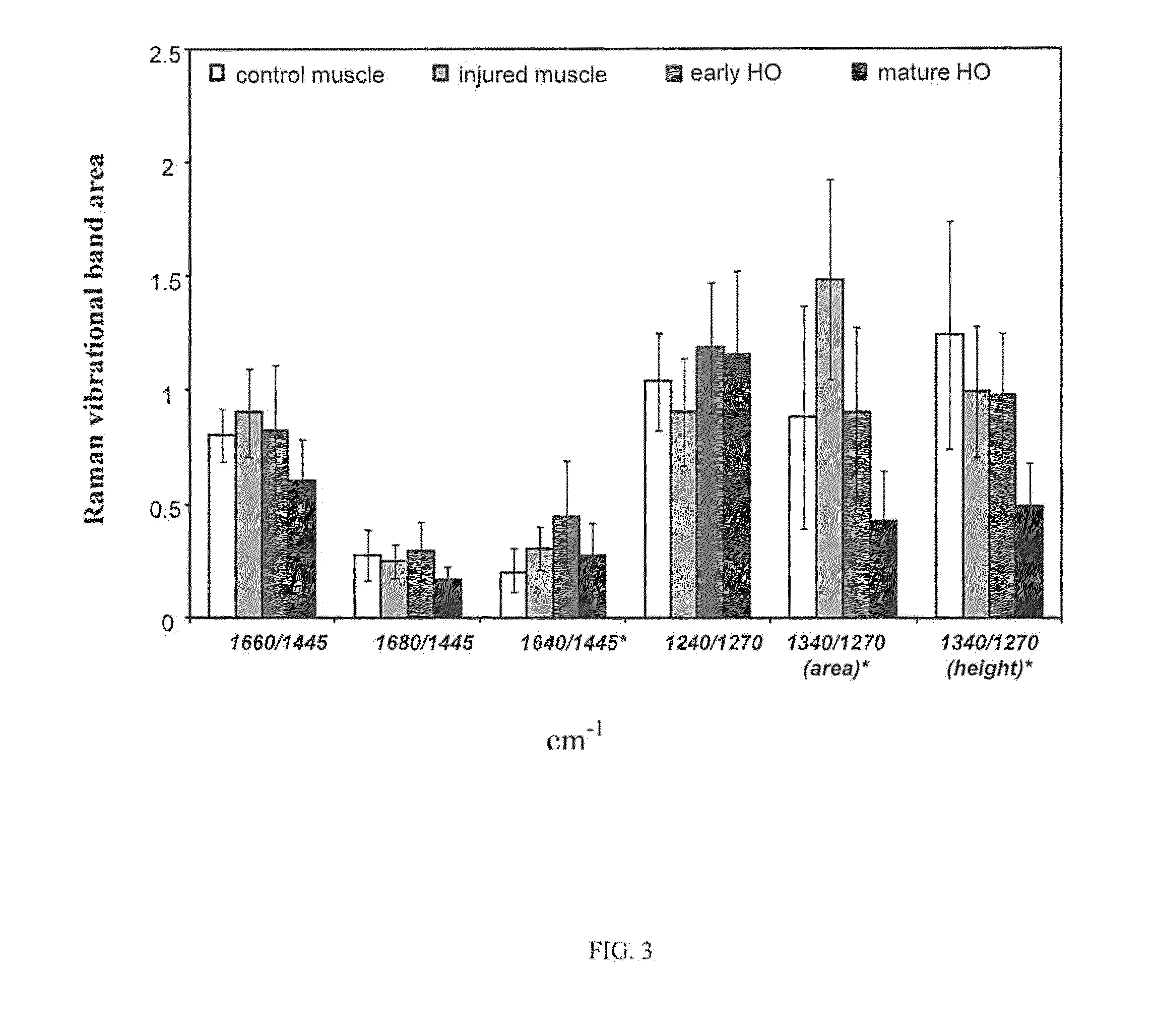
[0018]Raman spectral parameters can be used to determine the presence or maturity of hetertopic ossification (HO), which is defined as the aberrant formation of mature, lamellar bone in nonosseous tissue. Raman spectroscopy may be valuable in monitoring HO non-invasively (Potter, et al., J. Bone and Joint Surgery, Am., 92 Suppl 2: 74-89 (2010). Use of Raman spectroscopy for monitoring of HO has been suggested (Crane, et al., Proc. SPIE, 7895 (2011)) using band area ratios calculated from matrix bands 1660 / 1445, 1680 / 1445, 1640 / 1445, 1240 / 1270, and 1340 / 1270 cm−1. Similarly, band locations, but not band area ratios, has been suggested as a means of monitoring development of HO (Crane and Elster, J. Biomed. Opt. 17(1): 010902 (2012)).
[0019]One Raman spectral parameter, band area ratios (BARs), are calculated by dividing the band area of a Raman band (such as for 1070 cm−1) by another band area, for example 1445 cm−1. The association...
example 2
Determination of Progression of HO
[0035]When comparing muscle (normal or injured) to HO tissue (early or mature), there is a decrease in the 1660 / 1445 cm−1 (p−1 (p=0.03), and 1340 / 1270 cm−1 (α-helical structure, p−1 band area ratio (p−1 (protein order / disorder) band area ratio (p−1 and 1640 / 1445 cm−1 BARs as well as protein order / disorder BARs. In the transition from injured muscle to HO tissue, there is: 1) a decrease in the 1660 / 1445 cm−1 BAR, the α-helical structure BAR, and an increase in the protein order / disorder BAR.
[0036]Use of Raman spectroscopy to follow the development of HO progression is illustrated in FIG. 5. For FIG. 5, tissue samples were placed on an aluminum foil covered weighing dish prior to spectral acquisition. A 785 nm Raman PhAT system (Kaiser Optical Systems, Inc., Ann Arbor, Mich.) was used to collect spectra of the tissue biopsies. Final spectra were the accumulation of forty 5 second spectra, acquired using the 3 mm spot size. At least three dark-subtract...
example 3
Method for Monitoring HO Progress
[0039]Raman spectroscopy has distinct advantages over other techniques assessing tissue during surgery, such as histology or inspection by the surgeon. Frozen section and / or permanent pathologic analysis can be used to identify early stages associated with HO formation. However, this requires multiple biopsies, is time and labor intensive and may not be sufficiently precise. Also, the ability of the surgeon to identify early HO tissue during a debridement is subjective and relies on personal experience.
[0040]In a preferred embodiment, Raman spectral parameters, such as band areas, band area ratios, band height, ratio of band height, and decreases or increases from band center, are compared with other tissue, such as soft tissue (e.g., muscle) or bone in order to determine the composition of the measured tissue and determine the presence and maturity of HO. Shown in Table 4 are Raman spectral parameters associated with the determination of the presenc...
PUM
| Property | Measurement | Unit |
|---|---|---|
| spot size | aaaaa | aaaaa |
| volume | aaaaa | aaaaa |
| Raman | aaaaa | aaaaa |
Abstract
Description
Claims
Application Information
 - R&D
- Intellectual Property
- Life Sciences
- Materials
- Tech Scout
- Unparalleled Data Quality
- Higher Quality Content
- 60% Fewer Hallucinations
Browse by: Latest US Patents, China's latest patents, Technical Efficacy Thesaurus, Application Domain, Technology Topic, Popular Technical Reports.
© 2025 PatSnap. All rights reserved.Legal|Privacy policy|Modern Slavery Act Transparency Statement|Sitemap|About US| Contact US: help@patsnap.com


