
Inspection of dental roots and the endodontic cavity space therein

a technology of endodontic cavity space and endodontic root, which is applied in the field of dental microendoscope, can solve the problems of slowing down a clinical procedure, difficult to negotiate or clean, and insufficient conventional microscopes currently availabl

Inactive Publication Date: 2015-08-06
DENTSPLY SIRONA INC
View PDF8 Cites 14 Cited by
  • Summary
  • Abstract
  • Description
  • Claims
  • Application Information

AI Technical Summary

Benefits of technology

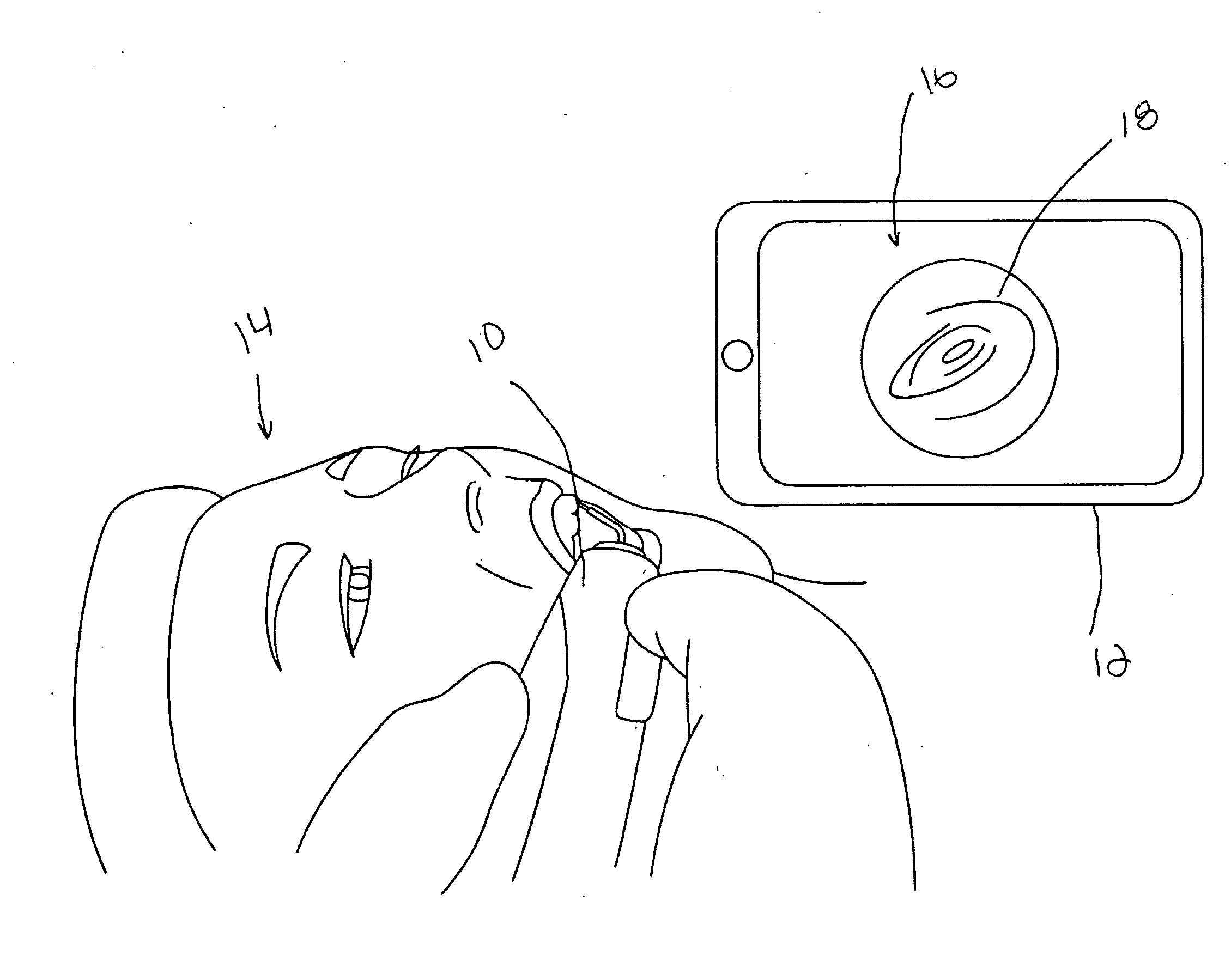
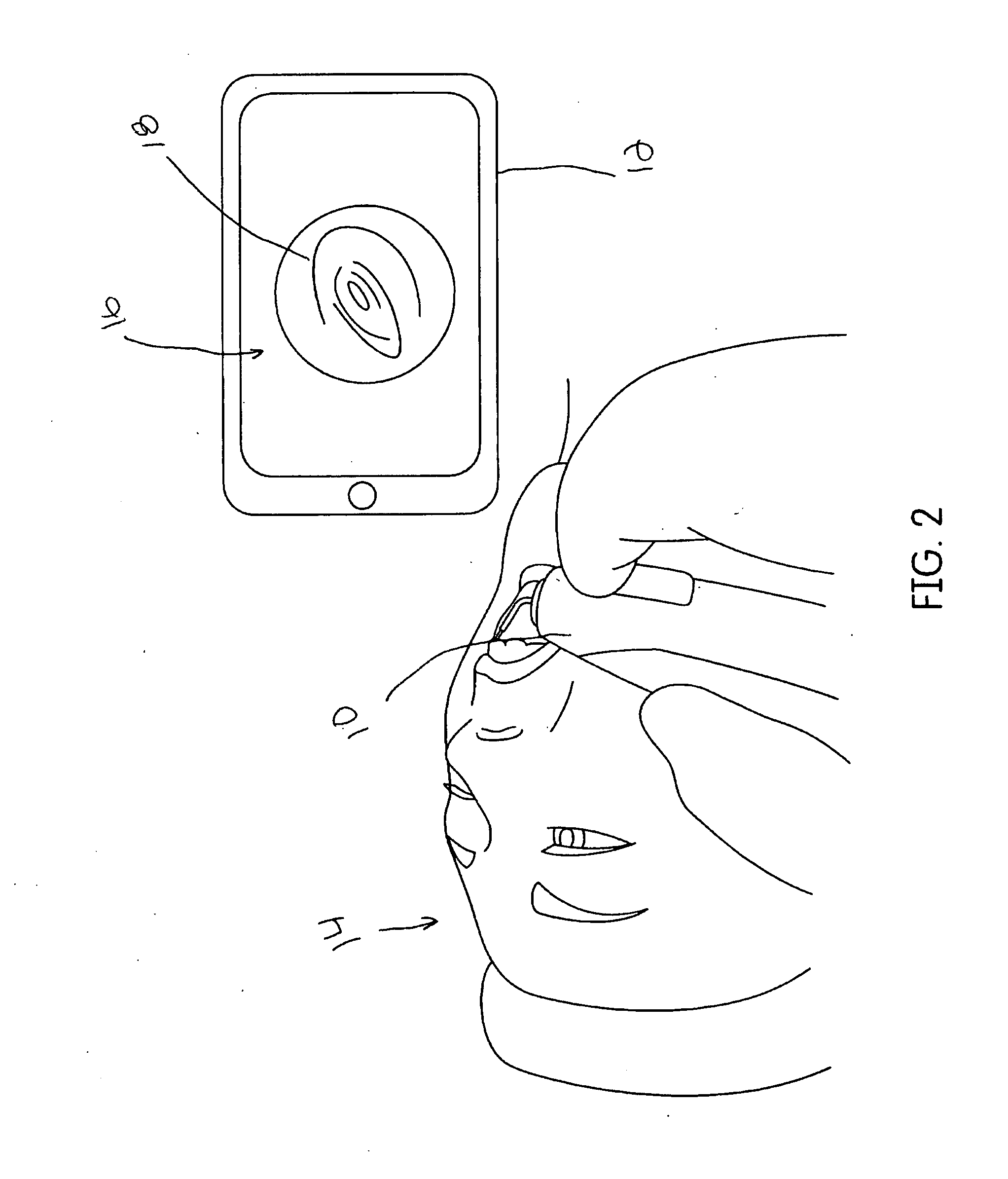
The present invention provides an improved micro-endoscope system for dental procedures that allows for real-time video and / or still images to be shared with a tooth or tooth root during a dental procedure. The system includes an endoscope with a fiber optic material, a heads-up display, and a micro-fiberscope for imaging root canal walls. The endoscope can also be attached to a disposable sheath for protection. The invention also includes a method for acquiring and sharing video and / or still images with a tooth or tooth root during a dental procedure. The images can be transmitted to a display, such as a television or computer monitor, through a wired or wireless network. The invention can also be used to identify bacteria, molds, or other molecules in the root canal by applying fluorophores or dyes that fluoresce under specific wavelengths.

Problems solved by technology

The root canal system of a human tooth is often narrow, curved and calcified, and can be extremely difficult to negotiate or clean.
Indeed, the conventional microscopes currently available are frequently inadequate in the complete observation of removal of the pulp and the efficient enlargement of the ECS.
Furthermore, they are usually predisposed to one observing angle at a time, further slowing a clinical procedure.
Often a mirror must be used for observation angles that cannot be seen by the standard dental microscopes and that those views are limited to the top portion of the canal for complex or curved canals.
Broken instruments are usually difficult, if not impossible to remove, often necessitating the removal of the root or tooth.
Injury to the tooth, which occurs as the result of a frank perforation or alteration of the natural anatomy of the ECS, can also lead to failure of the root canal and tooth loss.
Broken files are often out of the focal points of the dental microscope.
Viewing the canals themselves is difficult because of the root canal complex anatomy.
However, the technique is limited because viewing angles between teeth is nearly impossible depending upon the anatomy.
While dentists currently use a dental microscope to view images of teeth during examination, consultation with specialists normally requires that still images be sent to the consultant, delaying completion of the case.
The current methods of determining the level of cleaning the canal has reached in an operational clinical setting are inadequate; inadequate cleaning is thought to be a leading cause of RCT failure and results either in retreatment or more serious dental procedures.
This may cause a significant delay in treatment as well as reduced customer satisfaction.
The latter two are limited in use mainly for the top ⅓ of the canal or at the top of the canal due to the size.
This limits the image quality and the capability of discerning the cleanliness of the canal, objects or material in the canal, or canal defects or morphology.
At this time, there appears to be no product that has been accepted by the market for normal use.
As mentioned previously, there is literature describing the current technology, but it is not in normal use due to the above limitations.
The current methods of determining the level of cleaning the canal has reached in an operational clinical setting are inadequate; inadequate cleaning is thought to be a leading cause of RCT failure and results either in retreatment or more serious dental procedures.
The devices described previously for root canal inspection use white light for normal vision inspection and do not allow detection of microbial infections such as bacteria or mold.
Hand-eye motion between the tip placement and watching the still images or video on the monitor is not ideal in that the clinician's focus is on a screen away from the patient's mouth.

Method used


Image

  • Inspection of dental roots and the endodontic cavity space therein
  • Inspection of dental roots and the endodontic cavity space therein
  • Inspection of dental roots and the endodontic cavity space therein

Examples


Embodiment Construction

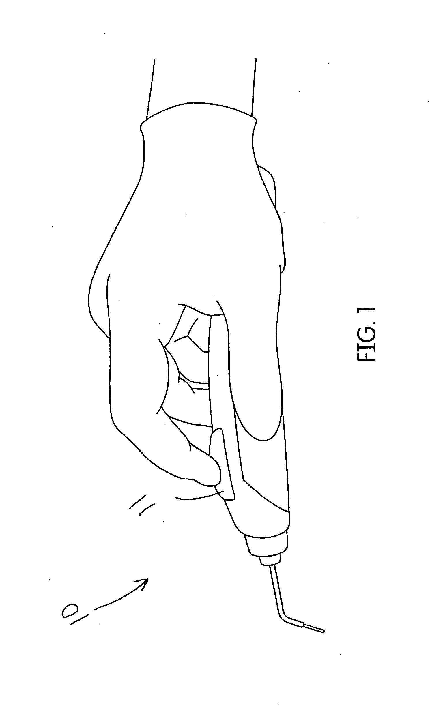
[0042]The present invention may provide an endoscopic handpiece assembly. More particularly, the present invention may provide a mini endoscopic handpiece assembly with a removable protective end portion (e.g., disposable guide tube) for illuminating and / or displaying and / or recording (either simultaneously, periodically, in combination, or separately) a root canal and / or cavity within a tooth or patient's oral cavity through a fiber optic cable or otherwise (e.g., micro-fiberscope attached to the body of the endoscopic handpiece assembly. It is appreciated that the endoscopic handpieces of the present invention may simultaneously illuminate and display inner cavity spaces of root canals during, in between, and / or after treatment of a root canal (e.g., cleaning and / or shaping of a root canal).

[0043]It is contemplated that in a live treatment case, the tooth of a patient undergoing an endodontic root canal treatment may be prepared using standard procedures whereby the infected pulp ...


PUM


Abstract

A system and method for acquiring and sharing in real time video and / or still images with a tooth or tooth root during a dental procedure using an endoscope.

Description

CROSS-REFERENCE TO RELATED APPLICATIONS[0001]This patent application claims the benefit of and priority to U.S. Provisional Application Ser. No. 61 / 936,513, filed on Feb. 6, 2014 and U.S. Provisional Application Ser. No. 62 / 049,624, filed Sep. 12, 2014, which are herein incorporated by reference for all purposes.FIELD OF INVENTION[0002]The present invention relates to the use of a dental micro-endoscope combined with an imaging camera, multiple optical fibers for coupling illumination and excitation energy, a wired or wireless transmission system for sending the still image or video information to a heads-up display of the dental root canals and other intra-oral areas in real time to patients, practitioners, consultants, and archives. In addition, dyes and fluorphores for staining and tagging various root canal native materials well as foreign material can be used with different excitation energies than in current dental scopes to fluoresce and visualize these materials; these image...

Claims


Application Information

Patent Timeline
IPC IPC(8): A61B5/00A61B1/24
CPCA61B5/0088A61B5/742A61B5/0071A61B1/24A61B1/00165A61B1/0017A61B1/247
Inventor AMMON, DANRIGSBY, STEPHENKEVIN, WILKINSONATKINSON, BRIAN
Owner DENTSPLY SIRONA INC
Learn More