Method for recovering rare cells and method for detecting rare cells

a rare cell and recovery method technology, applied in the field of rare cell recovery and detection, can solve the problems of insufficient cell recovery and extremely difficult detection, and achieve the effect of reducing the loss of rare cells that may exist in the cell suspension and high recovery ra

Active Publication Date: 2015-10-01
KONICA MINOLTA INC
View PDF4 Cites 5 Cited by
  • Summary
  • Abstract
  • Description
  • Claims
  • Application Information

AI Technical Summary

Benefits of technology

[0011]According to the present invention, even various kinds of cells contained in a cell suspension such as blood can be recovered into the microchamber(s) at an extremely high recovery rate, that is, almost all of the cells tha

Problems solved by technology

Although it is obvious that detection of such rare cells is clinically useful, the detection is extremely difficult.
In cases where this microdevice for cellular tissue body is used to planarly spread a cell suspe

Method used


Image

  • Method for recovering rare cells and method for detecting rare cells
  • Method for recovering rare cells and method for detecting rare cells
  • Method for recovering rare cells and method for detecting rare cells

Examples


example 1

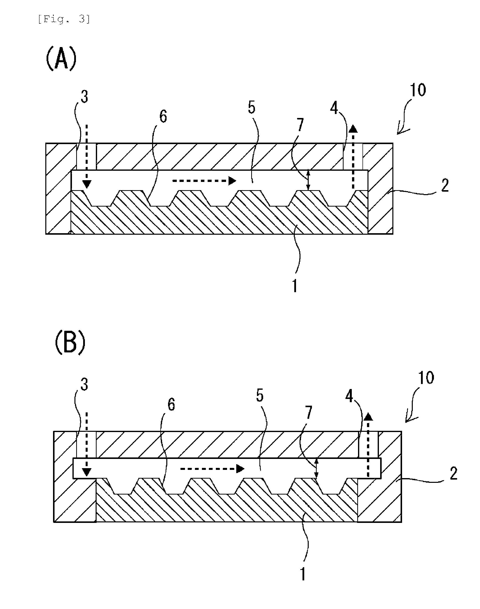
[0132]As a microchamber chip, a chip on which microchambers of 100 μm in diameter and 50 μm in depth were arranged in a lattice form (the pitch, which is the distance between the bottom centers of adjacent microchambers, was 210 μm) was used. This microchamber chip was made of polystyrene and had a bottom-flat inverted conical shape.

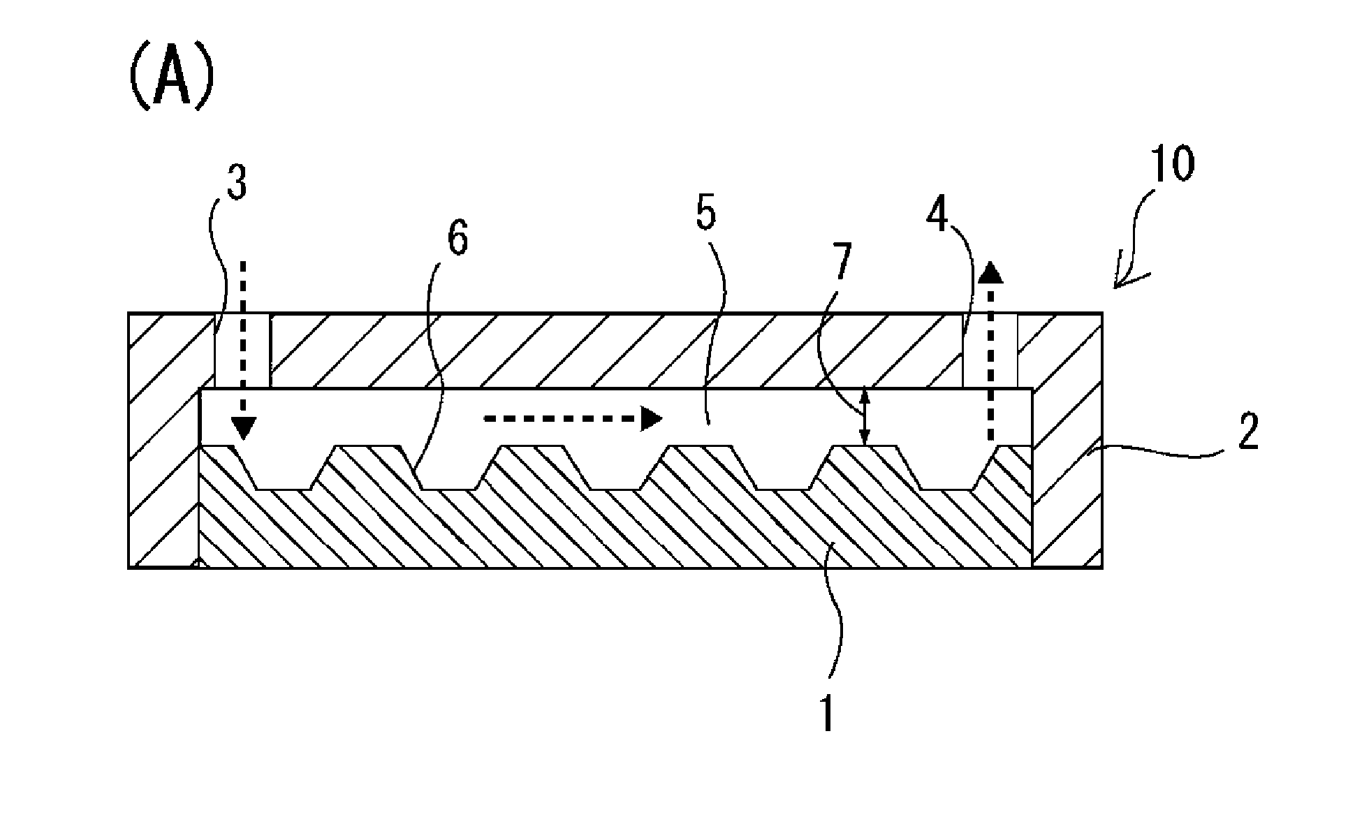
[0133]On the microchamber chip surface on which the microchambers were formed, a polystyrene-made flow path-forming frame was arranged such that a flow path of 15 mm in length, 26 mm in width and 100 μm in height was formed, and the microchamber chip and the flow path-forming frame were integrated with each other. The flow path of the thus obtained cell-spreading device had a cross-section of 1.5 mm2 and a volume of 39 mm3 (=39 μL). As a liquid-feeding device, KDS200 dual syringe pump (manufactured by KD Scientific Inc.) was used.

[0134]On the flow path-forming frame, an inlet port and an outlet port were each formed in an integrated manner, which inlet p...

example 2

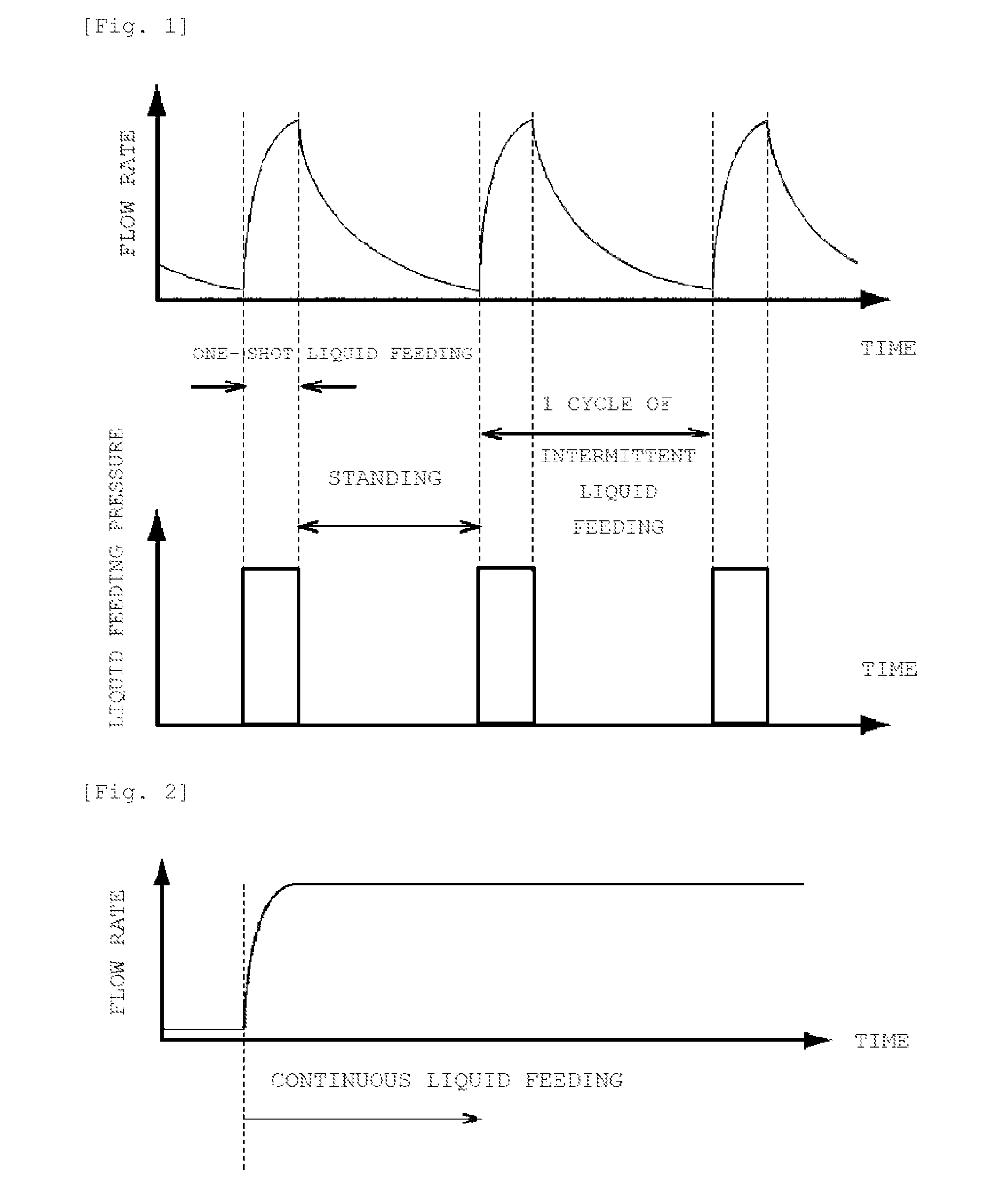
[0141]The step (Z) was carried out after the step (Y) of Example 1. That is, as the step (Z), 200 μL of PBS was continuously fed at a flow amount of 0.1 mL / min. As shown in FIG. 6, after the step (Y) of Example 1, the cells stored in some of the microchambers were in a layered form (FIG. 6(A); however, these cells were made into a monolayer by performing the step (Z).

example 3

[0142]After the step (Z) of Example 2, 200 μL of Hoechst 33342 stain was continuously fed at a flow amount of 0.1 mL / min. Three minutes later, the cells were observed under a microscope. The result thereof is shown in FIG. 7. This Hoechst 33342 stain was prepared by diluting “Hoechst 33342 10 mg / mL Solution in Water” manufactured by Molecular Probes Inc. in accordance with the manufacturer's instructions.

[0143]Although FIG. 7 shows only one microchamber, the number of stained cells corresponded to 90% of all cells introduced to the cell-spreading device.


PUM


Abstract

An object of the present invention is to provide a method of detecting rare cells in which, upon spreading a cell suspension in a flow path formed on a microchamber chip, the loss of rare cells is reduced by improving the cell recovery rate. The present invention provides a method of detecting rare cells from a cell suspension by using a cell-spreading device 10 comprising a microchamber chip 1, a flow path-forming frame 2, an inlet port 3, and an outlet port 4; and a method of recovering rare cells comprising: the step (X) of introducing a cell suspension to a flow path 5 via the inlet port 3 so as to spread cells in the flow path 5 on the microchamber chip 1; and the step (Y) of storing the cells spread in the flow path 5 on the microchamber chip 1 in microchamber 6s by intermittent liquid feeding.

Description

TECHNICAL FIELD[0001]The present invention relates to a method of recovering and detecting rare cells from cells contained in a cell suspension spread in a flow path on a microchamber chip by using a cell-spreading device comprising the microchamber chip.BACKGROUND ART[0002]Circulating tumor cells [CTCs], circulating vascular endothelial cells [CECs], circulating vascular endothelial precursor cells [CEPs], various stem cells and the like (hereinafter, these cells are collectively referred to as “rare cells”) are cells that extremely rarely exist in the whole blood depending on the pathological condition. Although it is obvious that detection of such rare cells is clinically useful, the detection is extremely difficult. In recent years, various separation methods have been applied in an attempt to perform the detection and various products have been commercialized; however, in any of these cases, because of the rarity of the subject, it is important to evaluate the results and effec...

Claims


Application Information

Patent Timeline
IPC IPC(8): G01N1/30
CPCG01N1/30B01L3/502761B01L2200/0668B01L2300/0816B01L2300/0877B01L2400/0487C12M47/04B01L2400/0409
Inventor ARAKI, JUNGOHOSHI, KUMIKO
Owner KONICA MINOLTA INC
Why Eureka
Try Eureka