Enhancing ultrasound images

Active Publication Date: 2015-10-15
KONINKLJIJKE PHILIPS NV
View PDF2 Cites 7 Cited by
  • Summary
  • Abstract
  • Description
  • Claims
  • Application Information

AI Technical Summary

Benefits of technology

[0003]There may thus be a need to provide image data showing the current situation, for example in a region of interest of a patient, wherein the image data requires reduced and minimum interpretation and combination skill on the side of the user.

Problems solved by technology

However, it has also been shown that providing fluoroscopic images and ultrasound images next to each other requires attention and awareness of the user mentally combining the two different contents shown in the respective images.

Method used


Image

  • Enhancing ultrasound images
  • Enhancing ultrasound images
  • Enhancing ultrasound images

Examples


Embodiment Construction

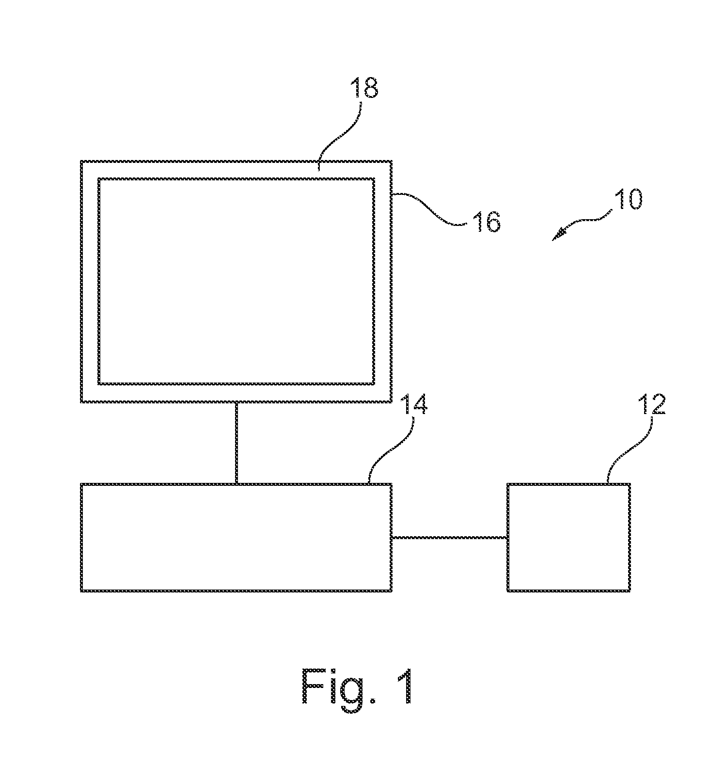
[0035]FIG. 1 shows an image processing device 10 for enhancing ultrasound images, comprising an image data input unit 12, a central processing unit 14, and a display unit 16. The image data input unit 12 is configured to provide an ultrasound image of a region of interest of an object, and to provide an X-ray image of the region of interest of the object.

[0036]The central processing unit 14 is configured to select a predetermined image area in the X-ray image, to register the ultrasound image and the X-ray image, to detect the predetermined area in the ultrasound image based on the registered selected predetermined image area, and to highlight at least a part of the detected area in the ultrasound image to generate a boosted ultrasound image. The display unit is configured to provide the boosted ultrasound image as guiding information on a display area 18.

[0037]In a further example (not further shown), the central processing unit is configured to project the selected area into the u...


PUM


Abstract

The present invention relates to image processing for enhancing ultrasound images. In order to provide image data showing the current situation, for example in a region of interest of a patient, an image processing device (10) for enhancing ultrasound images is provided that comprises an image data input unit (12), a central processing unit (14), and a display unit (16). The image data input unit is configured to provide an ultrasound image of a region of interest of an object, and to provide an X-ray image of the region of interest of the object. The central processing unit is configured to select a predetermined image area in the X-ray image, to register the ultrasound image and the X-ray image, to detect the predetermined area in the ultrasound image based on the registered selected predetermined image area, and to highlight at least a part of the detected area in the ultrasound image to generate a boosted ultrasound image. The display unit is configured to provide the boosted ultrasound image as guiding information on a display area (18).

Description

FIELD OF THE INVENTION[0001]The present invention relates to an image processing device for enhancing ultrasound images, to a medical imaging system, and to a method for enhancing ultrasound images, as well as to a computer program element and to a computer-readable medium.BACKGROUND OF THE INVENTION[0002]In medical examinations, such as endovascular interventions, different types of medical imaging can be used, for example the positioning of a mitral clip is performed under fluoroscopic X-ray imaging and ultrasound imaging for guidance purposes. The X-ray images are used for visualizing tools, such as catheters, clips, or the like, and also bone structures, whereas ultrasound imaging can be used for visualizing soft tissues. WO 2011 / 070492 A1 describes the combination of ultrasound image data and X-ray image data. An ultrasound probe is detected in the X-ray image, and acquisition settings of the ultrasound probe are visualized within the X-ray image. It has been shown that ultraso...

Claims


Application Information

Patent Timeline
Patent Type & Authority Applications(United States)
IPC IPC(8): G06T7/00G06T7/60A61B6/12A61B8/08A61B8/00A61B6/00G06T1/00G06T15/08
CPCG06T7/004G06T2215/12G06T7/0024G06T7/602G06T15/08A61B8/5261A61B8/463A61B8/4416A61B6/4417A61B8/469A61B6/469A61B6/12A61B8/0841G06T2207/10121G06T2207/10132G06T2207/30004G06T2207/20221G06T1/0007A61B6/4441A61B6/487A61B6/5247A61B8/0833A61B8/461A61B8/466G06T7/30G06T7/70G06T7/62
Inventor NEMPONT, OLIVIER PIERRECATHIER, PASCAL YVES FRANCOISFLORENT, RAOUL
Owner KONINKLJIJKE PHILIPS NV
Why Eureka
Try Eureka