

MRI with motion correction using navigators acquired using a dixon technique
a navigational technology and motion correction technology, applied in the field of magnetic resonance (mr), can solve the problems of blurring, misregistration, motion artifacts in the reconstructed mr image, etc., to improve the segmentation of relevant structures, improve the precision and robustness of motion descriptions, and improve segmentation. the effect of robustness
- Summary
- Abstract
- Description
- Claims
- Application Information
AI Technical Summary
Benefits of technology
Problems solved by technology
Method used
Image

Examples
Embodiment Construction
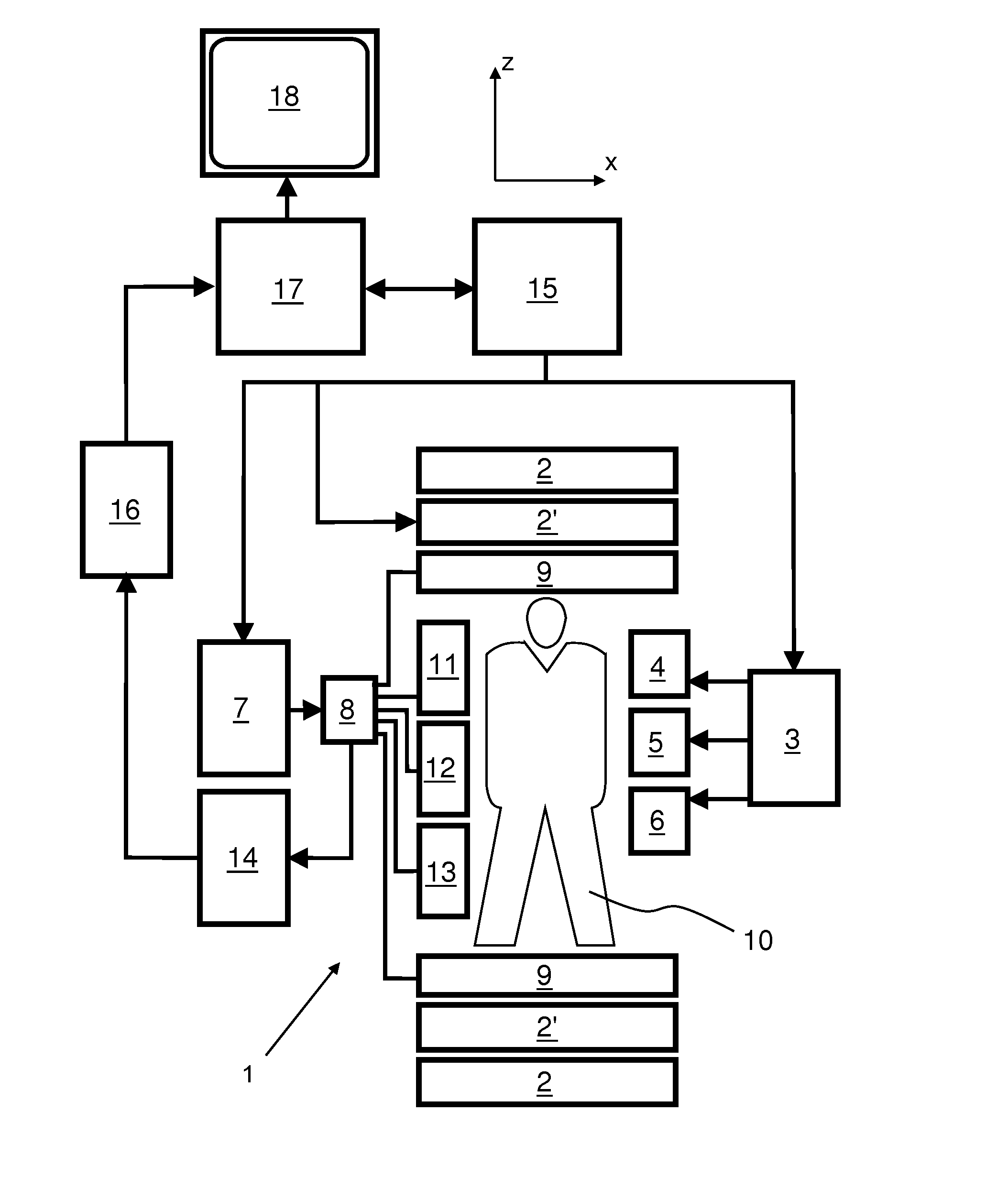
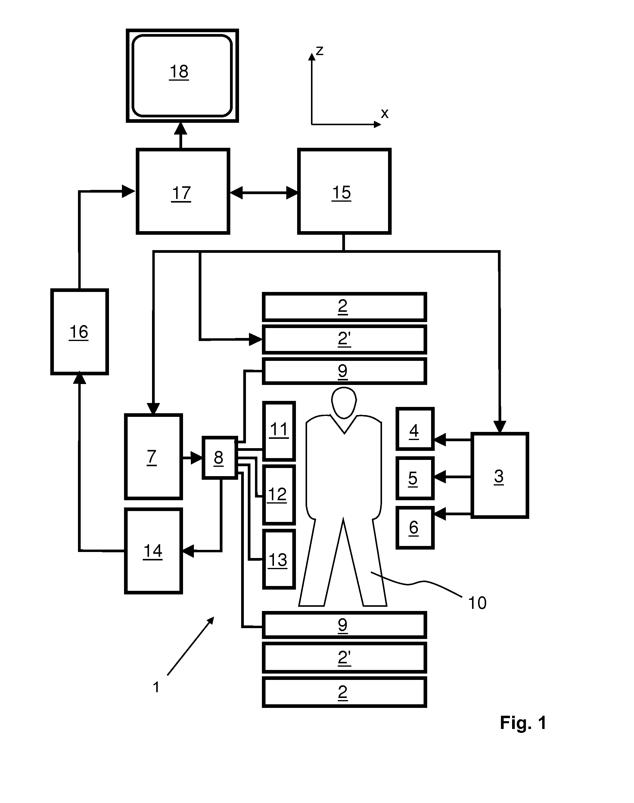
[0037]With reference to FIG. 1, a MR device 1 is shown. The device comprises superconducting or resistive main magnet coils 2 such that a substantially uniform, temporally constant main magnetic field B0 is created along a z-axis through an examination volume. The device further comprises a set of (1st, 2nd, and—where applicable—3rd order) shimming coils 2′, wherein the current flow through the individual shimming coils of the set 2′ is controllable for the purpose of minimizing B0 deviations within the examination volume.
[0038]A magnetic resonance generation and manipulation system applies a series of RF pulses and switched magnetic field gradients to invert or excite nuclear magnetic spins, induce magnetic resonance, refocus magnetic resonance, manipulate magnetic resonance, spatially and otherwise encode the magnetic resonance, saturate spins, and the like to perform MR imaging.
[0039]Most specifically, a gradient pulse amplifier 3 applies current pulses to selected ones of whole-...
PUM
 Abstract
Description
Claims
Application Information