

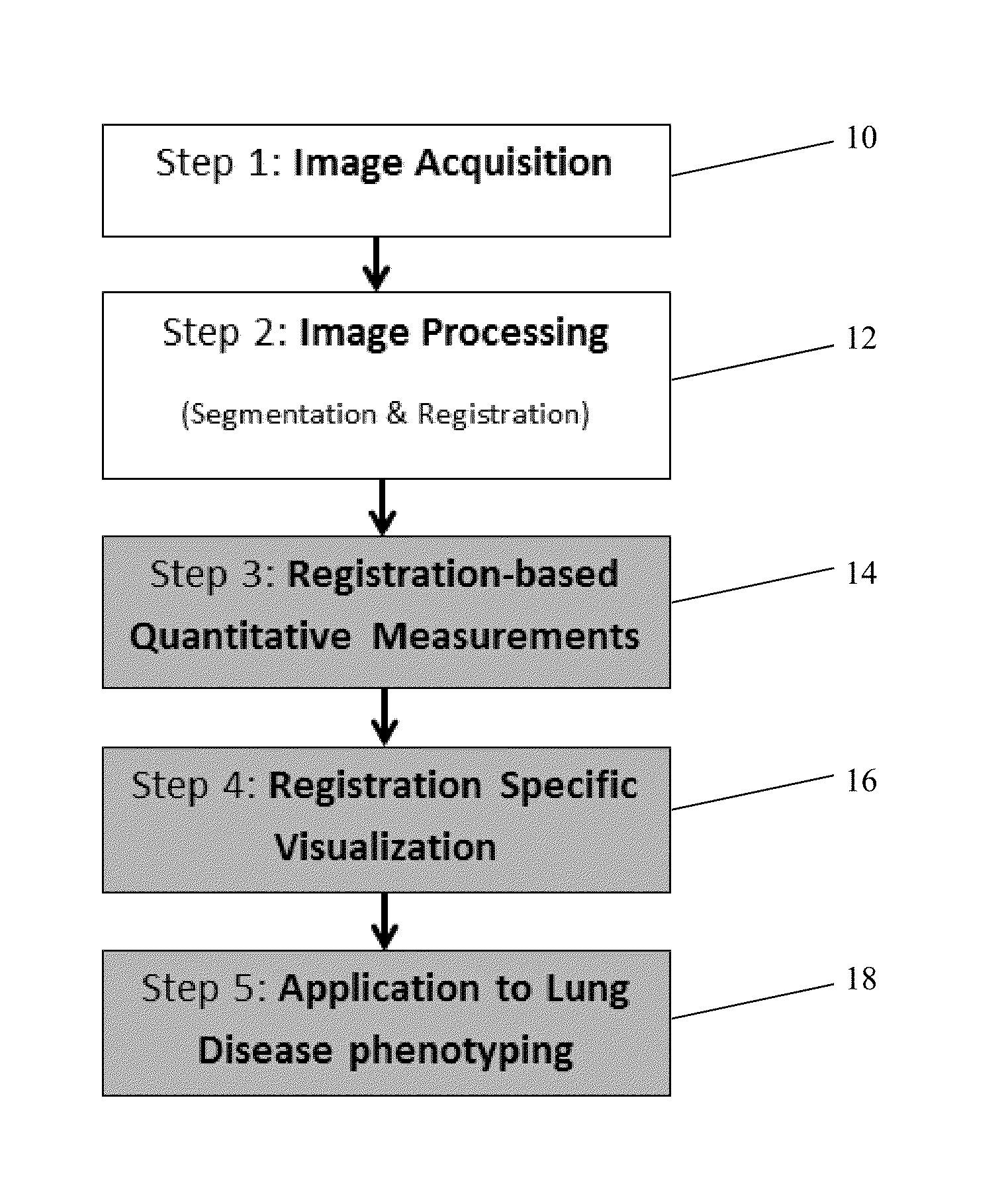
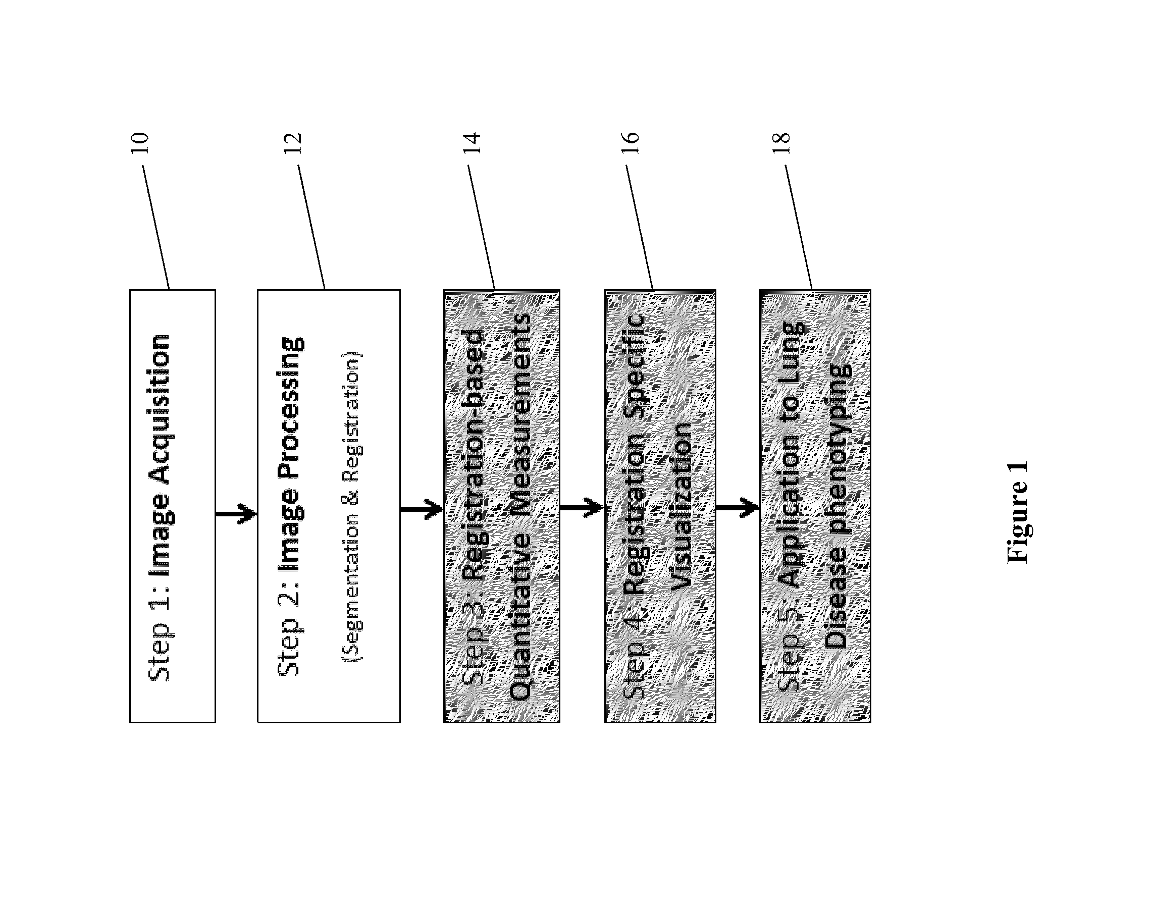
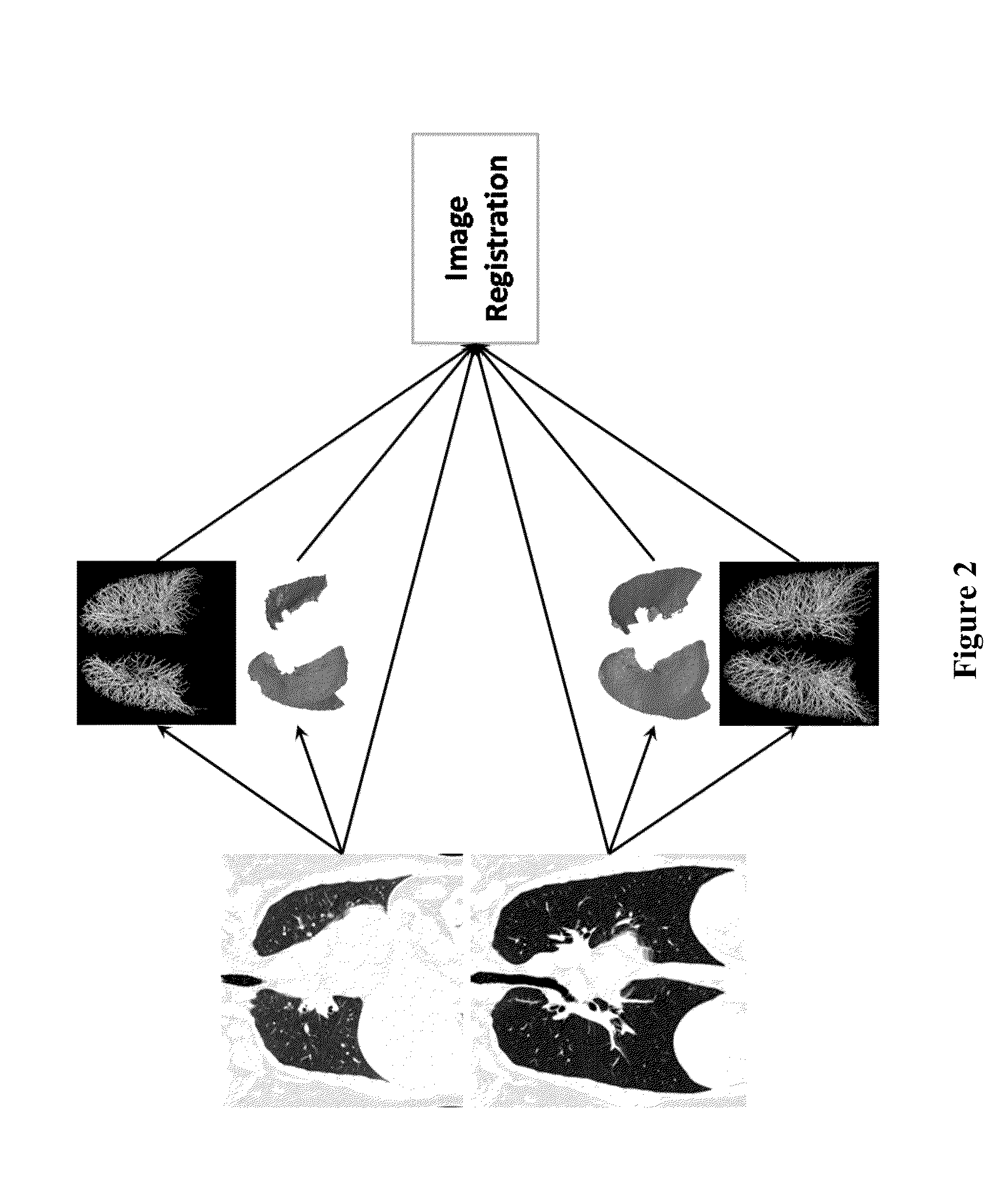
Visualization and quantification of lung disease utilizing image registration
a technology of image registration and lung disease, applied in image enhancement, tomography, instruments, etc., can solve the problems of loss of elasticity, high invasiveness, risky and uncomfortable for patients, and difficulty in understanding the disease within each patien
- Summary
- Abstract
- Description
- Claims
- Application Information
AI Technical Summary
Benefits of technology
Problems solved by technology
Method used
Image

Examples
example 1
[0090]Various examples were performed to compare the diagnostic capabilities of the registration methods described herein with other methods. A group of 188 subjects previously diagnosed with COPD at various GOLD stages were used for these examples. The gender, age, weight, and height of the patients in each GOLD stage are shown in FIG. 20. For each subject, full inspiration and full expiration CT images were registered and DPM values were calculated as described above.
[0091]CT based lung function measurements were calculated using the CT images of the subjects, including the EI ratio, the relative volume change between −860 HU and −950 HU (referred to as RVC), gas trapping measurements (referred to as Exp_LAA—856), and percentage of normal tissue computed using a threshold-based registration method (referred to as Galban NormalPerc). Each of these values, as well as the DPM derived normal tissue percentage (DPM_NormalPerc) value, was compared to clinical measurements of lung functi...
example 2
[0098]In another example, the CT images of current and former smokers were analyzed to determine whether the functional assessments described herein could be used to refine existing lung cancer screening guidelines through the use of independent quantitative CT (QCT) biomarkers of lung cancer.
[0099]Full inspiration and expiration CT images were obtained from 322 current or former smokers between the ages of 40 and 89 years old. Of these subjects, 42.5% (n=137) had proven lung cancer, and cancer staging was available for 101 subjects, which included 27.7% stage I (IA=17; IB=11), 18.8% stage II (IIA=11; IIB=8), 27.7% stage III (IIIA=18; IIIB=10), and 25.7% stage IV (IV=26). The images of each patient were processed and registered using nonlinear lung registration, CT functional measurements were calculated, and disease probability measurements were determined as described herein, including DPM_AirTrapPerc, DPM_EmphPerc, and DPM_NormalPerc.
[0100]A logistic regression analysis was perfo...
PUM
 Abstract
Description
Claims
Application Information