Ultrasound assisted catheter placement system

a technology of ultrasound assisted catheter placement and placement system, which is applied in the direction of catheters, applications, guide wires, etc., can solve the problems of catheter itself not being able to visualize, catheters are placed incorrectly, and the possibility of misplacement of guide wires is increased, so as to achieve convenient use and reliable and convenient use

Inactive Publication Date: 2015-12-10
SONOSTIK LLC
View PDF13 Cites 3 Cited by
  • Summary
  • Abstract
  • Description
  • Claims
  • Application Information

AI Technical Summary

Benefits of technology

[0015]Accordingly, it is an object of the invention to provide a needle / catheter introduction apparatus that can be operated with one hand. It is a further object to provide an introduction apparatus that is reliable and easy to use. It is a further object of the invention to provide a catheter that can be visualized with ultrasound. It is yet another object of the invention to provide a probe cover that is easy to use, reliable, and sterile. It is still a further object of the invention to provide an assembly or kit with components needed for ultrasound guided catheter introduction to peripheral veins or arteries.
[0016]A needle / catheter introducer is provided that can be operated by one hand. The introducer has a wheel located toward the front end of the device that is rotated by the index finger of the user. After placement of the needle in the lumen of the vessel, the user rotates the wheel, which advances the wire guide through the center of the needle and into the patient. Once the guide wire is advanced into the vessel lumen the catheter can be advanced over the guide wire with a hub or finger tab on the catheter close to the index finger. The introducer fits in the palm of the user's hand and the wheel can be operated by the user's index finger. Because the actions needed to advance the guide wire and advance the catheter can both be comfortably performed by one hand without moving the hand from its initial position, the catheter introducer is easy to use and allows the user to also operate an ultrasound detection device during insertion without the assistance of another person.
[0018]In addition, a probe cover is provided that is pre-sterilized and packaged in a cardboard with the top sides of the cover folded down over the cardboard. The cover is placed over the probe end, the sides of the cover are folded up, and the cardboard is removed. The sterile probe cover can be quickly and efficiently placed over the ultrasound's transducer end.

Problems solved by technology

However, these catheter introduction sets require the user to use two hands to operate the device.
This means that one hand has to be taken off the ultrasound probe to feed the guide wire.
Consequently, a single user cannot visualize the guide wire directly while threading the vessel, which increases the possibility of misplacement of the guide wire.
The guide wire and the introducer needle can be visualized under ultrasound, but the catheter itself cannot be visualized.
Because the catheter cannot be visualized, sometimes catheters are placed incorrectly.
Arteries flow next to veins and can be easily catheterized accidentally when aiming for a vein.
Accidental placement of a catheter in an artery instead of a vein can lead to destruction of tissues receiving blood from that artery due to application of medications that damage arteries and are intended only for use in veins.
Without the use of a directional marker the physician can become disoriented to the longitudinal direction.
However, currently available catheter introduction sets do not incorporate a means to make directional markings on the skin.
Currently there are no kits available for catheter introduction in the anti-cubital veins of the upper arm.
Because no such kits are available, practitioners do not have readily available means to stabilize the arm for catheterization.
Likewise, additional body parts that are catheterized besides the arm, also do not have kits available that provide body part stabilizing means.
Often the speed of vascular access can be a matter of life or death.
This procedure is invasive, meaning that there is the chance of bacteria being introduced from the skin into the vessel, which is why the procedure must be performed under sterile conditions.
Ultrasound probes used repeatedly have a propensity to be contaminated.
Current probe covers for probes used in ultrasound guided vascular access are large, bulky and time consuming to put on.
However, the necessary equipment is often scattered throughout the department or not available in the department, making the procedure even more difficult or impossible to perform.
With the deep and small nature of the vessels used in the ultrasound guided IV catheter technique it can take more time and attention to place these catheters.
A slight slip of the catheter can cause displacement of the catheter from the lumen.
In addition, sterile lubrication used for the ultrasound makes it difficult for tape and tegaderm to adhere to the skin.
All of these factors make it difficult and essential to secure an ultrasound guided IV catheter.
The placement of an ultrasound guided IV catheter is often painful to the patient.
The large gauge is necessary for the rapid transfusion of medicines, fluids and IV contrast, and this can be quite painful, especially because the placement of ultrasound guided IV catheters usually takes longer than the conventional method with a sharp needle being slowly manipulated beneath the skin.
Ultrasound guided catheter introduction kits are currently available for central venous access, but no kits are available for ultrasound guided peripheral venous access.

Method used


Image

  • Ultrasound assisted catheter placement system
  • Ultrasound assisted catheter placement system
  • Ultrasound assisted catheter placement system

Examples


Embodiment Construction

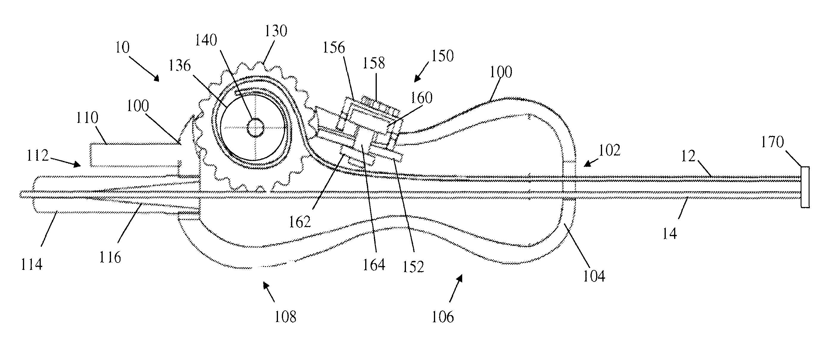
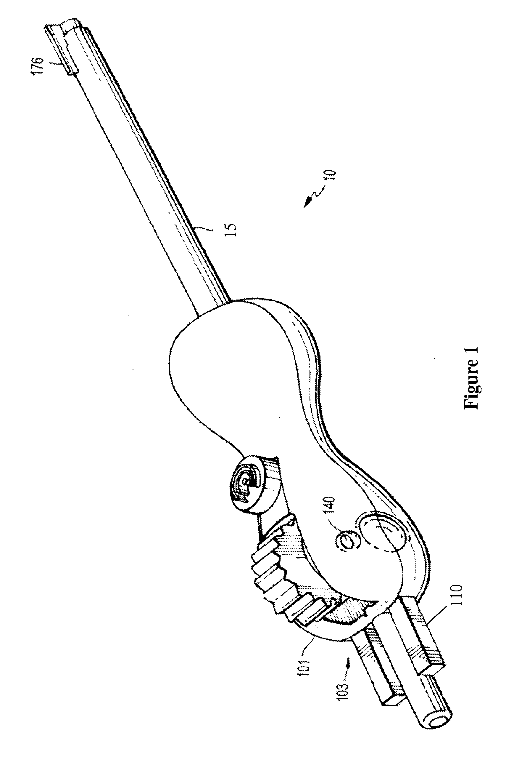
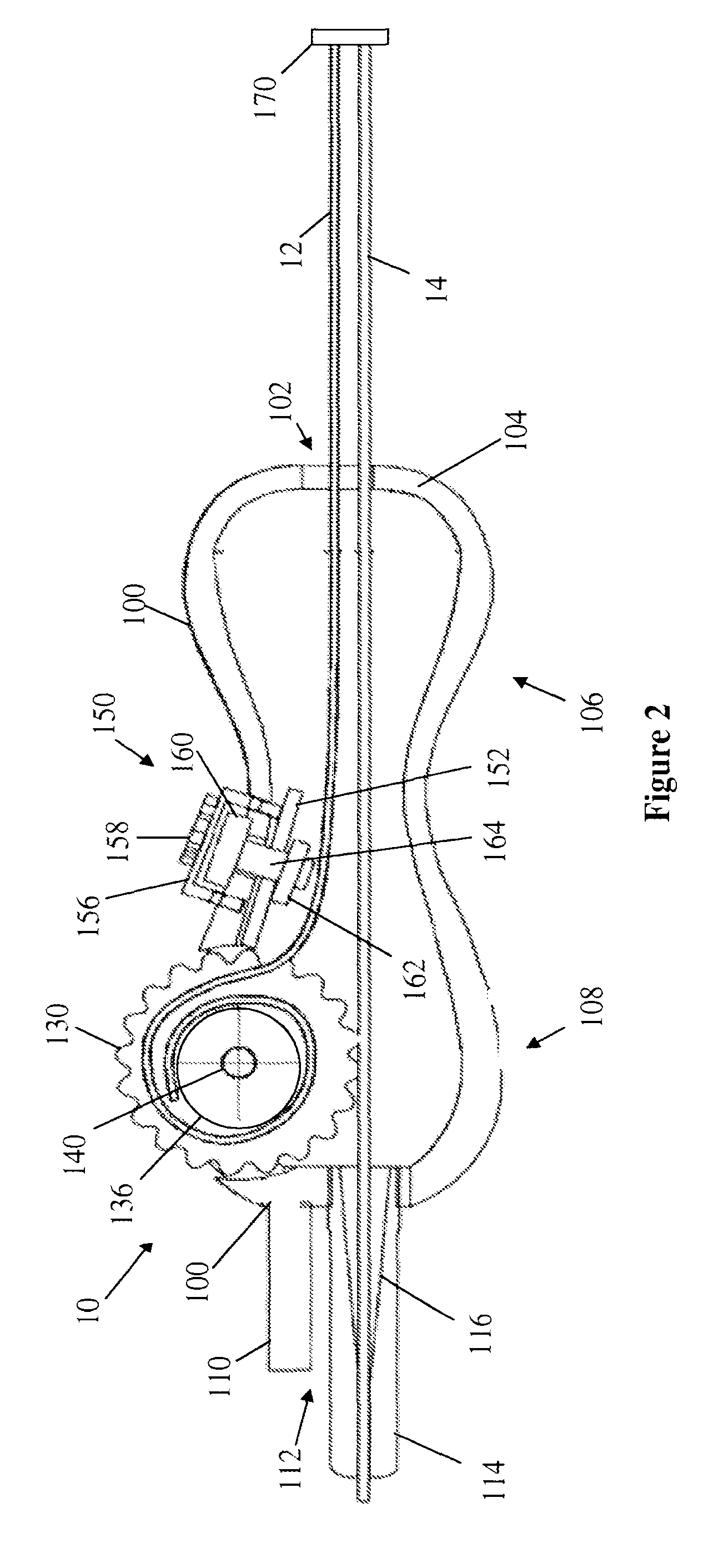
[0042]In describing a preferred embodiment of the invention illustrated in the drawings, specific terminology will be resorted to for the sake of clarity. However, the invention is not intended to be limited to the specific terms so selected, and it is to be understood that each specific term includes all technical equivalents that operate in similar manner to accomplish a similar purpose. Several preferred embodiments of the invention are described for illustrative purposes, it being understood that the invention may be embodied in other forms not specifically shown in the drawings.

[0043]This catheter has several features which will improve medical treatment. With these added features, the catheter makes IV placement with ultrasound easier, more efficient, and more accurate, thus significantly decreasing pain experienced by the patient by reducing the number of IV sticks currently performed.

[0044]As shown in the drawings the invention comprises of multiple elements that have separa...


PUM

PropertyMeasurementUnit
diameteraaaaaaaaaa
widthaaaaaaaaaa
widthaaaaaaaaaa

Abstract

A deep vein intravenous introducer has a wheel located toward the front end of the device that can be rotated by the index finger of the user. After placement of the needle in the lumen of the vessel, the user rotates the wheel, which advances the wire guide through the center of the needle and into the patient. Once the guide wire is advanced into the vessel lumen the catheter can be advanced over the guide wire with a hub or finger tab on the catheter close to the index finger. The operation can be performed by one hand without moving the hand from its initial position. The user can then simultaneously use the other hand to operate an ultrasound detection device during insertion without the assistance of another person. A catheter is provided that can be visualized by ultrasound to enable confirmation of correct placement in a vessel. A probe cover is provided that is pre-sterilized and packaged in a cardboard with the top sides of the cover folded down over the cardboard. The cover is placed over the probe end, the sides of the cover are folded up, and the cardboard is removed. The sterile probe cover can be quickly and efficiently placed over the ultrasound's transducer end. Finally, a kit is provided that contains all of the medical devices needed to perform cannulation.

Description

RELATED APPLICATIONS[0001]This application claims the benefit of U.S. Provisional Application No. 61 / 770,052, filed Feb. 27, 2013, the entire contents of which are incorporated herein by reference.BACKGROUND OF THE INVENTION[0002]1. Field of the Invention[0003]This invention relates to a system, device and method for the introduction of catheters and more particularly to the introduction of catheters into small diameter blood vessels, deep vessels, central veins, arteries, and those visualized by ultrasound.[0004]2. Background of the Related Art[0005]Catheter introduction sets are generally known. For instance, U.S. Pat. No. 4,417,886 discloses a catheter introduction set having a needle, catheter, wire guide and wire guide feed device in which when the needle is positioned into a lumen of a blood vessel, a wire guide is first inserted into the vessel and the catheter is fed over the wire guide from the use of a radially extended handle into the lumen. This arrangement is similarly ...

Claims


Application Information

Patent Timeline
Patent Type & Authority Applications(United States)
IPC IPC(8): A61M25/00A61B8/00A61G13/12A61M25/09A61M25/06
CPCA61M25/002A61M25/09041A61M25/0606A61M25/0662A61G13/1235A61B8/0841A61G13/126A61M25/0108A61M2210/12A61M2025/0681A61B8/4422Y10T29/49828A61M2025/09116A61M2210/005
Inventor ALMANSOURI, HAWAACASTRO, NATHAN J.CORMAN, ADAMHOLMES, BENJAMINSIKKA, NEAL K.
Owner SONOSTIK LLC
Why Eureka
Try Eureka