Optogenetic probes for measuring membrane potential
a technology of optogenetic probes and membrane potentials, applied in the direction of instruments, peptides, fluorescence/phosphorescence, etc., can solve the problems of slow electrophysiological experiment setup, inability to access deep buried tissues, and difficult to measure membrane potentials, etc., to improve sensitivity, improve properties, and increase brightness
- Summary
- Abstract
- Description
- Claims
- Application Information
AI Technical Summary
Benefits of technology
Problems solved by technology
Method used
Image
Examples
example 1
Directed Evolution and Engineering of an Arch-Based Voltage Indicator
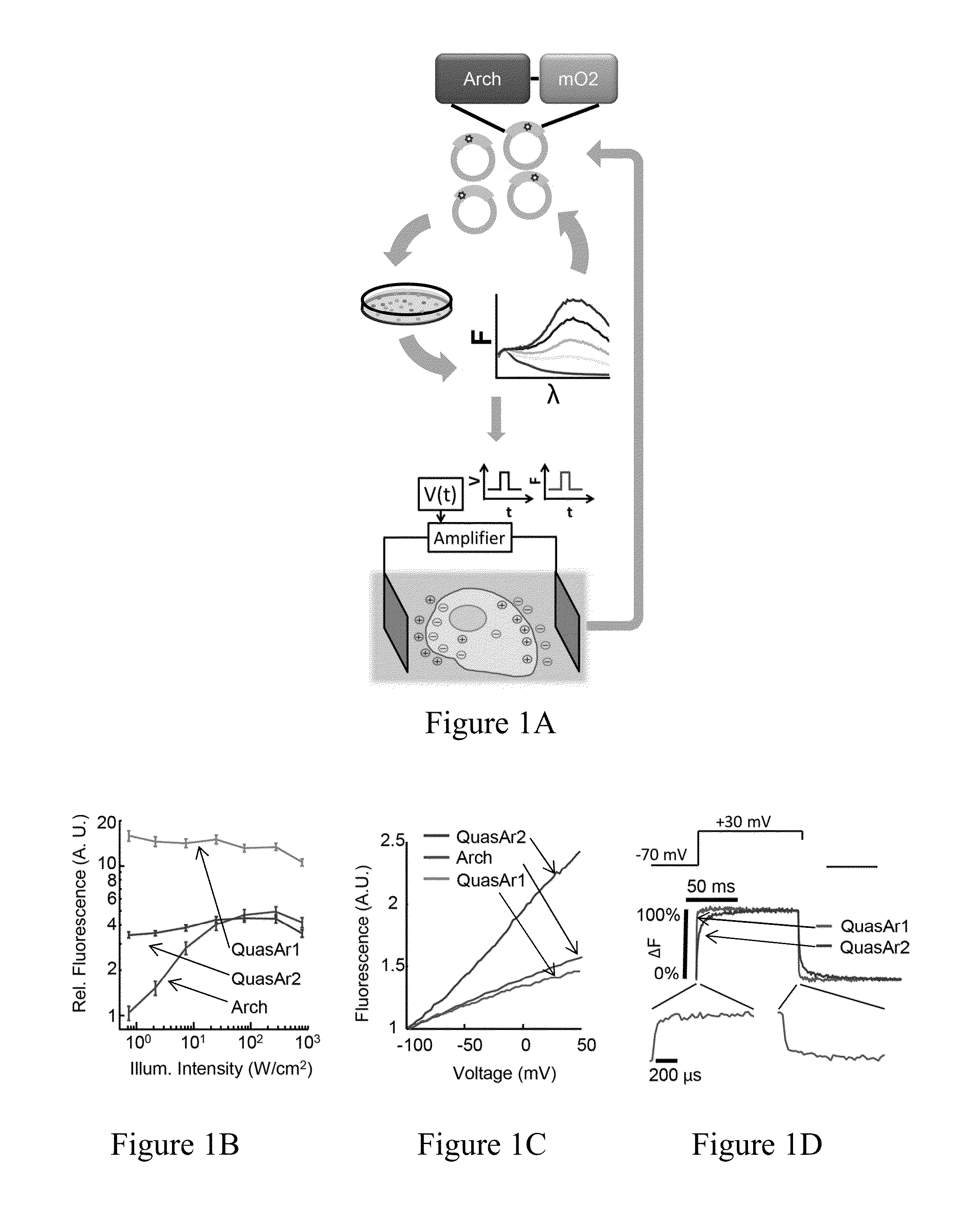
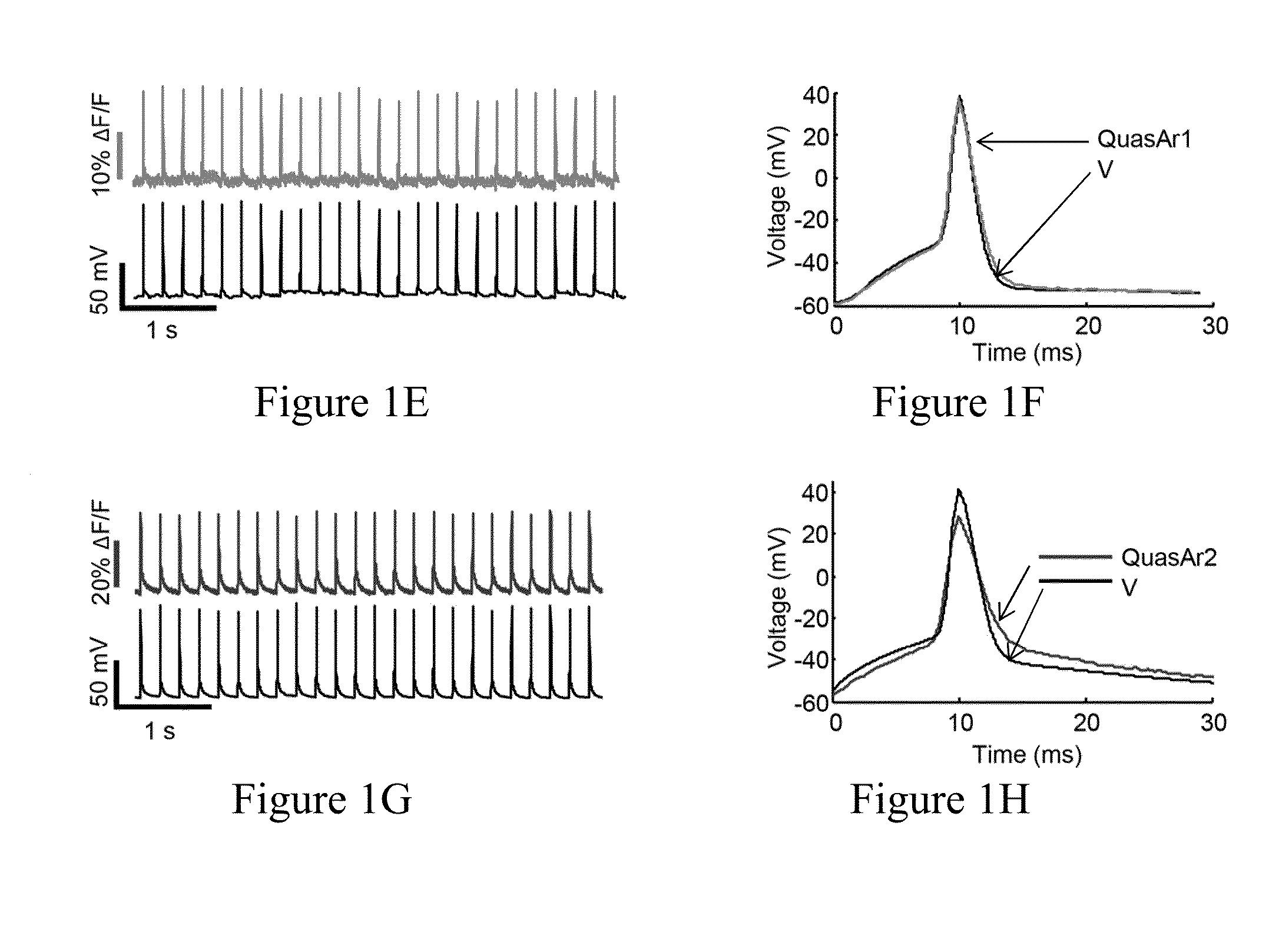
[0391]We previously showed that Archaerhodopsin 3 (Arch) functions as a fast and sensitive voltage indicator.10 Arch has the furthest red-shifted spectrum of any GEVI, giving it the unique property of little spectral overlap with channel rhodopsin actuators and GFP-based reporters. Thus it is natural to pair Arch-based indicators with optogenetic actuators for crosstalk-free all-optical electrophysiology.
[0392]However, wild-type Arch had some undesirable attributes for a reporter: it was very dim, and the brightness was a nonlinear function of illumination intensity.24 Illumination for imaging generated a hyperpolarizing photocurrent, which partially suppressed neural firing. The mutant Arch(D95N) did not pump, but its step response was dominated by a 41 ms time constant, too slow to resolve action potential (AP) waveforms,
[0393]We sought to repair these defects in engineered mutants of Arch. To accommodate the mu...
example 2
Comparison of QuasArs to Firelight
[0401]We compared the QuasArs to Arclight A242, a recently introduced GFP-based GEVI.23 Photophysical comparisons were performed in HEK cells, and action potential comparisons were performed in matched neuronal cultures (Methods). Arclight showed voltage sensitivity of −32±3% ΔF / F per 100 mV (n=7 cells; FIG. 9), comparable in magnitude to QuasAr1 and 2.8-fold smaller than QuasAr2. Arclight showed hi-exponential kinetics in response to rising or falling voltage steps (FIG. 9, Table 6). Mean half-response times were 42±8 ms and 76±5 ms on rising and falling edges at 23° C. (n=6 cells) and 11±1 and 17±2 ms on rising and falling edges at 34° C. (n=7 cells). Under continuous illumination at standard imaging intensity (488 nm, 10 W / cm2)11 Arclight photobleached with a time constant of 70 s. In cultured neurons, Arclight reported action potentials with an amplitude of ΔF / F=−2.7±0.5% (n=5 cells) and a single-trial signal-to-noise ratio (SNR) of 8.8±1.6 when...
example 3
Electrochromic Fluorescence Resonance Energy Transfer (eFRET)-Based GEVIs
[0403]Importance of Spectral Tunability in GEVIs.
[0404]The spectral range of GEVIs is important when combining GEVIs with other GEVIs, other optical reporters, or optogenetic actuators. Having GEVIs of multiple colors enables multiplex voltage imaging when distinct structures cannot be spatially resolved. For instance, to study separately excitatory and inhibitory neurons in intact tissue would require two colors of GEVIs. Furthermore, GFP-based GEVIs cannot be paired with other GFP-based reporters, e.g. gCaMP reporters of Ca2+,9 iGluSnFR reporter of glutamate,80 Perceval reporter of ATP,81 Clomelion reporter of Cl−,82 Pyronic reporter of pyruvate.83 GFP-based reporters also experience severe optical crosstalk with all optogenetic actuators. Even the reddest channel rhodopsin variants retain ˜20% activation with blue light used for GFP excitation.84
[0405]Spectral range is also important for imaging in intact t...
PUM
| Property | Measurement | Unit |
|---|---|---|
| Fraction | aaaaa | aaaaa |
| Length | aaaaa | aaaaa |
| Fluorescence | aaaaa | aaaaa |
Abstract
Description
Claims
Application Information
 - R&D
- Intellectual Property
- Life Sciences
- Materials
- Tech Scout
- Unparalleled Data Quality
- Higher Quality Content
- 60% Fewer Hallucinations
Browse by: Latest US Patents, China's latest patents, Technical Efficacy Thesaurus, Application Domain, Technology Topic, Popular Technical Reports.
© 2025 PatSnap. All rights reserved.Legal|Privacy policy|Modern Slavery Act Transparency Statement|Sitemap|About US| Contact US: help@patsnap.com


