Device and method for processing tomographic data

Active Publication Date: 2015-12-31
DRAGERWERK AG
View PDF4 Cites 15 Cited by
  • Summary
  • Abstract
  • Description
  • Claims
  • Application Information

AI Technical Summary

Benefits of technology

The present invention provides a method for processing and visualizing data obtained from a medical device that can image the lung and heart. This method allows for a quantitative analysis of perfusion in specific regions of the lung and heart. The data is processed using a combination of methods, including CPRS and VRS, to determine the phase position of the heart and compare it to the data based on CPRS. The method also takes into account the difference in frequency ranges between the signals associated with perfusion and ventilation. The resulting data provides a valuable tool for analyzing the condition of the lung and heart regarding perfusion and ventilation.

Problems solved by technology

Such a contrast medium, especially when a radioactive isotope is used thereby, represents an extraordinary physical burden for the body and thus makes a continuous imaging monitoring of vital functions of the heart and lung impossible.
A drawback in both methods is that the computer tomograph (CT) additionally needs an ECG signal as an external signal for synchronization; moreover, it is especially disadvantageous in “ECG gating” that the patient may not breathe during the tomographic examination by means of spiral CT and that the reprocessing of the acquired data is very time-consuming, and thus the result is not available during the examination itself, but rather only delayed in time.
As for sonographic devices for cardiological, angiological, prenatal or neonatal imaging, it should be noted that these devices are limited in use to temporary examinations, since the transducer must be guided in connection with the contact gel by the user and the alignment of the transducer, as well as the function of the contact gel must be continually visually observed by the user during the examination.
The result of the overlay between VRS and CPRS, or PRS is that in the sequence of images it is not possible to visually recognize how the constellation regarding perfusion (PRS) and ventilation (VRS) in a local lung region is in each case.
In addition, the visual recognizability and quantitative analysis of the perfusion is made more difficult by the phase shifting of the blood flow due to the pulse transit time between heart and lung tissue.
Therefore, adding up the quantities of a plurality of signals without including the phase information at a point in time over a plurality of regions does not produce a quantitative overall image of the perfusion over a plurality of regions of the lung.
Also, an adding up over time of a plurality of perfusion signals at a single location or a plurality of defined locations, for example, over a breath or a plurality of breaths, parts of a breath, such as inspiration and expiration or a plurality of breaths is not suitable to generate local images, from which differences in the perfusion between various regions of the lung are clearly and unambiguously obvious.

Method used


Image

  • Device and method for processing tomographic data
  • Device and method for processing tomographic data
  • Device and method for processing tomographic data

Examples


Embodiment Construction

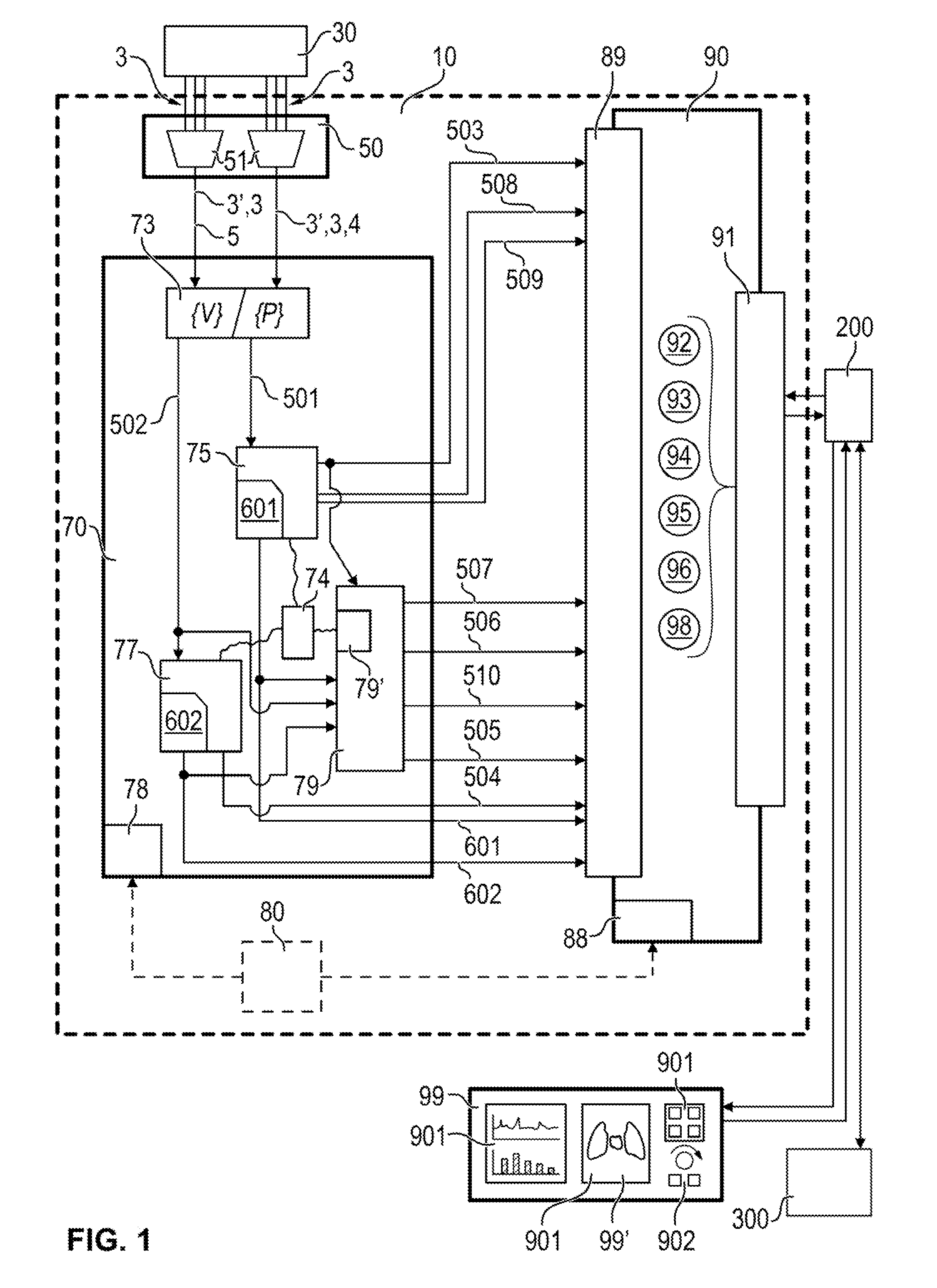
[0138]FIG. 1 schematically shows an arrangement 10 of functional elements for a processing of EIT data 3. This arrangement 10 comprises a data input unit 50, a calculation and control unit 70 and a data output unit 90 as basic components. Moreover, a display device 99 connected with the data output unit 90 is shown in this FIG. 1. The display device 99 comprises a visualization means 901, such as display elements, display screens, displays for visualization of graphs, curve courses, diagrams or images or even numerical value displays for the reproduction of numerical values. Moreover, the display device 99 comprises input and control elements 902, such as switches, buttons, knobs, and rotary knobs. A contact-sensitive display (touchscreen) with combination of input and visualization functionalities represents a special embodiment variant. The data input unit 50 reads in data from an EIT device 30 as EIT data 3 at at least one data input 51 or a plurality of data inputs 51. The EIT d...


PUM


Abstract

A device and a method (100) for the processing of data (501), which were obtained by an imaging method, make possible an improvement in a location-specific visualization of the perfusion of the lung. With a reference to a comparison variable, a location-specific variable (503), characteristic of a period of observation, regarding the perfusion of the lung and heart region, is determined and provided as an output signal.

Description

CROSS REFERENCE TO RELATED APPLICATIONS[0001]This application claims the benefit of priority under 35 U.S.C. §119 of German Patent Application DE 10 2014 009 439.1 filed Jun. 25, 2014, the entire contents of which are incorporated herein by reference.FIELD OF THE INVENTION[0002]The present invention pertains to a device and a method for the processing and visualization of data, obtained by means of a medical device suitable for generating data for imaging, on a perfusion and a ventilation of the lung or areas of the lung, as well as on the perfusion of the heart.BACKGROUND OF THE INVENTION[0003]Devices for electroimpedance tomography (EIT) are known from the state of the art. These devices are designed and provided for generating an image, a plurality of images or a continuous sequence of images from signals obtained by means of electroimpedance measurements and by means of data and data streams obtained therefrom. These images or sequences of images show differences in the conducti...

Claims


Application Information

Patent Timeline
IPC IPC(8): G06T7/00G06T11/00A61B8/13A61B5/053A61B6/03
CPCG06T7/0016G06T11/206G06T2207/10116G06T2207/10132G06T2207/30104G06T2207/30061G06T2207/30048G06T2207/10081G06T2211/40G06T7/0012A61B8/13A61B6/032G06T11/003A61B5/0536A61B5/02028A61B5/08G01R33/56366A61B2576/02G06T2207/10072G16H30/40
Inventor LEONHARDT, STEFFENBAIER-LÖWENSTEIN, TIMMERSMANN, STEFANPIKKEMAAT, ROBERTTESCHNER, ECKHARD
Owner DRAGERWERK AG
Why Eureka
Try Eureka