Perfused three-dimensional cell/tissue disease models

a three-dimensional, cell/tissue technology, applied in the field of arrays of perfused bioreactors, can solve the problems of difficult control using standard processing techniques, device limitations, and inability to use tissue as diagnostic or screening tools, and achieve the effect of facilitating perfusion

Active Publication Date: 2005-11-24
MASSACHUSETTS INST OF TECH +1
View PDF48 Cites 180 Cited by
  • Summary
  • Abstract
  • Description
  • Claims
  • Application Information

AI Technical Summary

Benefits of technology

[0013] A system has been constructed that recapitulate the features of a capillary bed through normal human tissue. The system facilitates perfusion of three-dimensional (3D) cell monocultures and heterotypic cell co-cultures at the length scale of the capillary bed. The system also allows the addition of a second cell type such as a tumor cell after the tissue has been formed. A major feature is that the system can be utilized within a “multiwell plate” format amenable to high-throughput assays compatible with the type of robotics commonly used in pharmaceutical development. The system provides a means to conduct assays for toxicology and metabolism and as a model for human diseases such as hepatic diseases, including hepatitis, exposure-related pathologies, and cancer. Cancer applications include primary liver cancer as well as metastases from other cancers, and for linking drug metabolism with anti-tumor activity. The system is particularly useful for complex and chronic diseases such as cancer, viral infection, and chronic liver fibrosis. The system can also be used as a means of testing gene therapy approaches for treating disease and inborn genetic defects.

Problems solved by technology

Factors such as pore size, shape, and tortuosity can all affect tissue ingrowth but are difficult to control using standard processing techniques.
However, the devices still have a major limitation: ingrowth of new tissue to form blood vessels which sustain the implanted cells must occur at the right time relative to the increasing cell density within the matrix to sustain the implanted cells, and other tissues must not encapsulate or infiltrate the matrix to choke out or otherwise destroy the implanted cells.
None of this technology, however, provides a means to maintain the tissue in vitro, nor to use the tissue as a diagnostic or screening tool.
Thus, while cultured cells may be adequate for certain applications, for example, in detection of toxins and pathogens, they are certain to fail in other applications, for example, screening of drug which are metabolized by the tissues, or drugs which are cleared through interaction with a complex organ, not just a single isolated cell type.
Similarly, drugs that must be bound by cell specific receptors to be taken up by the cells to be active, also cannot be tested in such systems.
Although the hepatic enzymes render most exogenous compounds less toxic, other molecules (as a common example, the pain-relieving drug acetaminophen) can actually become more toxic when metabolized by the liver.
Currently, no in vitro models or animal models adequately capture the complex responses of human tissues to drugs and environmental agents.
Furthermore, no in vitro model captures the complex biology of tumor cell interactions with adjacent normal tissues.
Each year, many new drugs fail in early clinical trials due to unanticipated toxicity, especially liver toxicity, or due to failure to account for liver metabolism of anti-tumor agents.
Thus assessing long-term toxicity of drugs on human liver tissue in vitro is not possible.
Further, sources of human liver cells are scarce, and are not able to meet the demand for cell and tissue-based assays in the pharmaceutical industry.
The current state-of-the art 2D and 3D culture methods do not enable perfusion through the tissue mass in a manner that replicates physiological flow.

Method used


Image

  • Perfused three-dimensional cell/tissue disease models
  • Perfused three-dimensional cell/tissue disease models
  • Perfused three-dimensional cell/tissue disease models

Examples


Embodiment Construction

I. System

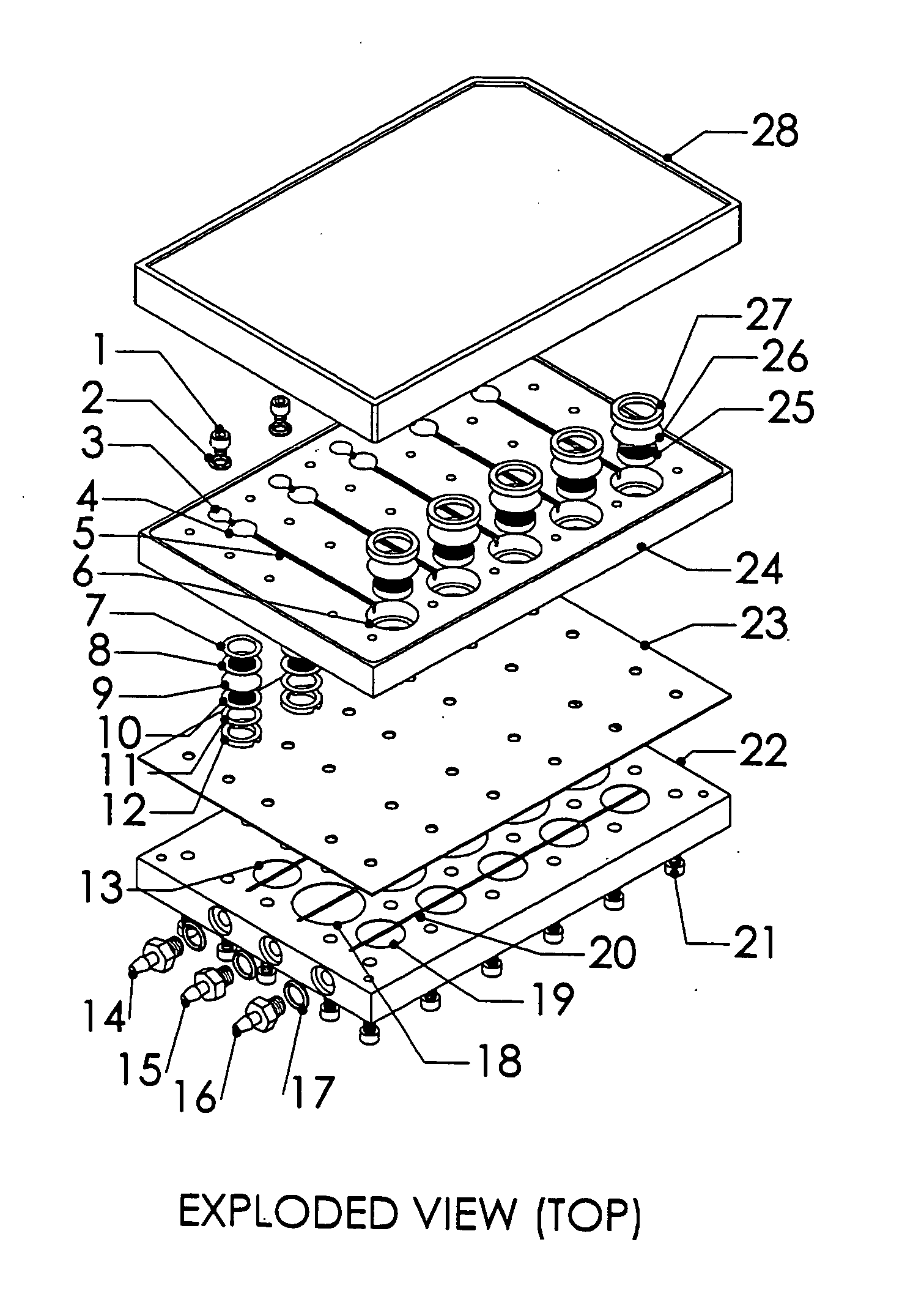
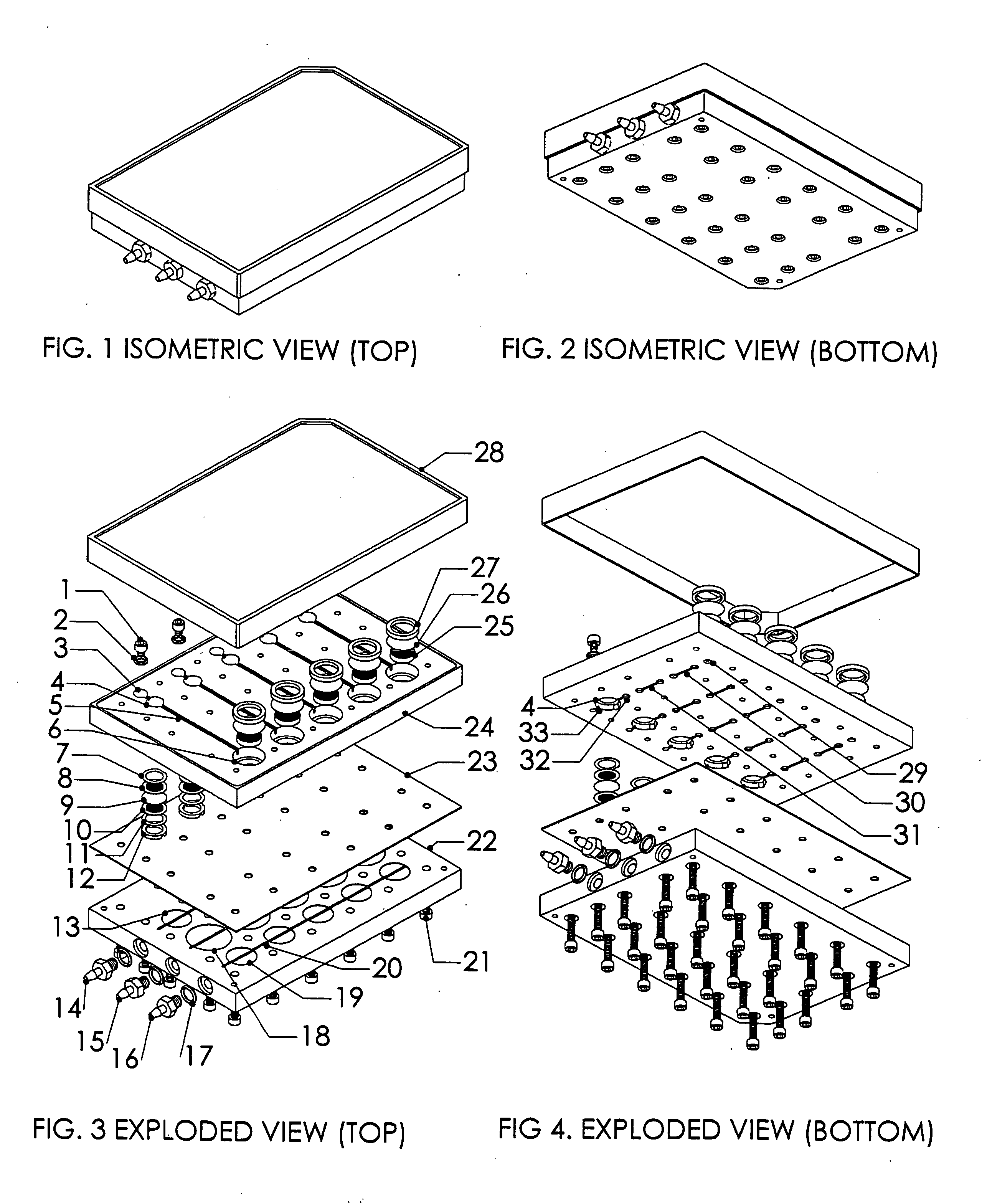
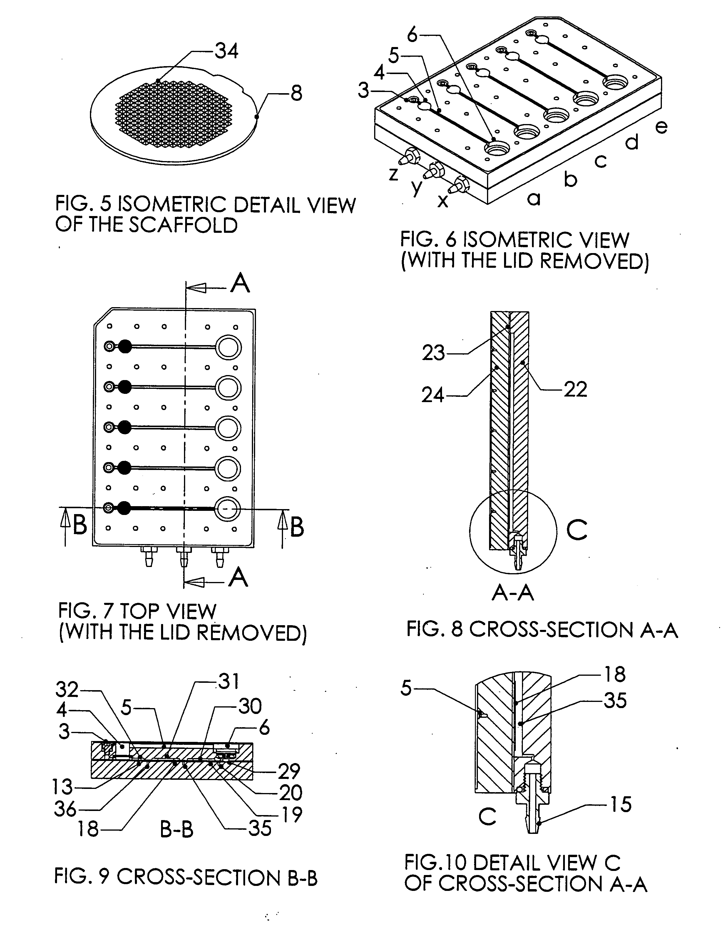
[0024] A system, and methods of manufacture and use thereof, have been developed based on a perfused micromatrix approach modified to utilize highly parallel means for circulating cell culture medium through the micromatrices of tissue or organ structures, making the technology exceedingly suitable for high throughput assays. This system has applications in testing drug toxicity, models of cancer metastasis, stem cell culture, and other human disease models. The system has as its basic components a dense array of perfusion bioreactor and reservoir pairs for cell or tissue culture, and valves and pumps actuated in parallel via common control channels and re-circulating medium through the array of bioreactor and reservoir pairs. Each bioreactor of the array includes a bioreactor well and its own reservoir well. The bioreactor wells and reservoir wells are connected by fluidic channels allowing re-circulation of cell culture medium. Each bioreactor / reservoir pair is fluidicall...


PUM

PropertyMeasurementUnit
sizeaaaaaaaaaa
thicknessaaaaaaaaaa
flow ratesaaaaaaaaaa

Abstract

A system has been constructed that recapitulate the features of a capillary bed through normal human tissue. The system facilitates perfusion of three-dimensional (3D) cell monocultures and heterotypic cell co-cultures at the length scale of the capillary bed. A major feature is that the system can be utilized within a “multiwell plate” format amenable to high-throughput assays compatible with the type of robotics commonly used in pharmaceutical development. The system provides a means to conduct assays for toxicology and metabolism and as a model for human diseases such as hepatic diseases, including hepatitis, exposure-related pathologies, and cancer. Cancer applications include primary liver cancer as well as metastases. The system can also be used as a means of testing gene therapy approaches for treating disease and inborn genetic defects.

Description

CROSS-REFERENCE TO RELATED APPLICATIONS [0001] This application claims priority to U.S. Provisional Application Ser. No. 60 / 572,583 filed in the U.S. Patent and Trademark Office on May 19, 2004.STATEMENT REGARDING FEDERALLY SPONSORED RESEARCH OR DEVELOPMENT [0002] The United States government has certain rights in this invention by virtue of National Science Foundation ERC-8803014 and a supplement to National Institutes of Health grant (3R01 CA088865-02S1) to Douglas A. Lauffenburger (P.I) of MIT. On this latter grant supplement, Alan Wells acted as site PI for the University of Pittsburgh portion of the work, and Linda Griffith was an Investigator.BACKGROUND OF THE INVENTION [0003] The present invention relates to an array of perfused bioreactors in a “multiwell plate” format, wherein each bioreactor of the array consists of a microscale matrix seeded with cells which forms a microtissue, multiple tissues, and / or cell aggregate and methods for using the array in high throughput ass...

Claims


Application Information

Patent Timeline
Patent Type & Authority Applications(United States)
IPC IPC(8): B01L3/00C12M1/00C12M1/32C12M1/34C12Q1/68
CPCB01J2219/00315C12M29/10B01L3/50255B01L3/502715B01L3/50273B01L3/502738B01L2300/0681B01L2300/0829B01L2300/0861B01L2400/0481B01L2400/0638C12M23/12C12M23/40C12M25/14B01J2219/00743B33Y80/00
Inventor DOMANSKY, KARELGRIFFITH, LINDA G.TANNENBAUM, STEVEN R.WELLS, ALAN
Owner MASSACHUSETTS INST OF TECH
Why Eureka
Try Eureka