Improved method of mapping glycans of glycoproteins

a technology of glycoproteins and mapping methods, applied in the direction of material testing goods, biochemistry apparatus and processes, and post translational modification detection, etc., can solve the problems of reducing sensitivity, difficult discrimination, and limited mass spectrometry information gained from this kind of analytical methods, and achieves sensitivity and selectivity. high, strong retention

Inactive Publication Date: 2016-01-21
HEXAL AG
View PDF1 Cites 8 Cited by
  • Summary
  • Abstract
  • Description
  • Claims
  • Application Information

AI Technical Summary

Benefits of technology

The present invention describes a method for analyzing anthranilic acid labeled glycans, specifically N-glycans, with high sensitivity and selectivity. Using this method, researchers can analyze both acidic and neutral N-glycans simultaneously by utilizing a single mobile phase for liquid chromatography coupled with negative or positive ionization modes depending upon the needs of the study. Additionally, the use of formic acid in the mobile phase results in improved separation and efficiency of detecting these important molecules. Overall, this technology offers greater accuracy and precision for studying the structure and function of various glycans involved in biological processes.

Problems solved by technology

The technical problem addressed in this patent text is how to effectively characterize the glycan structure of proteins and other biologically important compounds like N-glycans without being affected by the small amount of samples available for analysis. The challenge is made difficult when trying to determine if a new glycan attachment impacts the function or effectiveness of a protein or glycan analogue. The inventor proposes a novel method called RP-nano-LC-MS that offers a simplified way to prepare and detect such attachments while also allowing for untargeted identification of specific N-glycans.

Method used


Image

  • Improved method of mapping glycans of glycoproteins
  • Improved method of mapping glycans of glycoproteins
  • Improved method of mapping glycans of glycoproteins

Examples


example 1

Analysis of Glycans Using the 2-AA Method and the 2-AB Method

Materials

[0116]PNGase F was from New England Biolabs. Acetonitrile (ACN) and acetic acid was from Merck. Formic acid and Sodiumcyanoborohydride was from Fluke. PD MiniTrap™ Sephadex® G-10 columns were from GE Healthcare. DMSO was from Sigma. Amicon Ultra 30K filter devices were from Milipore. The mAb glycan standard was prepared at Sandoz. Monoclonal antibodies 1-3 were obtained from in-house development at Sandoz.

Methods

Enzymatic N-Glycan Release Using PNGaseF

[0117]1 mg of desalted mAb was used. The N-glycans (15 nmol) were released using PNGaseF by incubating the samples overnight (˜17 hours) at 37° C. N-glycans were separated from the proteins using Amicon 30K filter devices and were brought to dryness using a speedvac.

Fluorescence Labeling of Released N-Glycans

[0118]Na[BH3(CN)] and either 2-AA or 2-AB were dissolved in 70% DMSO: 30% acetic acid to obtain concentrations of 63 mg / ml and 50 mg / ml, respectively. 15 μl of t...

example 2

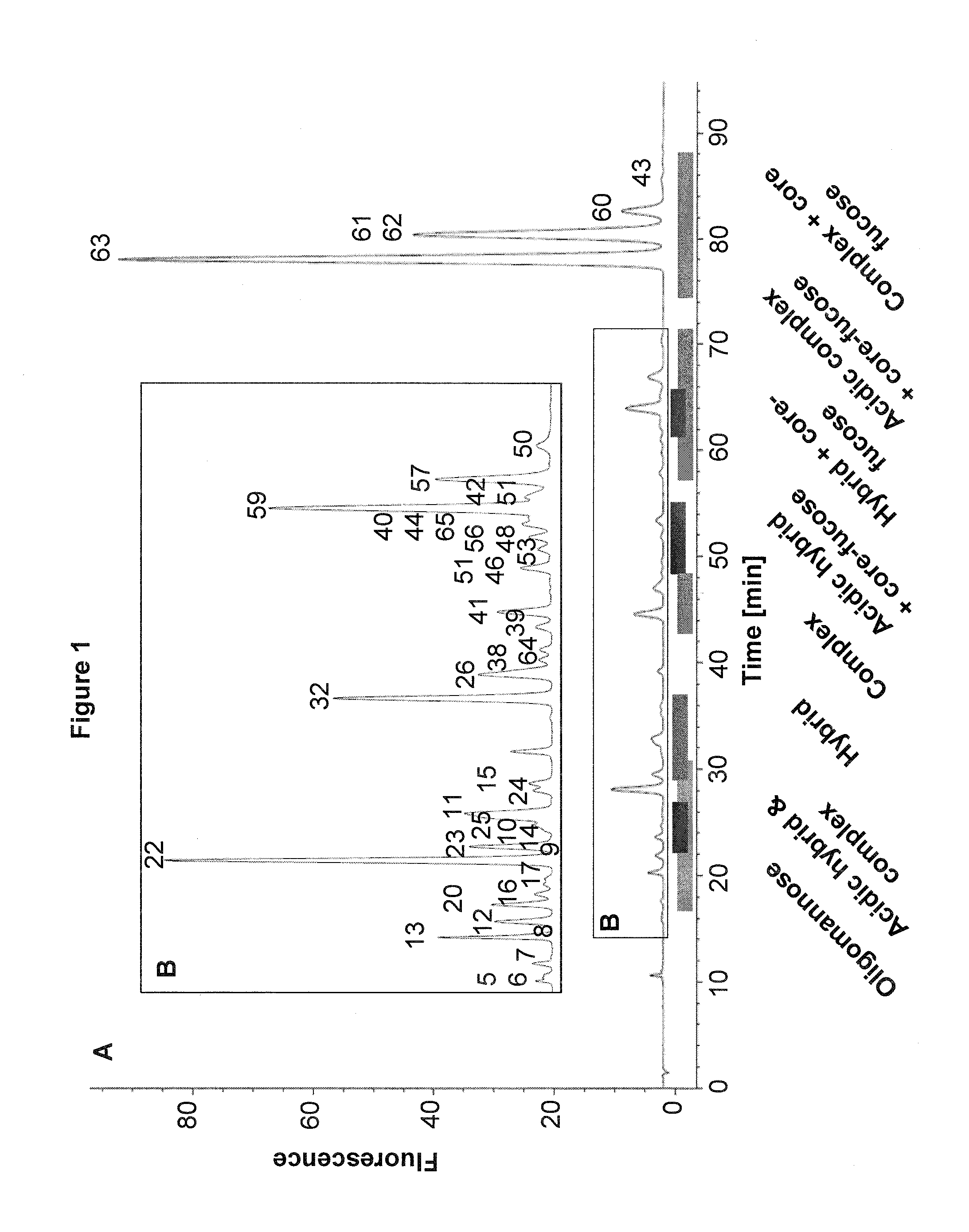
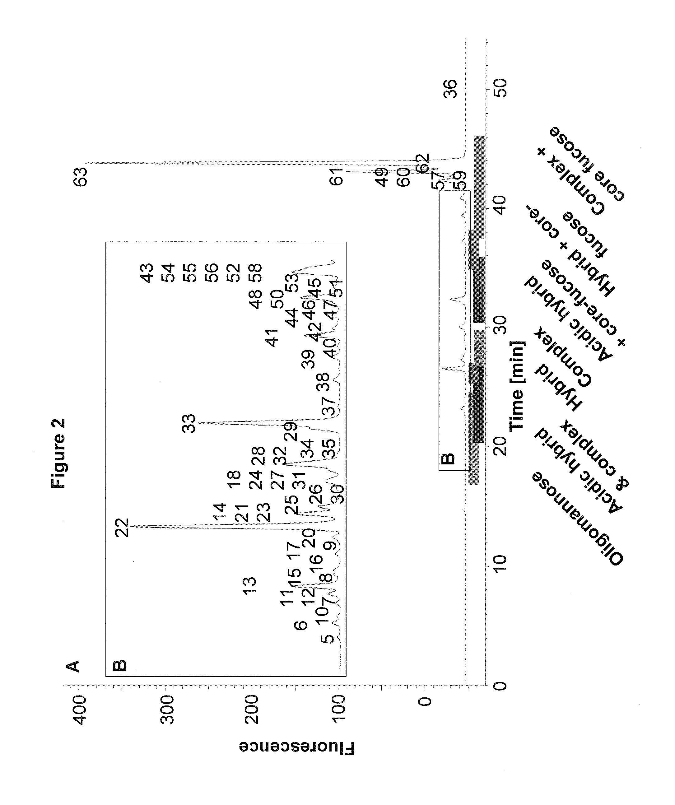
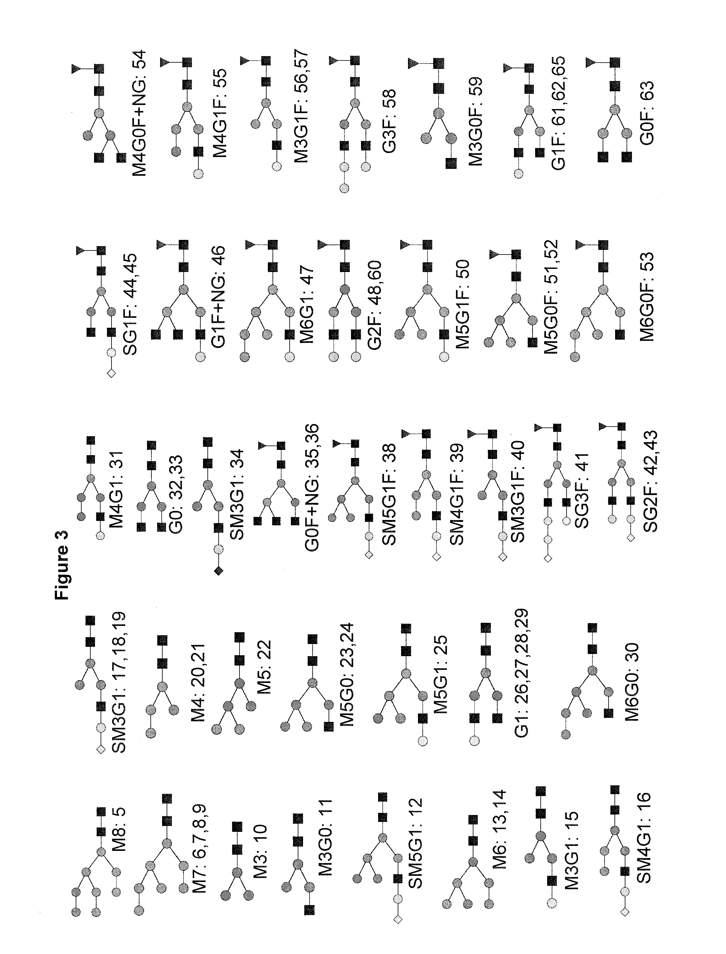
Use of the 2-AA Method Together with Nano-LC-MS

Materials

[0137]PNGase F was from Roche (Penzberg, Germany). Acetonitrile (ACN) and acetic acid was from Merck (Darmstadt, Germany). Formic acid and Picolineborane were from Fluka (Sigma, Munich, Germany). Protein A sepharose, Protein G sepharose and Sephadex® G-10 96-well plates (custom made) were from GE Healthcare (Munich, Germany). RNase B and DMSO were from Sigma (Munich, Germany). AcroPrep™ Advance Omega™ 10K 96-well filter plates were from Pall (Dreieich, Germany). Human serum was from Lonza. Monoclonal antibody A and cell culture supernatants were obtained from in-house development at Sandoz. G0F glycan standard was from Dextra (MoBiTech, Goettingen, Germany). Complex and acidic N-glycan standards were from TheraProtein (Barcarena, Portugal).

Methods

[0138]Purification of IgGs from Human Serum or Cell Culture Supernatant and N-Glycan Release

[0139]Protein G and Protein A sepharose were used to purify IgG from human pooled serum or c...


PUM


Abstract

The use of anthranilic acid (2-AA) to label N-glycans prior to separation using a reversed-phase liquid chromatography (RP-LC) column under acidic conditions using formic acid is described herein. Negatively charged 2-AA offers stronger retention on the reversed phase column than 2-aminobenzamide (2-AB) in RP-LC and allows efficient ionization and detection of 2-AA labeled N-glycans. The acidic conditions used for the RP-LC leads to an efficient separation of acidic 2-AA N-glycans carrying terminal sialylation without the need for an ion-pairing reagent. The method and compositions described herein may be used with RP-nano-LC-MS and a 96-well plate sample preparation, which allows attomolar sensitivity and high throughput.

Description


Claims


Application Information

Patent Timeline
Owner HEXAL AG
Why Eureka
Try Eureka