Anatomical site relocalisation using dual data synchronisation

anatomical site and data synchronisation technology, applied in the field of anatomical site relocalisation using dual data synchronisation, can solve the problems of limiting the ability of the endoscopy and the optical biopsy probe to accurately reposition and thus to effectively, not providing any spatial relations of the extracted segments, and not sufficiently clarifying, so as to achieve the effect of increasing the accuracy of the relocation

Active Publication Date: 2016-01-28
INST HOSPITALO UNIV DE CHIRURGIE MINI INVASIVE GUIDEE PAR LIMAGE +1
View PDF16 Cites 37 Cited by
  • Summary
  • Abstract
  • Description
  • Claims
  • Application Information

AI Technical Summary

Benefits of technology

[0022]In connection with this embodiment, one can consider using feature descriptor to improve synchronisation.
[0042]Second step: adding an interactive virtual tagging of one of more images of the first or reference exploration flexible endoscopic video indicating specific points of interest in the body of the explored subject, this virtual tagging being realized from a specific first or reference exploration video reader allowing to add tags in any selected image of the video through any interaction or human interface device allowing to precisely select a pixel or group of pixels in a video, such as by mouse, pad, trackball, joystick or optical tracking.
[0053]To increase the accuracy of the relocation, the referential points can be used to define and attach a frame or referential to the subject in the first and subsequent explorations.

Problems solved by technology

Typically, the GI specialist uses the printed markings on the endoscope (commonly one mark every 5 or 10 cm along the flexible body), which can be highly unreliable and which limit his or her ability to accurately re-position the endoscope and the optical biopsy probe and hence to effectively track the disease.
Due to the lack of deterministic tools for providing such re-localisation inter-operatively, the GI specialist has to survey or biopsy the entire affected oesophagus region, which prevents targeted treatments.
However, they do not provide any spatial relations of the extracted segments inter-operatively, and so have not sufficiently clarified the application of their result in a clinical context for re-localisation.
The inventors are of the opinion that, relying only on image based information for information extraction, that has to be mapped across multiple interventions can be highly unreliable; especially, due to temporal changes in tissue texture over multiple procedures, coupled with a highly deformable endoscopic scene, where repeatability of feature extraction, matching and tracking poses a significant challenge.
These solutions thus require an important preliminary data treatment and cannot take changes in tissue texture or deformation of the targeted scene or of the environment of said scene into account.

Method used


Image

  • Anatomical site relocalisation using dual data synchronisation
  • Anatomical site relocalisation using dual data synchronisation
  • Anatomical site relocalisation using dual data synchronisation

Examples


Embodiment Construction

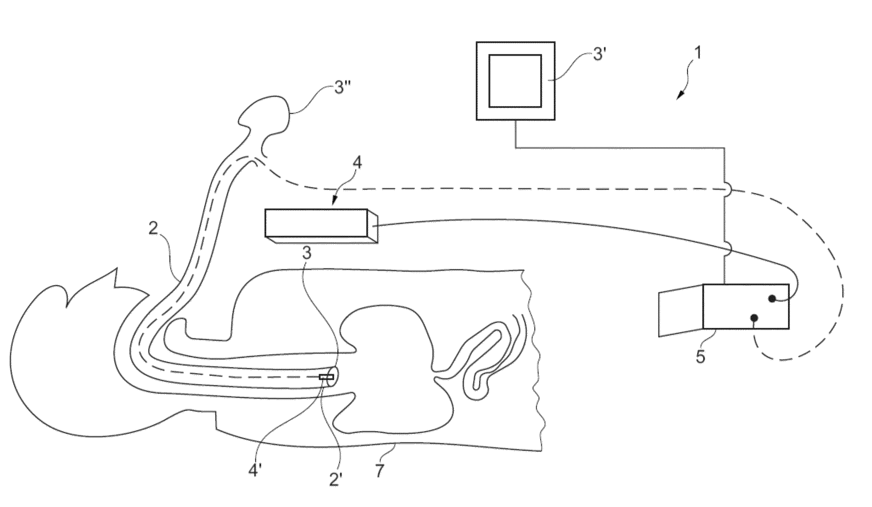
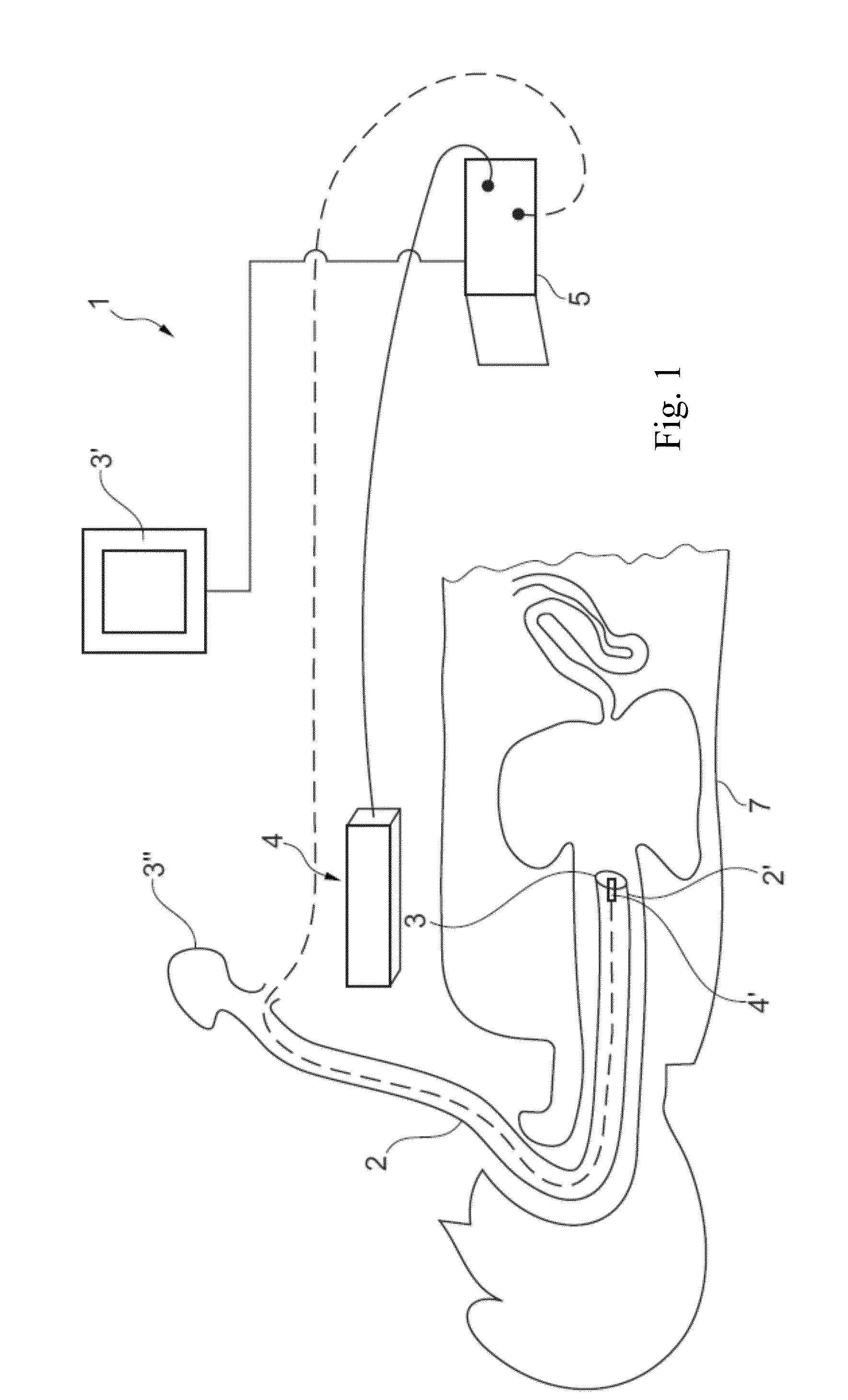
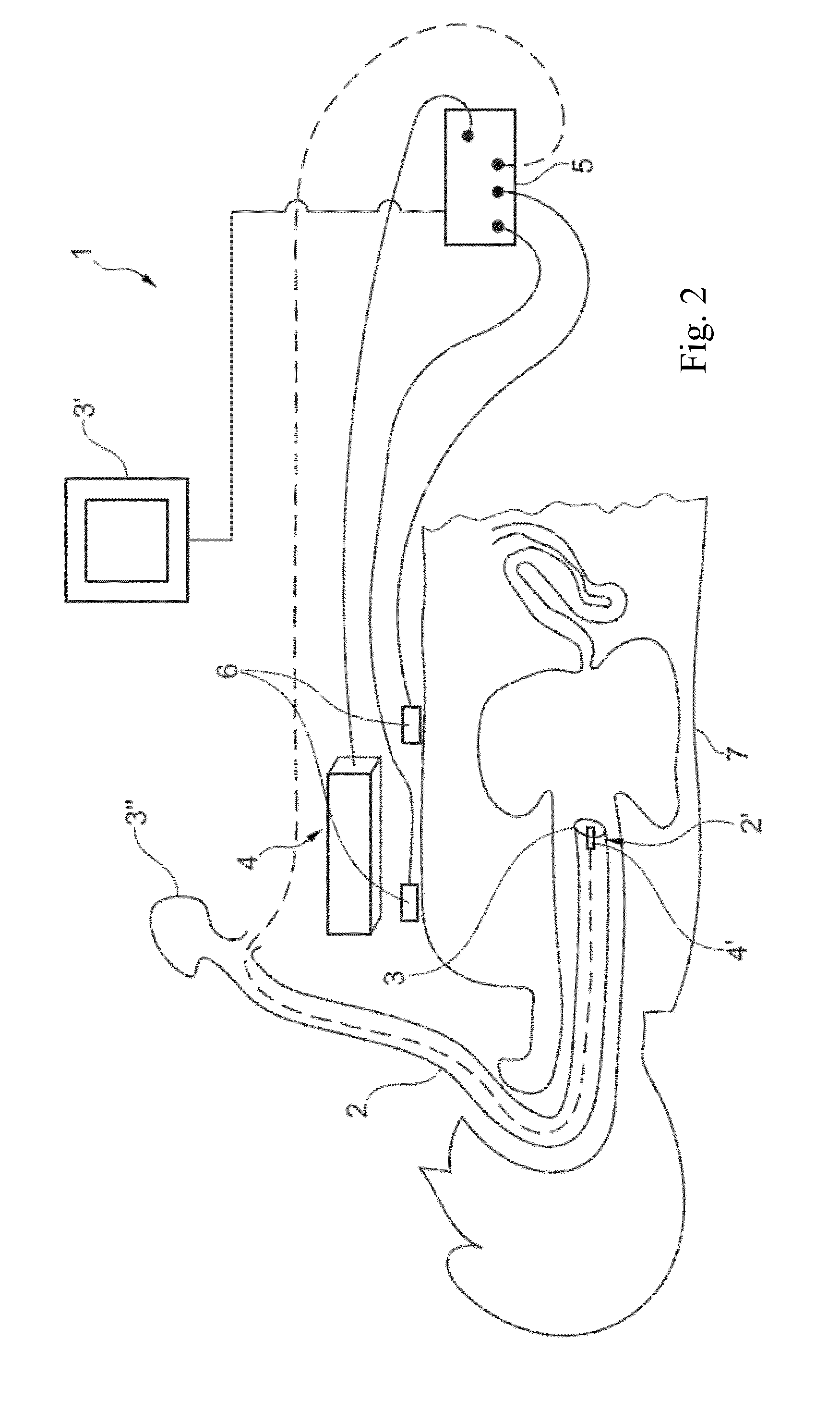
[0074]FIGS. 1 and 2, and partially FIG. 9, show a medical system 1 according to the invention, comprising a flexible endoscope 2 which can be introduced in a tubular organ of a subject 7, and able to be used for performing the method according to the invention.

[0075]More precisely, said medical system 1 mainly comprises:

[0076]i) a flexible endoscope 2 at least equipped with an image taking device 3, and possibly at least one tool or instrument,

[0077]ii) display 3′ and movement control 3″ means associated with said endoscope 3,

[0078]iii) a tracking device or system 4 providing the location and orientation of the end tip 2′ of the endoscope 2, such as an electromagnetic tracking device or a flexible optical tracking device,

[0079]iiii) computer means 5 and associated storing means,

[0080]wherein the frontal or end tip 2′ of the endoscope 2 is provided with an electromagnetic sensor 4′ or a similar tracking device and said medical system 1 further comprises means, in particular software ...


PUM


Abstract

A method and a system for anatomical site relocalisation using dual data synchronisation. Method for repositioning, possibly several times, at a specific location, which has been already explored during a first or “reference exploration”, a flexible endoscope during one or more successive endoluminal or extraluminal subsequent or “new exploration”, or similar procedures, wherein the repositioning is realized either manually, by a human user, or automatically, by a robotic system, from or by way of a synchronisation process between a “new exploration” flexible endoscope video and the “reference exploration” flexible endoscope video displayed in parallel on or in two different windows, on one or more screens.

Description

FIELD OF THE INVENTION[0001]The present invention concerns a method and a system for repositioning quickly and precisely at a specific location already explored a flexible endoscope (or a similar medical device) during successive endoluminal or extraluminal exploration or similar procedures (after partial or total extraction) performed on a body of a subject.[0002]The localisation and tracking of biopsy sites inter-operatively poses a significant challenge for providing targeted treatments. This invention provides a guided navigation to the medical operator or surgeon for accurate re-positioning of a flexible endoscope at previously targeted sites.[0003]The invention concerns more specifically target sites located in more or less flexible tubular organs of subjects, or at least tube like elongated passages within the bodies of such subjects.[0004]Primarily, but not limitatively, envisaged organs are the oesophagus and the colon.BACKGROUND OF THE INVENTION[0005]In relation to the fie...

Claims


Application Information

Patent Timeline
Patent Type & Authority Applications(United States)
IPC IPC(8): A61B1/04A61B1/00G06K9/00G06K9/46G06T7/00G06T19/00H04N5/225
CPCA61B1/04G06T2207/30004A61B1/0016A61B1/00009H04N5/2251G06K9/00758G06T7/0044G06K9/4671G06T19/006G06T7/0014A61B2019/5251A61B2019/5255A61B2019/5265A61B2019/5289A61B19/5244A61B5/062A61B2034/2055A61B34/20A61B2090/364A61B2034/2051A61B2034/2065G06T7/74G06V20/48A61B1/000094
Inventor NICOLAU, STEPHANEVEMURI, ANANT SURAJSOLER, LUCMARESCAUX, JACQUES
Owner INST HOSPITALO UNIV DE CHIRURGIE MINI INVASIVE GUIDEE PAR LIMAGE
Why Eureka
Try Eureka