
Tracheobronchial implantable medical device and methods of use

a technology of tracheobronchial and implantable medical devices, which is applied in the field of tracheobronchial implantable medical devices and methods of use, can solve the problems of granulation tissue formation, mucous plugging, and stent migration, and achieve the effects of reducing the risk of infection

Inactive Publication Date: 2016-01-28
ABBOTT CARDIOVASCULAR
View PDF9 Cites 11 Cited by
  • Summary
  • Abstract
  • Description
  • Claims
  • Application Information

AI Technical Summary

Problems solved by technology

In adults, primary cancer of the tracheobronchial tree or cancer of the head, neck or chest that extends into the tracheobronchial tree frequently causes lumen compromise and airway obstruction.
Some problems associated with known stent types delivered to the tracheobronchial region include inflammation, stent migration, epithelial damage, granulation tissue formation and mucous plugging.
In addition, it is believed that known bioabsorbable stents designed for placement in the tracheobronchial region are not able to adequately combat inflammation caused by stent placement.
Mucous plugging can cause interference with breathing.
Excessive granulation tissue formation can cause a stent to be permanently lodged within a passageway complicating removal if required.
Metal stents are especially susceptible to granulation tissue formation.

Method used


Image

  • Tracheobronchial implantable medical device and methods of use
  • Tracheobronchial implantable medical device and methods of use
  • Tracheobronchial implantable medical device and methods of use

Examples


Embodiment Construction

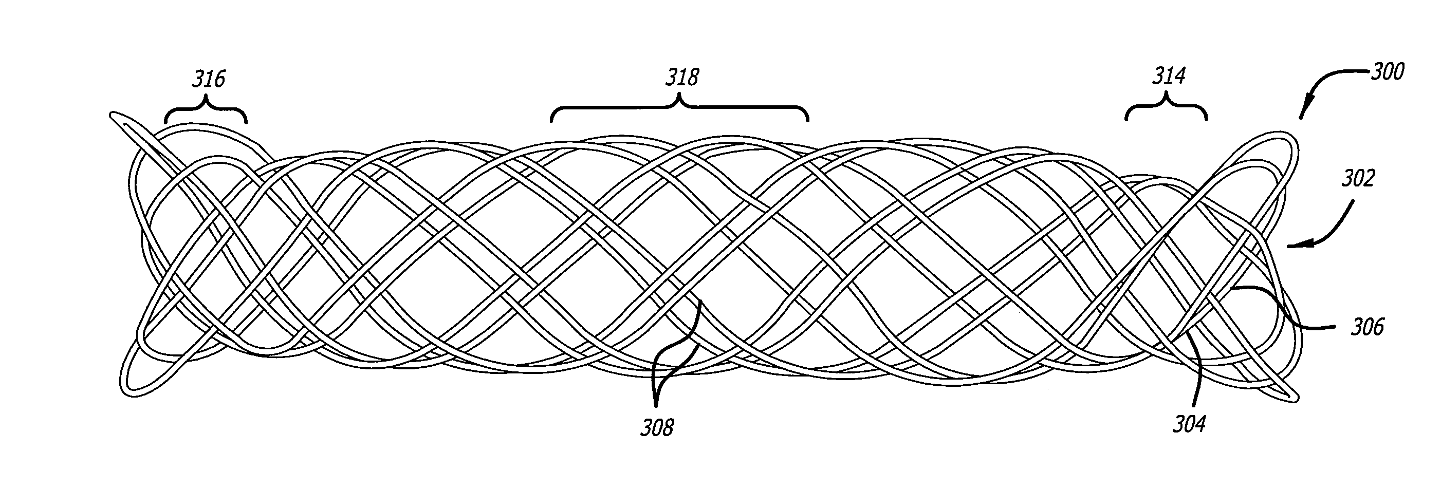
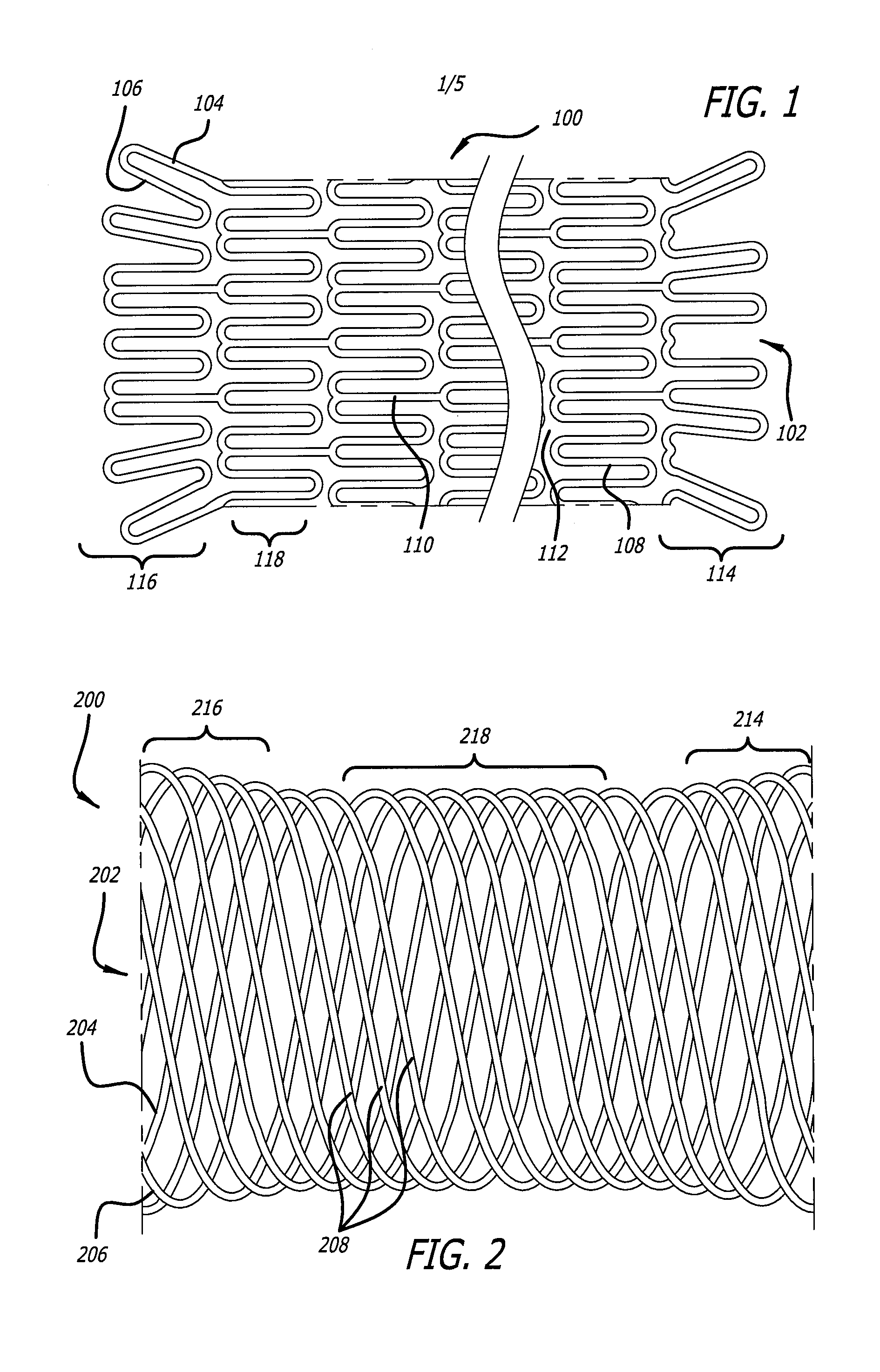
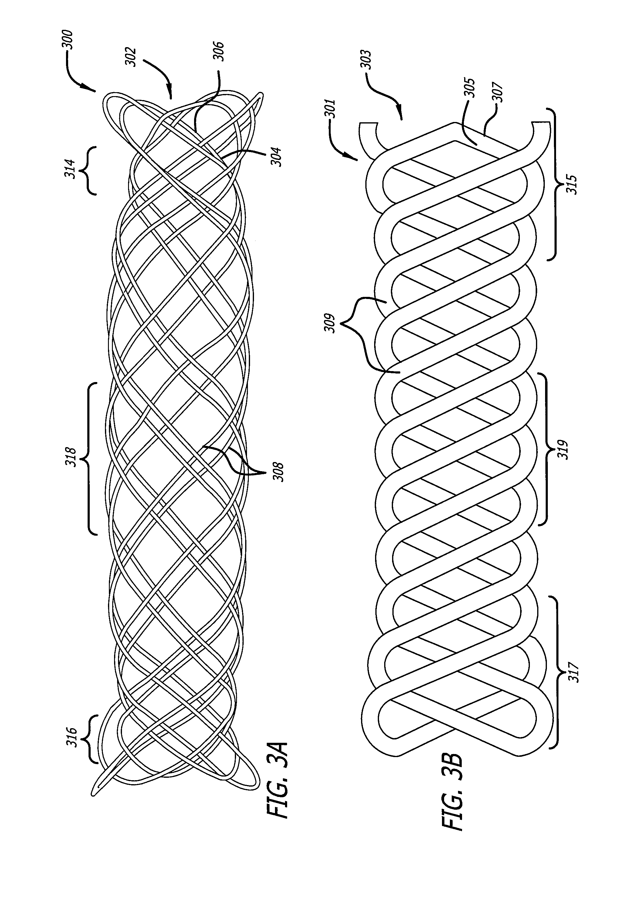
[0018]Embodiments of devices and methods for treating a diseased tracheobronchial region in a mammal, including, but not limited to, humans, are herein disclosed. In some embodiments, the device can be an implantable medical device such as a stent. Representative examples of implantable medical devices include, but are not limited to, self-expandable stents, balloon-expandable stents, micro-depot or micro-channel stents and grafts. In some embodiments, a treatment method can be delivery of a stent to a tracheobronchial region by a delivery device such as a catheter assembly.

[0019]In some treatment applications, a stent may only be required to be present in the tracheobronchial region for a limited period of time. To accommodate this, a stent can be made of a biodegradable, bioerodable or bioabsorbable polymer, hereinafter used interchangeably. A stent can also be made of a biostable or biodurable (hereinafter used interchangeably) or a combination of a biostable and biodegradable po...


PUM

PropertyMeasurementUnit
pitch of angleaaaaaaaaaa

Abstract

Devices and methods for treating a diseased tracheobronchial region in a mammal. The device can be a stent which can include a sustained-release material such as a polymer matrix with a treatment agent. The stent can be bioabsorbable and a treatment agent can be incorporated therewith. A treatment method can be delivery of a stent to a tracheobronchial region by a delivery device such as a catheter assembly.

Description

CROSS-REFERENCE TO RELATED APPLICATION[0001]This application is a divisional application of co-pending U.S. patent application Ser. No. 11 / 507,913, filed Aug. 21, 2006, the disclosure of which is incorporated herein by reference.FIELD OF INVENTION[0002]Bioabsorbable implantable medical devices for the treatment of lesions caused by cancer of the tracheobronchial tree or cancer of the head, neck or chest.BACKGROUND OF INVENTION[0003]This invention relates generally to radially expandable endoprostheses which are adapted to be implanted in a physiological lumen. An “endoprosthesis” corresponds to an artificial device that is placed inside of a physiological lumen. A “lumen” refers to a cavity of a tubular organ such as a blood vessel or other physiological passageway. A stent, or implantable medical device, is an example of an endoprosthesis. Stents are generally cylindrically shaped devices which function to hold open or expand a physiological lumen, or to compress a lesion. A stent ...

Claims


Application Information

Patent Timeline
Patent Type & Authority Applications(United States)
IPC IPC(8): A61F2/88A61F2/844A61F2/90A61F2/958
CPCA61F2/885A61F2/844A61F2/88A61F2250/0067A61F2/958A61F2210/0004A61F2230/0069A61F2/90A61F2/04A61F2/962A61F2002/046A61F2210/0076A61F2220/0008A61F2230/0091A61F2250/0015A61F2250/0018A61F2250/0028A61F2250/0039A61F2250/0098A61F2/186A61F2/20A61F2/95A61F2002/043A61F2002/044
Inventor LIM, FLORENCIASUH, MARYSTAMBERG, BARBARAHUANG, BIN
Owner ABBOTT CARDIOVASCULAR
Learn More