Methods and media for differentiating eye cells

a technology of eye cells and media, applied in the field of eye treatment and eye research, can solve problems such as deterioration or total loss of vision, unstable bowman's membrane, and use of a medium which requires donated limbal cells,

Inactive Publication Date: 2016-03-03
UNIVERSITY OF TAMPERE
View PDF2 Cites 4 Cited by
  • Summary
  • Abstract
  • Description
  • Claims
  • Application Information

AI Technical Summary

Benefits of technology

[0014]It is thus an object of the present invention to provide means and methods effective to differentiate stem cells into eye precursor cells and further into corneal epithelial or early pigmented RPE precursor cells, which may then be maturated into mature corneal epithelial cells and mature RPE cells, respectively.

Problems solved by technology

The Bowman's membrane is known being unstable, which casts uncertainty to the procedure.
Use of a medium which requires donated limbal cells can be considered problematic.
Dysfunctional RPE causes impairment and death of the photo-receptor cells, leading to deterioration or total loss of vision.
However, the method is very complex with at least four different stages and four different culture media, each characterized by a unique combination of active supplement.
Xeno-products and undefined factors used in many differentiation processes pose further challenges, because animal-derived components may carry factors such as sialic acid or Neu5Gc, causing unwanted immunogenicity of the cells or even animal pathogens.

Method used


Image

  • Methods and media for differentiating eye cells
  • Methods and media for differentiating eye cells
  • Methods and media for differentiating eye cells

Examples


Embodiment Construction

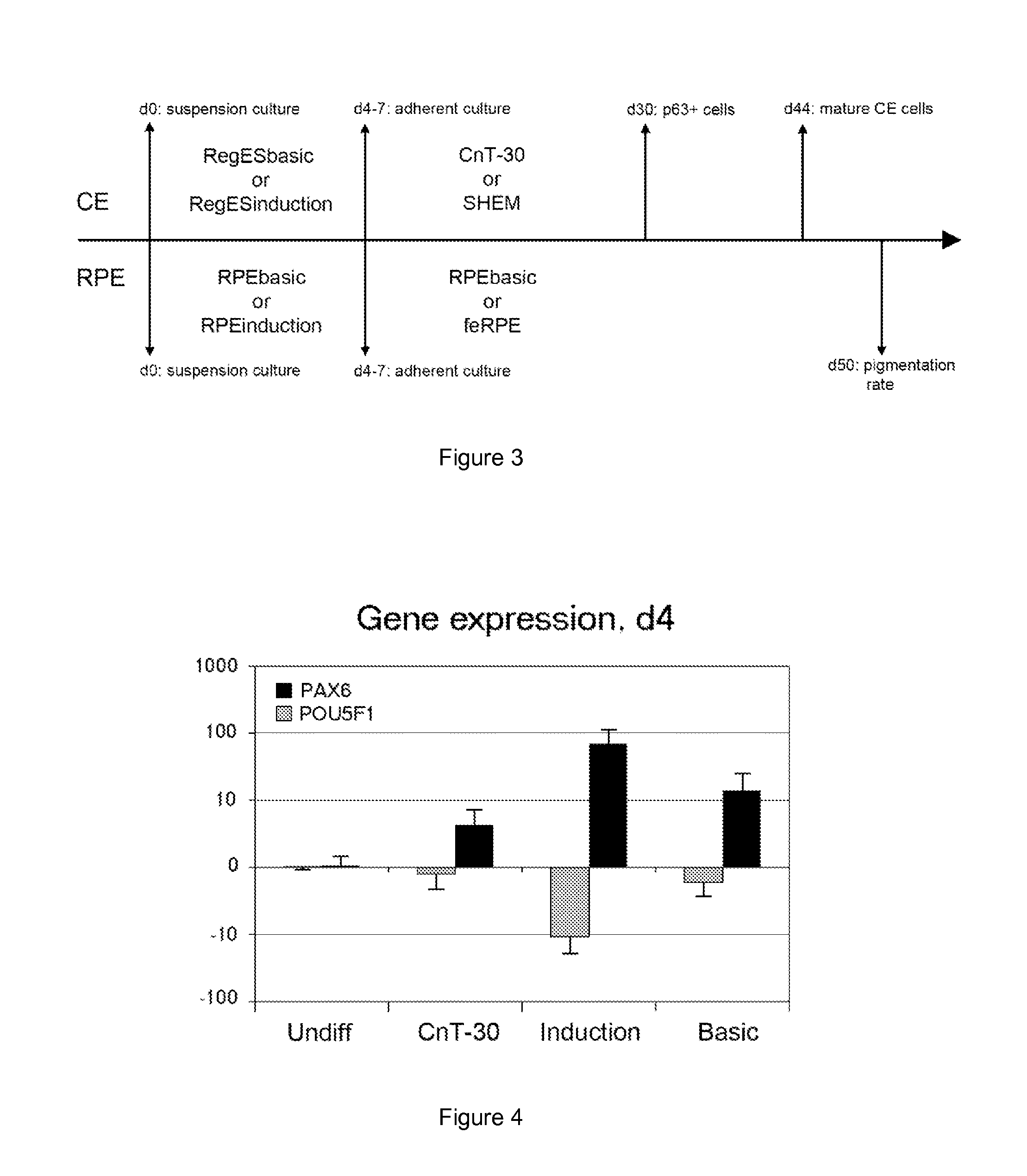
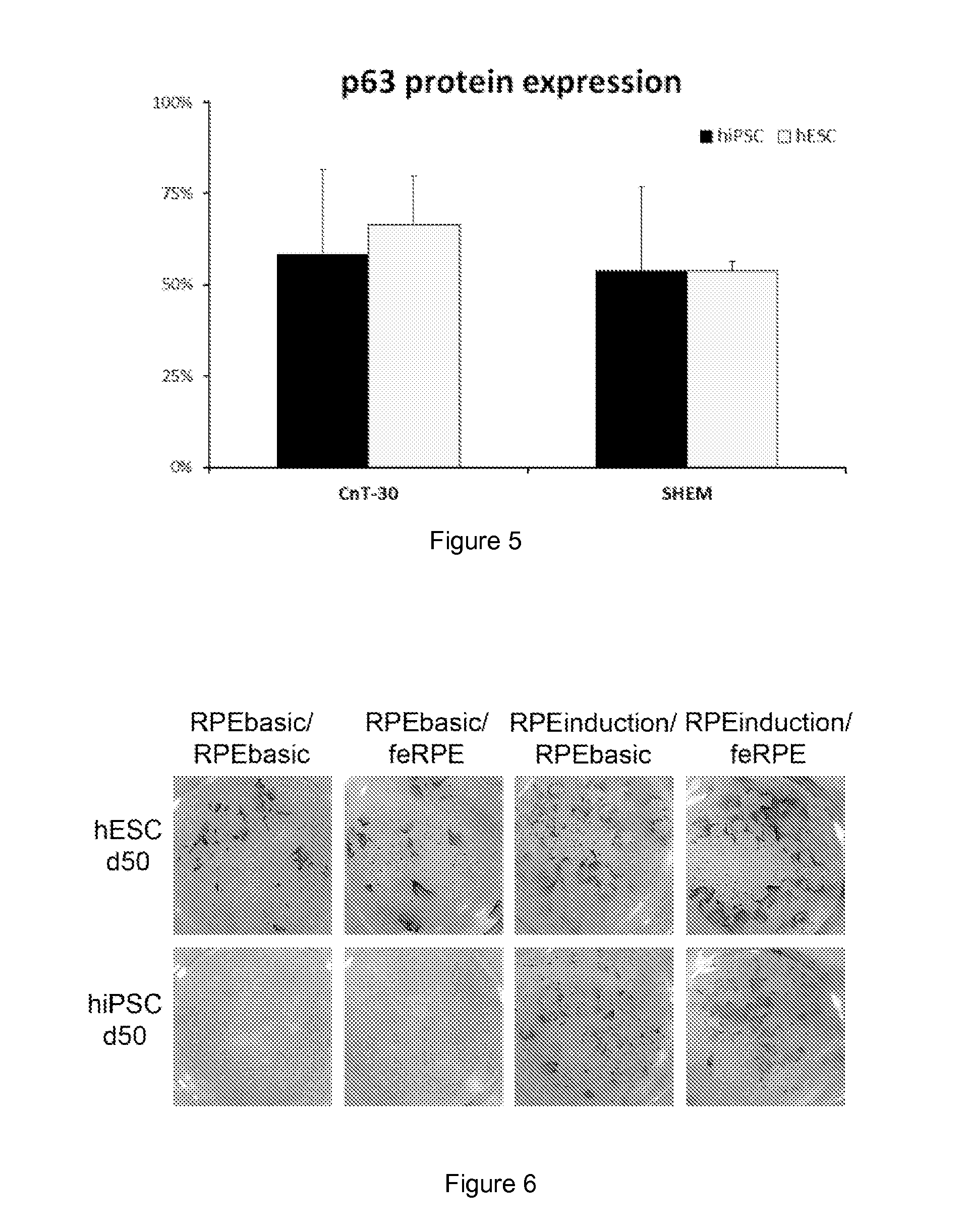
[0031]In some aspects, the present invention provides a one-stage induction method for producing eye precursor cells, and a two-stage differentiation method comprising said induction method and a further differentiation and maturation stage for differentiating said eye precursor cells into differentiated eye cells, such as corneal epithelial precursor cells or early pigmented retinal pigment epithelial (RPE) precursor cells, and further into mature corneal epithelial cells or mature RPE cells, respectively. As used herein, the terms “pre-cursor” and “progenitor” may be used interchangeably unless otherwise indicated.

Induction Method and Medium

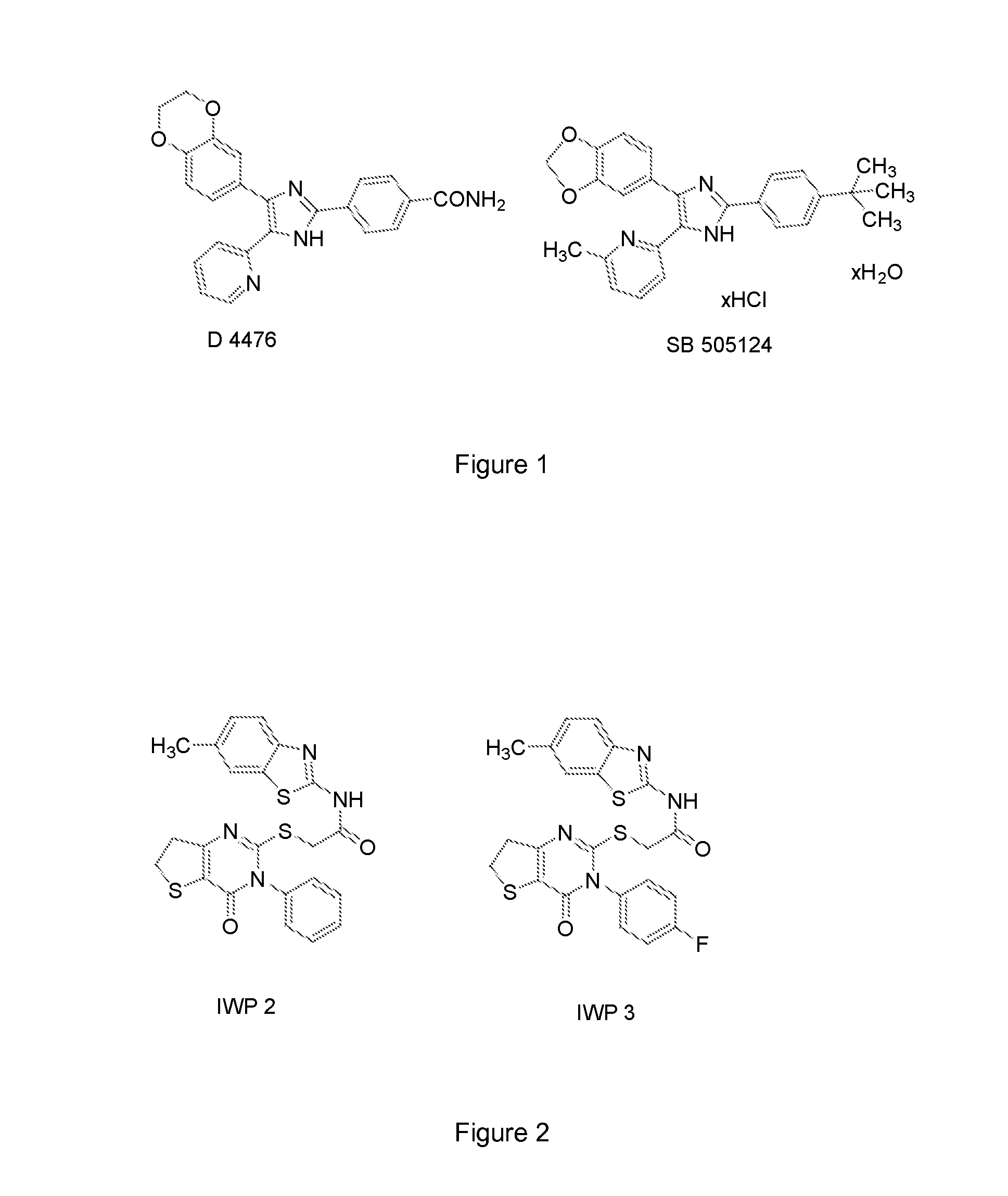
[0032]The present induction method is aimed at inducing pluripotent stem cells towards surface ectoderm and eye precursor cells by subjecting said stem cells, preferably in a suspension culture, to an induction medium comprising active “induction supplements”, i.e. a TGF-beta inhibitor, a Wnt inhibitor and a fibroblast growth factor. Both said ...


PUM

PropertyMeasurementUnit
molar massaaaaaaaaaa
timeaaaaaaaaaa

Abstract

Here is provided a novel differentiation protocol, which was experimentally shown to give rise to corneal epithelial precursor cells or early pigmented RPE precursor cells in defined and xeno-free conditions. The early precursor cells may be further maturated towards corneal epithelium cells, stratified corneal epithelium or mature RPE cells. Such cells may contribute to treatment and research of corneal and retinal conditions, diseases, pathologies as well as toxicology and drug development.

Description

FIELD OF THE INVENTION[0001]The technical field concerned is treatment and study of the eye, more specifically corneal and retinal diseases and disorders. The present description is related to differentiation of stem cells into eye precursor cells and further into differentiated eye cells, such as corneal epithelial cells or retinal pigment epithelial cells. Accordingly, here is provided means and methods contributing to fast and effective induction, maturation and differentiation.BACKGROUND OF THE INVENTION[0002]The cornea is located at the front surface of the eye and is multi-layered, transparent and avascular in structure. The main functions of the cornea are to protect the eyeball and its contents, while allowing accurate focusing of light to produce a sharp image on the retina. The cornea consists of three cellular layers, namely epithelium, stroma and endothelium, separated by two acellular layers—Bowman's layer, and Descemet membrane. As the outermost layer, corneal epitheli...

Claims


Application Information

Patent Timeline
Patent Type & Authority Applications(United States)
IPC IPC(8): C12N5/079
CPCC12N5/0621C12N2501/115C12N2501/40C12N2501/11C12N2501/415C12N2506/03C12N2500/98C12N2501/15C12N2506/02
Inventor MIKHAILOVA, ALEXANDRAILMARINEN, TANJASKOTTMAN, HELI
Owner UNIVERSITY OF TAMPERE
Why Eureka
Try Eureka