

Intra- Abdominal Lightfield 3D Endoscope and Method of Making the Same
a technology of intraabdominal light and endoscope, which is applied in the field of design of intraabdominal three-dimensional (3d) imaging systems, can solve the problems of increasing surgeons' mental workload, affecting surgeons' ability to establish a stable horizon and perceive depth, and instruments falling o
- Summary
- Abstract
- Description
- Claims
- Application Information
AI Technical Summary
Benefits of technology
Problems solved by technology
Method used
Image

Examples
embodiment # 1
Embodiment #1
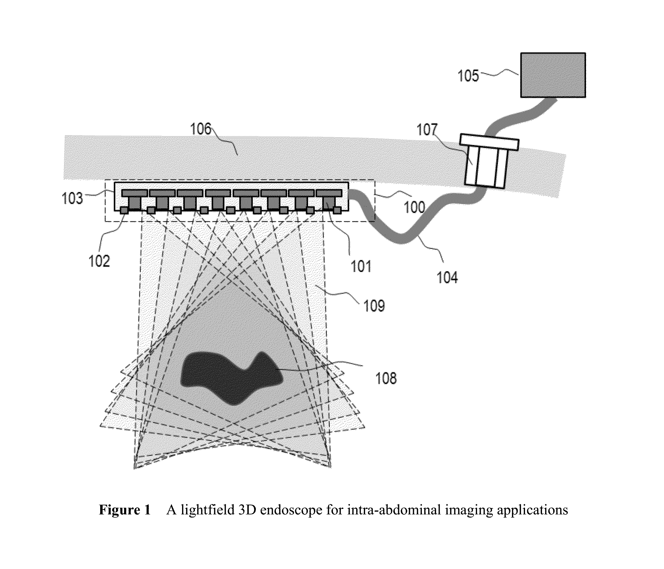
[0035]FIG. 1 illustrates an example design of the disclosed lightfield 3D endoscope 100. It consists of an array of imaging sensors 101, illumination devices 102, outer housing 103, connection cable 104 and extra-peritoneal control unit 105. Typical imaging sensors include charge-coupled device (CCD) or complementary metal-oxide-semiconductor (CMOS) sensors, but any other types of imaging sensor can be used. Both analog and digital version of CCD / CMOS sensor modules can be used. In an exemplary design, we select a CMOS chip from OmniVision, which has image resolution of 672×492 pixels, image area 4.032 mm×2.952 mm, and pixel size 6×6 μm. The high quality miniature optical lens are used which offers proper field of view (FOV) (for example 120-degree FOV). The geometric locations of all sensors are arbitrary but are known or can be obtained via calibration techniques. Sensors in the array can be all the same or differ in optical, mechanical and / or electronic characteristi...
embodiment # 2
4.3. Embodiment #2
Structured Light Lightfield 3D Endoscope
[0041]FIG. 3 discloses a design of the lightfield 3D endoscope with active structured light illumination mechanism. The structured light projector 110 generates spatially varying illumination pattern 111 on the surface of target 108. Structured light is a well-known 3D surface imaging technique. In this invention, we apply the structured light illumination technique to the lightfield 3D endoscope.
[0042]With projected surface pattern in by the structured light projector 110, one can easily distinguish surface features in the captured lightfield images. Reliable 3D surface reconstruction can be performed based on multiview 3D reconstruction techniques. This type of computation does not require calibrated geometric position / orientation of the structured light projector. The projected surface pattern only serve the purpose of enhancing surface features thus improve the quality and reliability of the 3D reconstruction results.
[004...
embodiment # 3
4.4. Embodiment #3
Multi-Spectral and / or Polarizing Lightfield 3D Endoscope
[0047]Given multiple imaging sensors on the lightfield 3D endoscope, one can configure some of sensors to acquire images in different spectral bands or different polarization directions. For example, narrow band filters can be used to enhance contrast (signal to noise ratio) of issue imaging. Polarizing imaging acquisition can suppress the effect of surface reflection on imaging quality.
[0048]FIG. 6 illustrates an example of a mixed sensor platform with both spectral and polarization image capture channels. Note that the spectral imaging and polarization imaging are entirely independent imaging modalities. They can be used simultaneously, or separately, depending on specific application needs.
[0049]As shown in FIG. 6, there are eight optical channels; each has its unique spectral and polarization properties. They are used to acquire multi-spectrum composite images of target surface and sub-surface. The 3D surf...
PUM
 Abstract
Description
Claims
Application Information