
Ompa and asp14 in vaccine compositions and as diagnostic targets

a technology of anaplasmosis and vaccine composition, which is applied in the field of anaplasma phagocytophilum outer surface protein a (ompa) epitopes, can solve the problems of insufficient tools for rapid and definitive diagnosis in any species other than dogs, high probability of actual cases, and inability to detect anaplasmosis in humans and animals

Inactive Publication Date: 2016-05-26
VIRGINIA COMMONWEALTH UNIV
View PDF0 Cites 7 Cited by
  • Summary
  • Abstract
  • Description
  • Claims
  • Application Information

AI Technical Summary

Benefits of technology

The invention relates to a composition that includes isolated polypeptides linked to various components such as amino acid spanners, linkers, signal sequences, stop transfer sequences, transmembrane domains, protein purification ligands, and others. The composition also includes polypeptides that are fragments of SEQ ID NO:03 and SEQ ID NO:06. The polypeptides can be used to protect against or treat zoonotic diseases caused by Anaplasmataceae bacteria, such as human granulocytic anaplasmosis (HGA) and anaplasmosis. Additionally, the invention includes a method for detecting anti-Anaplasmataceae polypeptide antibodies in a sample using a composition that includes specific polypeptides.

Problems solved by technology

Since diagnostic tools for HGA are lacking, this number of actual cases is likely to be much higher.
Despite these teachings, there are currently no human or animal vaccines against anaplasmosis caused by any of the Anaplasmataceae family members.
Antibiotic treatments with doxycycline or tetracycline can be effective, but tools for rapid and definitive diagnosis in any species other than dogs are lacking.
However, IgM antibodies, which usually rise at the same time as IgG near the end of the first week of illness and remain elevated for months or longer, are even less specific than IgG antibodies and more likely to result in a false positive.
However, EIA tests are qualitative rather than quantitative, meaning they only provide a positive / negative result, and are less useful to measure changes in antibody titers between paired specimens.
Furthermore, some EIA assays rely on the evaluation of IgM antibody alone, which again may have a higher frequency of false positive results.
If only one sample is tested it can be difficult to interpret.

Method used


Image

  • Ompa and asp14 in vaccine compositions and as diagnostic targets
  • Ompa and asp14 in vaccine compositions and as diagnostic targets
  • Ompa and asp14 in vaccine compositions and as diagnostic targets

Examples


example 1

Neutravidin Affinity Purification of Biotinylated Aph DC Surface Proteins snd Two-Dimensional-Liquid Chromatography Tandem Mass Spectrometry (2D-LC / MS-MS) Proteome Analysis Identifies Novel Outer Membrane Protein Candidates

[0071]DC bacteria were purified to remove the majority of contaminating host cellular debris. DC surface proteins labeled by Sulfo-NHS-SS-Biotin were recovered by neutravidin affinity chromatography (data not shown). Aliquots of input host cell-free DC lysate, affinity-captured DC surface proteins, neutravidin beads plus unlabeled DC whole cell lysate (lane 3), and neutravidin beads alone were resolved by SDS-PAGE followed by silver staining.

[0072]Because the Aph DC is the adherent and infectious form and the complement of DC surface proteins is unknown, we set out to identify DC surface proteins. Aph infected HL-60 cells were sonicated to liberate the bacteria from host cells and destroy fragile RC organisms. Electron microscopic examination of sonicated samples ...

example 2

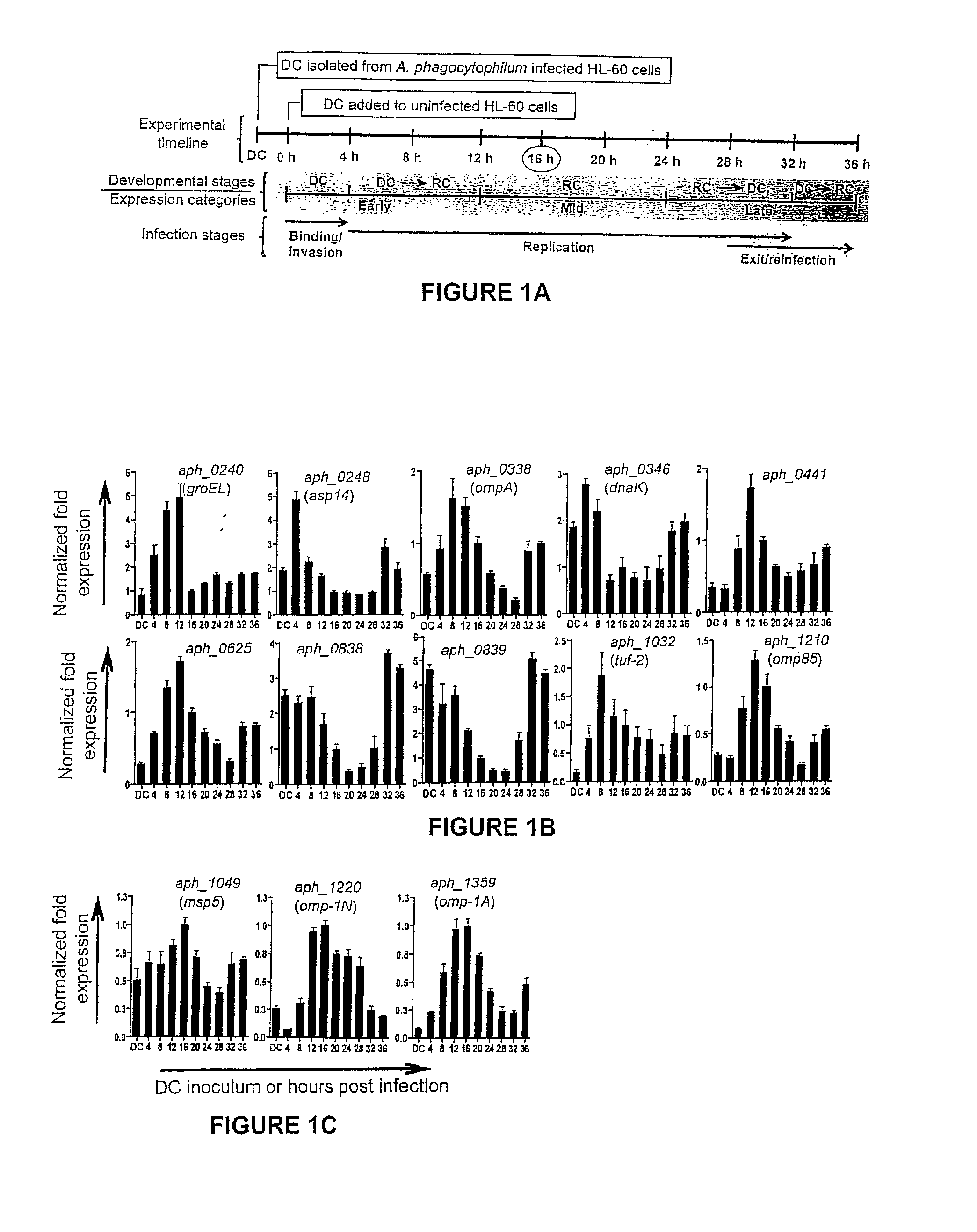
Selection of Aph OMP Candidates for Further Study. FIG. 1A Illustrates the Experimental Timeline Relative to the the Infection Cycle and Stages of Aph Organisms during Infection of a host

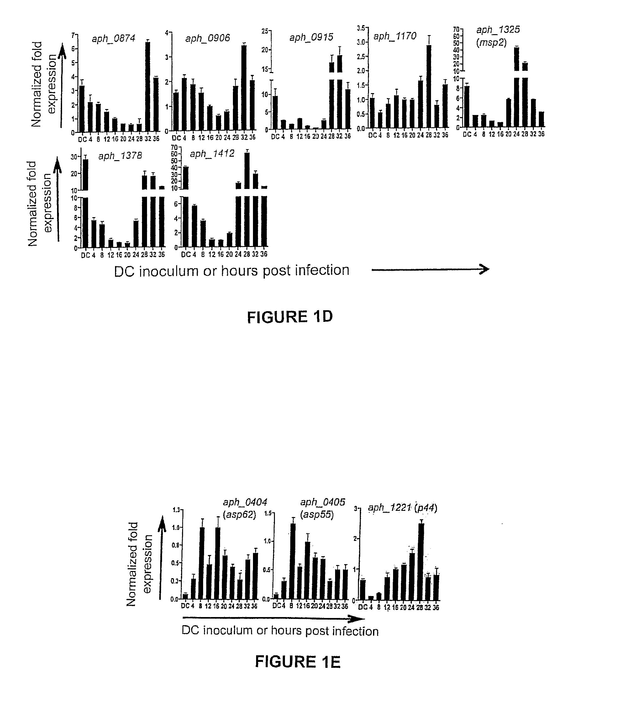
[0075]DC organisms were used to synchronously infect HL-60 cells and the infection proceeded for 36 h, a time period that allows for the bacteria to complete their biphasic developmental cycle and reinitiate infection. Total RNA was isolated from the DC inoculum and from infected host cells at several postinfection time points. RT-qPCR was performed using gene-specific primers. Relative transcript levels for each target were normalized to Aph 16S rRNA gene transcript levels using the 2−ΔΔCT method. To determine the relative transcription of OMP candidate genes between RC and DC organisms, normalized transcript levels of each gene per time point (shown in FIG. 1B-D) were calculated as the fold-change in expression relative to expression at 16 h (encircled in the experimental timeline in FIG. 1A), a t...

example 3

Differential Transcription Profiling of Omp Candidate Genes Throughout the Aph Infection Cycle

[0078]To gain insight into the transcription of the 20 genes of interest during the Aph infection cycle, we synchronously infected HL-60 cells with DC organisms and allowed the infection to proceed in order for the bacteria to complete their biphasic developmental cycle and initiate a second round of infection. We isolated total RNA from DC organisms used as the inoculum and from bacteria recovered at several post-infection time points. RT-qPCR was performed on total RNA using gene-specific primers. Relative transcript levels for each target were normalized to Aph 16S rRNA gene (aph_1000) transcript levels using the 2−ΔΔCT method. To facilitate identification of genes that are up-regulated in the infectious DC form compared to the non-infectious RC form, normalized transcript levels for each gene per time point were calculated as the fold-change in expression relative to expression at 16 h,...


PUM


Abstract

Anaplasma phagocytophilum surface proteins Asp14 and OmpA and homologous genes from Anaplasmatacaea family members are used in compositions suitable for vaccines to treat or prevent infections caused by tick-born bacteria of the Anaplasmatacaea family. Asp14 and / or OmpA proteins or peptide fragments may be used in combination with other Anaplasmatacaea surface proteins to elicit an immune response. Furthermore, antibodies to Asp14 and / or OmpA proteins can be used in diagnostic methods to determine whether an individual has contracted an Anaplasmatacaea infection. Because of the conserved invasin domains in the surface proteins, a wide range of Anaplasmatacaea infections may be diagnosed, treated or prevented using compositions of the invention.

Description

FIELD OF THE INVENTION[0001]The invention generally relates to a vaccine and diagnostic for anaplasmosis in animals and humans. In particular, the invention provides Anaplasma phagocytophilum outer surface protein A (OmpA) epitopes and / or Anaplasma phagocytophilum surface protein 14 (Asp14) epitopes.PRIORITY[0002]This application claims the benefit of U.S. application Ser. No. 61 / 665,223, filed Jun. 27, 2012, and U.S. application Ser. No. 61 / 698,979, filed Sep. 10, 2012. These applications are incorporated herein by reference in their entirety.SEQUENCE LISTING[0003]This document incorporates by reference an electronic sequence listing text file, which was electronically submitted along with this document. The text file is named 02940826TA_ST25.txt, is 87 kilobytes, and was created on Jun. 17, 2013.BACKGROUND OF THE INVENTION[0004]Anaplasma phagocytophilum (Aph) is a tick-transmitted obligate intracellular bacterium of the family Anaplasmataceae that can infect humans, livestock, com...

Claims


Application Information

Patent Timeline
Patent Type & Authority Applications(United States)
IPC IPC(8): G01N33/569
CPCG01N33/56911G01N2800/42G01N2333/195G01N2469/10A61K39/0233A61K39/40A61K2039/55566A61K2039/6031A61K2039/6081A61K2039/70A61P33/02C07K16/1246C07K2317/76Y02A50/30G01N33/56983G01N2333/29
Inventor CARLYON, JASON A.
Owner VIRGINIA COMMONWEALTH UNIV
Learn More