Clostridial neurotoxin fusion proteins, propeptide fusions, their expression, and use

a neurotoxin and fusion protein technology, applied in the field of clostridial neurotoxin fusion proteins, propeptide fusions, their expression, etc., can solve the problems of not being able to directly gain access to targets inside the cytoplasmic compartment of cells, limitations for therapeutic applications, and well-known limitations for use in therapeutic products

Pending Publication Date: 2016-06-09
NEW YORK UNIV
View PDF36 Cites 11 Cited by
  • Summary
  • Abstract
  • Description
  • Claims
  • Application Information

AI Technical Summary

Benefits of technology

[0034]The Examples set forth herein infra provide evidence regarding the successful delivery of single chain antibodies using atoxic derivatives of Clostridial neurotoxins. Fusion proteins of the present invention target neurons, can accumulate in the cytosolic fraction of neuronal

Problems solved by technology

Canonic antibodies are large multimeric proteins that cannot penetrate the cell membrane and, therefore, cannot directly gain access to targets inside the cytoplasmic compartment of the cell.
While all of these methodologies are demonstrated to be effective in vitro, they all have limitations for therapeutic application (Marschall et al., “Delivery of Antibodies to the Cytosol: Debunking the Myths,”mAbs 6(4):943-956 (2014); Yin et al., “Non-Viral Vectors for Gene-Based Therapy,”Nature Reviews Genetics 15(8):541-555 (2014)).
In particular, transfection based methods have well-known limitations for use in therapeutic products, primarily due to toxicity and lack of specificity.
Though viral-mediated transduction is being tested for clinical application, it is still considered to present significant risks for therapeutic intervention, and faces significant regulatory barriers.
Neuron specific viral carriers are not available for clinical use.
The use of protein transduction domains is not neuron-specific and likewise presents significant safety concerns.
The reconstituted BoNT holotoxin derivatives had a severely reduced ability to transport LC into the neuronal cytosol, probably resulting from the harsh conditions required for HC-LC separation and the difficulty of renaturing the protein and rec

Method used


Image

  • Clostridial neurotoxin fusion proteins, propeptide fusions, their expression, and use
  • Clostridial neurotoxin fusion proteins, propeptide fusions, their expression, and use
  • Clostridial neurotoxin fusion proteins, propeptide fusions, their expression, and use

Examples


example 1

Atoxic Derivative Devoid of SNAP-25 Activity to Deliver Single Chain Antibodies into the Cytosol of Neurons

[0224]Materials and Methods

[0225]Expression of Botulinum Neurotoxin a Atoxic Derivatives

[0226]The full-length single forms of the BoNT / A ad discussed below were bioengineered, expressed, and purified, and then converted to the di-chain by treatment with TEV protease as described before (U.S. Pat. No. 8,980,284 to Ichtchenko and Band, which is hereby incorporated by reference in its entirety).

[0227]Preparation and Maintenance of E19 Rat Hippocampal Neurons

[0228]Time pregnant Sprague-Dawley rats (Taconic) were used to isolate embryonic-day 19 (“E19”) hippocampal neurons. E19 rat hippocampal neurons were prepared from hippocampi according to the protocol of Vicario-Abejón (Vicario-Abejon, “Long-term Culture of Hippocampal Neurons,”Curr. Protoc. Neurosci. Chapter 3: Unit 32 (2004), which is hereby incorporated by reference in its entirety). Bilateral hippocampi were dissected from ...

example 2

BoNT / A Ad-0 as a Delivery Vehicle to Deliver Single Chain Antibodies to the Cytoplasm of Neurons

[0254]Introduction

[0255]It has previously been shown that BoNT / A ad-0 is found at the pre-synaptic region in neuromuscular junctions after systemic administration in vivo. In vitro, BoNT / A ad-0 is internalized into the cytosol of neurons at micromolar concentrations, where the BoNT / A ad-0 light chain co-localizes with synaptic proteins. Local intramuscular administration of BoNT / A ad-0 results in muscle weakness / paralysis, a hallmark of wild-type BoNT / A, demonstrating the pharmacological properties of BoNT / A ad-0 as a neuromodulator.

[0256]In this example, empirical evidence is provided regarding the successful delivery of single chain antibodies using botulinum neurotoxin atoxic derivatives with residual SNAP-25 catalytic activity (BoNT / A ad-0). The catalytic activity of the BoNT / A ad-0 light chain towards SNAP-25 was used as a readout to measure successful delivery of the cargo material;...

example 3

Atoxic Derivative of Botulinum Neurotoxin C (BoNT / C Ad) as a Molecular Vehicle for Targeted Delivery to the Neuronal Cytoplasm

[0278]Introduction

[0279]Methods that enable facile production of recombinant derivatives of botulinum neurotoxins (BoNTs) have been developed, which retain the structural and trafficking properties of wild type (wt) BoNTs. Atoxic derivatives of wt BoNT / A have been described supra. Here, an atoxic derivative of BoNT / C1 with three amino acid substitutions in the catalytic domain of the light chain (E446>A;H449>G;Y591>A), termed BoNT / C ad, was designed, expressed, purified, and evaluated.

[0280]Methods

[0281]The coding sequence for BoNT / C ad was designed to inactivate the light chain protease with minimal disruption of the light chain / heavy chain interactions within the protein heterodimer. Recombinant protein was secreted into culture media as a soluble propeptide. The protein was purified to homogeneity by tandem affinity chromatography and processed with TEV pr...


PUM

PropertyMeasurementUnit
Concentrationaaaaaaaaaa
Therapeuticaaaaaaaaaa
Stabilityaaaaaaaaaa

Abstract

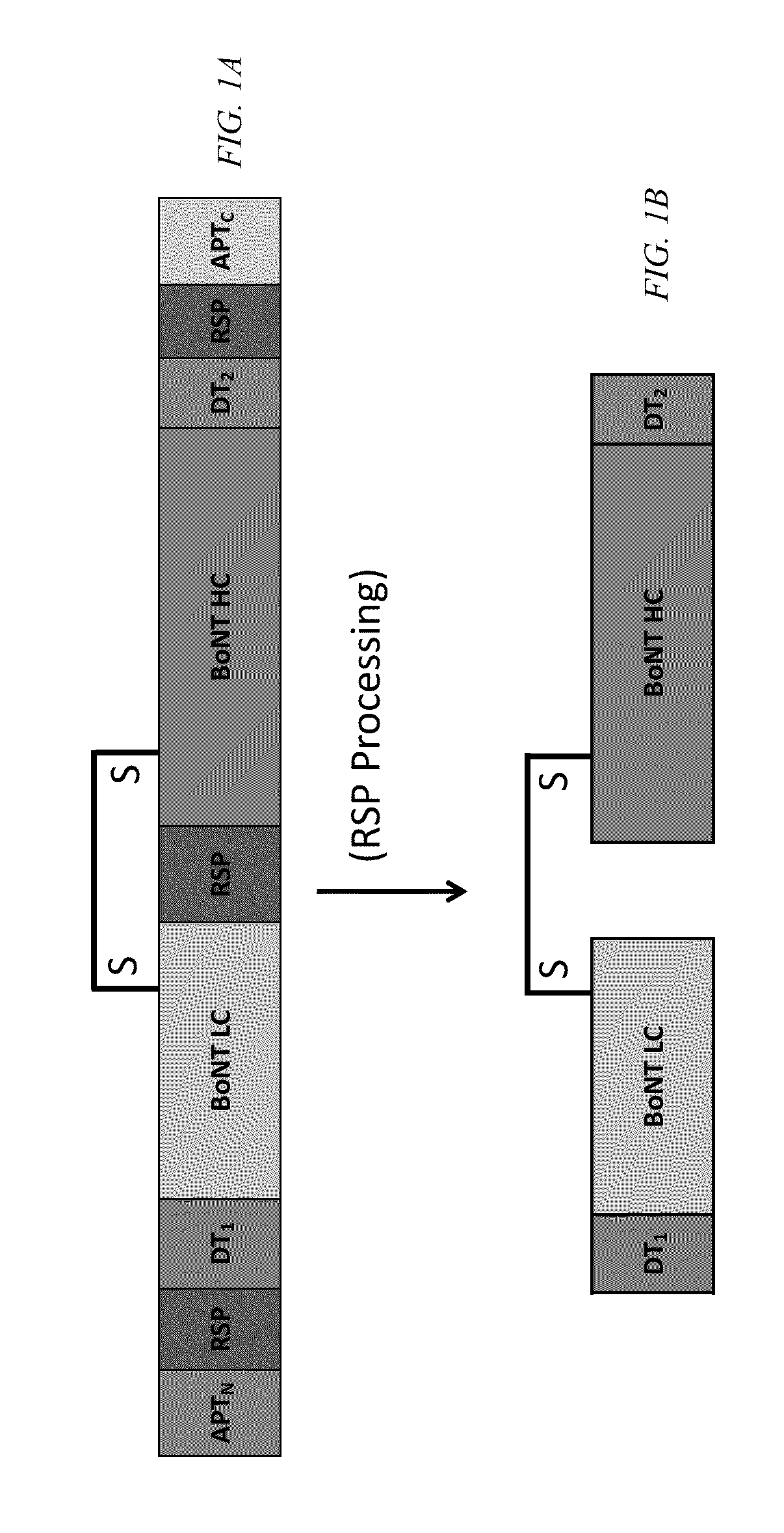
The present invention is directed to a fusion protein comprising a light chain region of a Clostridial neurotoxin and a heavy chain region of a Clostridial neurotoxin, where the light and heavy chain regions are linked by a disulfide bond. The fusion protein also has a single chain antibody positioned upstream of the light chain region, where the single chain antibody possesses antigen-binding activity. Also disclosed are therapeutic agents, treatment methods, propeptide fusions, isolated nucleic acid molecules, expression systems, host cells, and methods of expressing fusion proteins.

Description

[0001]This application claims the benefit of U.S. Provisional Patent Application Ser. Nos. 62 / 089,646, filed Dec. 9, 2014, and 62 / 118,970, filed Feb. 20, 2015, both of which are hereby incorporated by reference in their entirety.[0002]This invention was made with government support under grant number RO1AI093504 awarded by the National Institutes of Health. The government has certain rights in this invention.FIELD OF THE INVENTION[0003]The present invention relates to Clostridial neurotoxin fusion proteins containing a single chain antibody, propeptide fusions, and methods thereof.BACKGROUND OF THE INVENTIONDelivery of Antibodies to Intracellular Targets[0004]Canonic antibodies are large multimeric proteins that cannot penetrate the cell membrane and, therefore, cannot directly gain access to targets inside the cytoplasmic compartment of the cell. Because many pharmaceutically important targets are exclusively exposed in the cytoplasm of cells, multiple technological approaches have...

Claims


Application Information

Patent Timeline
IPC IPC(8): C07K14/33
CPCC07K2319/30C07K14/33C07K16/1282C07K2319/00C07K2319/21C07K2319/22C07K2319/41C07K2319/50A61K2039/505C07K2317/622A61P25/00A61P39/06Y02A50/30
Inventor ICHTCHENKO, KONSTANTINVAZQUEZ-CINTRON, EDWINBAND, PHILIP A.CARDOZO, TIMOTHY J.
Owner NEW YORK UNIV
Why Eureka
Try Eureka