Method and system for localizing body structures

a body structure and localization technology, applied in the field of body structure localization, can solve the problems of insufficient and inaccurate knowledge, prostate cancer, and almost all patients' urinary symptomatology, and achieve the effects of high localization accuracy, improved clinical outcomes, and reduced clinical workload and cos

Inactive Publication Date: 2016-06-30
KONINKLJIJKE PHILIPS NV
View PDF17 Cites 5 Cited by
  • Summary
  • Abstract
  • Description
  • Claims
  • Application Information

AI Technical Summary

Benefits of technology

[0008]Accordingly, the proposed localizing or tracking approach is based on at least one ultrasound sensor (or receiver) embodied on a medical tool to be used during an ultrasound-guided procedure. As the medical tool and ultrasound sensor enter the ultrasound field of view, the ultrasound sensor receives the ultrasound signals coming from the ultrasound probe as its beam(s) sweep the field of view. The position of the ultrasound sensor can then be determined and tracked in the frame of reference (i.e. field of view) of the ultrasound probe and the course of the critical body structure to be localized can be delineated based on the determined location of the ultrasound sensor. This enables precise outlining of the course or shape of the critical body structure (e.g. prostatic urethra).
[0009]Moreover, the proposed localization also allows continuous real-time tracking of the critical body structure during treatment, thereby enabling adaptive treatment planning and dose delivery for better clinical outcomes (e.g. fewer urethra injury-related complications).
[0010]In general, the proposed solution as claimed enables high accuracy in localization and segmentation of critical body structures without imposing more clinical workload and cost.
[0011]According to a first aspect, the controller may be adapted to segment and track the delineated course of the body structure and to calculate a radiation dose to be delivered to the target tissue or organ based on the tracking and segmentation. This segmenting and tracking enables real-time adjustment of radiation dose planning to minimize detrimental impact on the body structure.

Problems solved by technology

One major drawback of brachytherapy for treatment of prostate cancer is that almost all patients develop some degree of urinary symptomatology.
This can mainly be attributed to an unwanted incidental dose delivered to the urethra during brachytherapy procedures, which is the result of insufficient and inaccurate knowledge of the shape and pose of the urethra.
However, such approaches impose additional clinical workload and increases patient financial burden, or other benefit maybe diminishing due to patient motion and image distortion caused by a patient's physiological change.

Method used


Image

  • Method and system for localizing body structures
  • Method and system for localizing body structures
  • Method and system for localizing body structures

Examples


first embodiment

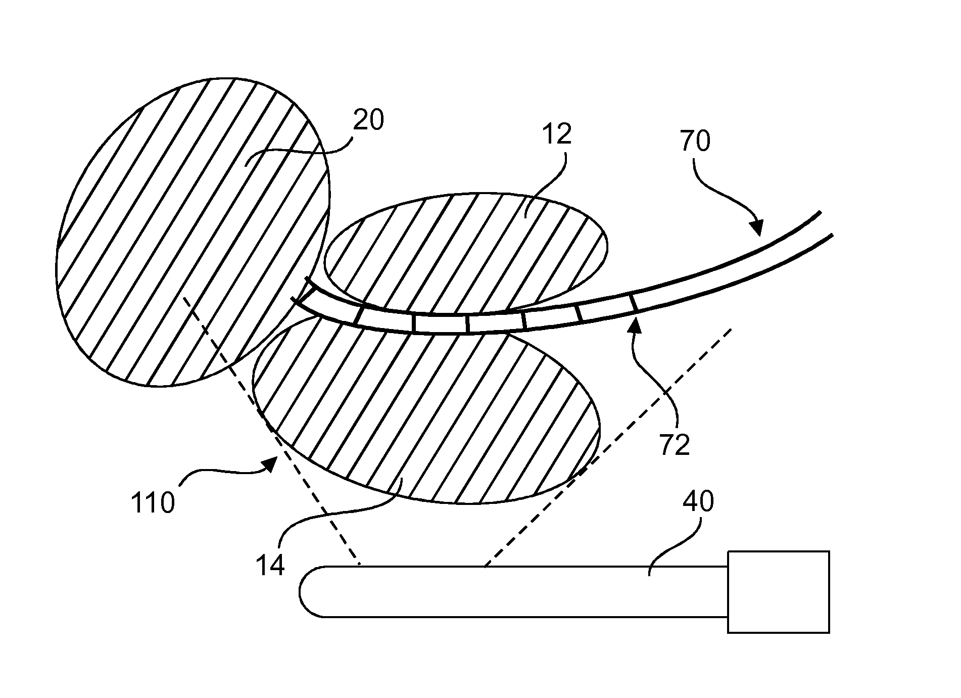
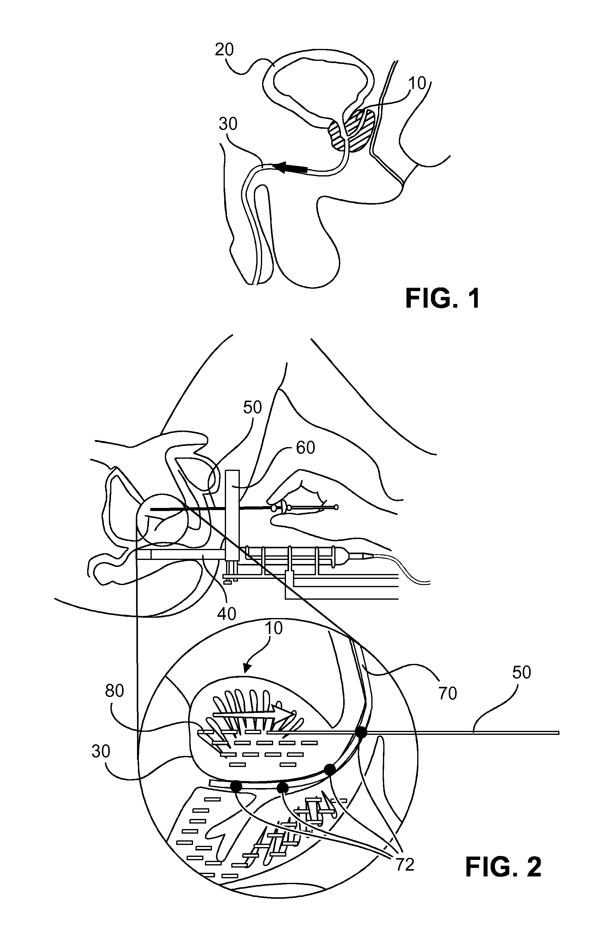
[0037]In the present embodiments, the use of such a Foley catheter 70 with embedded US sensors 72 enables real-time localization of the prostatic urethra 30 which is a crucial in the brachytherapy procedure to avoid unnecessary or excessive radiation dose to the prostatic urethra 30. The Foley catheter 70 is equipped with one or multiple US sensors 72 that are mounted at a fixed distance from one another in the

[0038]In the following, the word “catheter” is used as a generic term to denote a catheter equipped with sensors or a guide wire equipped with sensors inside a catheter.

[0039]During the brachytherapy procedure, a transrectal US (TRUS) probe 40 is used as an ultrasound signal source required for sensor-tracking The US sensors 72 on the Foley catheter 70 receive ultrasound waves emitted by the TRUS probe 40. In particular, the ultrasound signals received from appropriate A-line(s) of the TRUS probe 40 as the beam of the TRUS probe 40 sweeps the field of view can be analyzed for ...

third embodiment

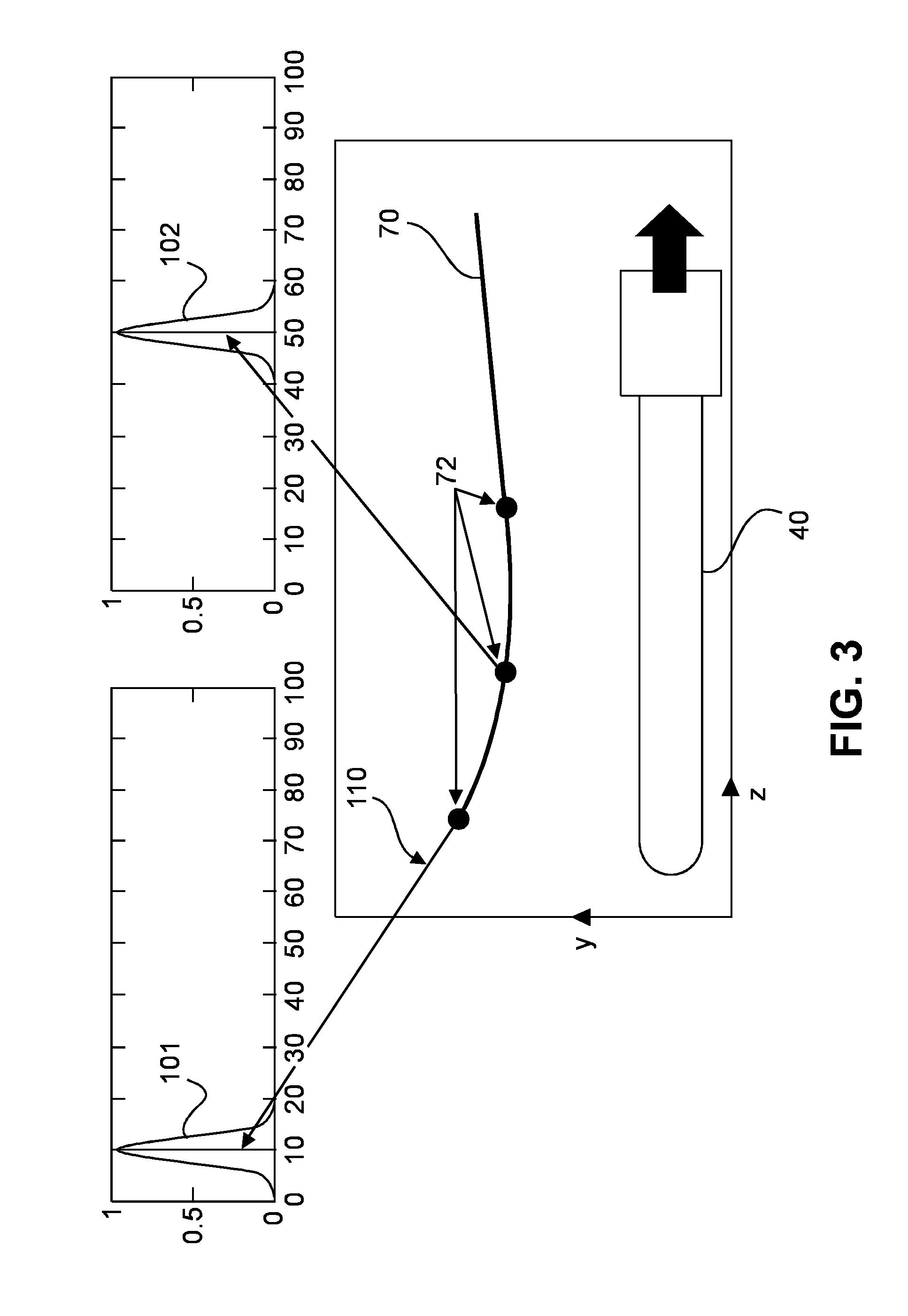
[0054]FIG. 4 shows a schematic arrangement of a localization system with a single-sensor catheter 70 in closed-loop control which can be used for 2D TRUS probes 40.

[0055]In the third embodiment, the catheter 70 has a single US sensor 72, wherein the output signal 720 of the single US sensor 72 is input to a computer or controller 200 that controls a retractor or retracting device 300 that retracts the catheter 70. The controller 200 may be controlled by a control procedure implemented as a software routine. Similarly, the encoded TRUS probe 40 may be retracted manually or automatically.

[0056]In the automatic system with closed-loop control, the retractor 300 can be controlled by the controller 200 based on the sensor output signal 720 and a probe position signal 420 received from the TRUS probe 40, in a way that the US sensor 72 is always kept in the 2D image plan of the moving TRUS probe 40. Then, the 2D position of the US sensor 72 in the TRUS image can be combined with the posit...

fourth embodiment

[0057] the controller 200 may control a robot (not shown) that advances or retracts the TRUS probe 40. Additionally, the sensor-equipped catheter 70 is retracted or advanced by the retracting device 300, as shown in FIG. 4. The probe-holding robot is controlled in a way that the US sensor 72 is always kept in the 2D image of the moving TRUS probe 40. Again, the 2D position of the US sensor 72 in the TRUS image can be combined with the position of the TRUS probe to reveal the 3D position of the US sensor 72.


PUM


Abstract

The invention relates to a system and method in which a Foley catheter (70) or other medical tool which is equipped with ultrasound (US) sensor(s) (72) is inserted into the prostatic urethra. Based on analysis of the US signal received by these US sensors (72) as the US beams from a transrectal US (TRUS) probe (40) or other ultrasound probe sweep the field of view, it is possible to precisely detect and track these US sensors (72) in the same frame of reference as the TRUS images, thereby precisely delineating the Foley catheter and the course of the prostatic urethra. During the procedure, before each seed is dropped, the delivered dose to the prostatic urethra can be computed based on real-time tracking and segmentation of prostatic urethra and dose radiation based on previously dropped seeds and if necessary, the procedure can be re-planned automatically.

Description

FIELD OF THE INVENTION[0001]The invention relates to the field of methods and systems for localizing body structures.BACKGROUND OF THE INVENTION[0002]Prostate cancer is the most common organ malignancy among American men. Several treatments have been proposed for treating prostate cancer. One of them is prostate brachytherapy which refers to placement of permanent radioactive seeds or temporary radioactive sources (within hollow catheters) inside the prostate gland with insertion through the perineum. Most often, treatment planning and needle or catheter placement relies on intensive use of transrectal ultrasound imaging (TRUS).[0003]One major drawback of brachytherapy for treatment of prostate cancer is that almost all patients develop some degree of urinary symptomatology. Urethra irritation and urinary obstruction are well documented short-term complications of the modern brachytherapy techniques. This can mainly be attributed to an unwanted incidental dose delivered to the ureth...

Claims


Application Information

Patent Timeline
Patent Type & Authority Applications(United States)
IPC IPC(8): A61B8/08A61B8/00A61B8/12
CPCA61B8/085A61B8/4245A61B8/12A61B5/065A61B8/0841A61B8/4254A61B2018/00547A61M25/0017A61M25/0108A61M25/04A61M27/00A61M2025/0166A61N5/1027A61N2005/1058A61B5/064
Inventor TAHMASEBI MARAGHOOSH, AMIR MOHAMMADVIGNON, FRANCOIS GUY GERARD MARIEBHARAT, SHYAMDEHGHAN MARVAST, EHSANJAIN, AMEET KUMAR
Owner KONINKLJIJKE PHILIPS NV
Why Eureka
Try Eureka