
Ultrasound imaging apparatus, ultrasound imaging method and ultrasound imaging program

a technology of ultrasound imaging applied in the field of ultrasound imaging methods and ultrasound imaging equipment, can solve the problems of not being able to discern the boundary between the tumor and the surrounding tissue, not being able to perform diagnostic image sequences, not being able to perform elasticity images, etc., and deteriorating the discerning degree of boundary

Inactive Publication Date: 2016-07-28
HITACHI LTD
View PDF5 Cites 5 Cited by
  • Summary
  • Abstract
  • Description
  • Claims
  • Application Information

AI Technical Summary

Benefits of technology

The present invention describes a method for generating an image that shows the boundaries of a test subject without generating a vector field. This is done by obtaining a value that represents the state of the norm distribution in the search region, and assuming this value as the pixel value of the region of interest associated with the search region. The technical effect of this invention is to improve the efficiency of generating an image that shows the boundaries of a test subject.

Problems solved by technology

However, in the case of peripheral zone of tumor, for instance, there is not found a large difference in acoustic impedance nor in elastic modulus, relative to surrounding tissue, and in this situation, it is not possible to discern the boundary between the tumor and the surrounding tissue, in the diagnostic image sequence nor in the elasticity image.
However, in a region of the image data including high noise, such as a marginal domain for signal penetration where echo signals become faint, an error vector may occur due to noise influence, upon obtaining the motion vector, and this may deteriorate the discerning degree of the boundary.

Method used


Image

  • Ultrasound imaging apparatus, ultrasound imaging method and ultrasound imaging program
  • Ultrasound imaging apparatus, ultrasound imaging method and ultrasound imaging program
  • Ultrasound imaging apparatus, ultrasound imaging method and ultrasound imaging program

Examples


first embodiment

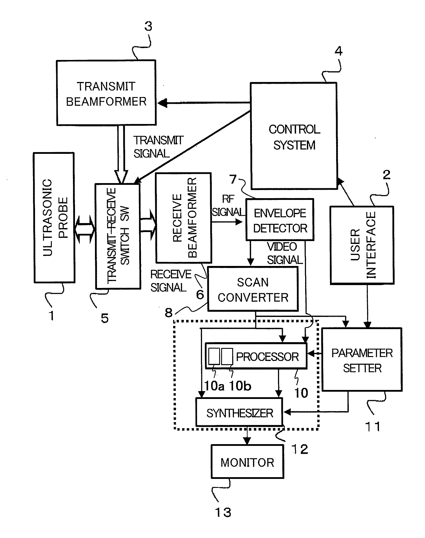
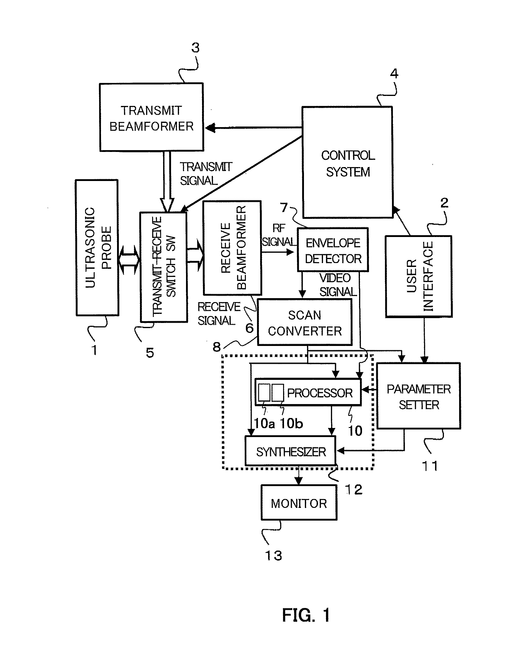
[0045]FIG. 1 illustrates a system configuration of the ultrasound imaging apparatus according to the present embodiment. This apparatus is provided with a ultrasound boundary detecting function. As illustrated in FIG. 1, this apparatus is provided with an ultrasound probe (probe) 1, a user interface 2, a transmit beamformer 3, a control system 4, a transmit-receive switch 5, a receive beamformer 6, an envelope detector 7, a scan converter 8, a processor 10, a parameter setter 11, a synthesizer 12, and a monitor 13.

[0046]The ultrasound probe 1 on which the ultrasound elements are provided in one-dimensional array, serves as a transmitter configured to transmit an ultrasound beam (an ultrasound pulse) to a living body. The ultrasound probe 1 serves as a receiver configured to receive an echo signal (a received signal) reflected from the living body. Under the control of the control system 4, the transmit beamformer 3 outputs a transmit signal having a delay time in accordance with a t...

second embodiment

[0076]In the second embodiment, if any virtual image occurs in the scalar field image obtained in the first embodiment, this virtual image may be removed, or the like. In other words, a degree of reliability of the image region is identified, and a region with a low reliability is removed, or the like, thereby eliminating the virtual image and enhancing the reliability of the entire image. This will be explained with reference to FIG. 9 and FIG. 10.

[0077]FIG. 9(a) illustrates the scalar field image obtained assuming p=1 in the formula (1), as described in the first embodiment, and the virtual image is generated in the boundary. FIG. 9(b) illustrates a histogram that is used for identifying the degree of reliability, and FIG. 9(c) illustrates a scalar field image in which the brightness in the low-reliability region is replaced by a dark color. FIG. 10 is a flowchart showing the operation of the processor 10 for removing the virtual image.

[0078]Upon receiving an instruction for remov...

third embodiment

[0081]In the first embodiment, statistics (the rate of divergence or the coefficient of variation) of the p-norm distribution is obtained to generate an image. In the third embodiment, an image is generated from the p-norm distribution where a tissue boundary is discernible, through the use of a different method. This processing method will be explained with reference to FIG. 11 and FIG. 12.

[0082]In the p-norm value distribution in the search region 32 as described in the first embodiment, the candidate region 33 along the boundary in the test subject forms a region with small p-norm values (a valley of p-norm values) along the boundary. Therefore, the distribution of p-norm values has the characteristics that the values of the candidate region 33 along the boundary indicate smaller values than the candidate region 33 in the direction orthogonal to the boundary. With the use of the characteristics, an image is generated in the present embodiment.

[0083]FIG. 11 illustrates a processin...


PUM


Abstract

The present invention is to provide an ultrasound imaging apparatus which generates a scalar field image directly without obtaining a motion vector, so as to make tissue boundaries discernible.A received signal is processed to generate images of two or more frames, two frames are selected from the images, multiple regions of interest are set in one frame, and in the other frame, search regions each wider than the region of interest are set respectively for the multiple regions of interest. In the search region, multiple candidate regions each in a size corresponding to the region of interest are provided. Then, a norm is obtained between a pixel value of the region of interest and a pixel value of the candidate region, for each of the multiple candidate regions, thereby obtaining a norm distribution within the search region. An image is generated assuming a value representing the state of the norm distribution as a pixel value of the region of interest that is associated with the search region.

Description

TECHNICAL FIELD[0001]The present invention is directed to a technique that relates to an ultrasound imaging method and an ultrasound imaging apparatus, allowing a tissue boundary to be clearly discerned, upon imaging a living body through the use of ultrasound waves.BACKGROUND ART[0002]In an ultrasound imaging apparatus used for medical diagnostic imaging, there is known a method for estimating a distribution of elastic modulus of tissues, based on an amount of change in a small area of a diagnostic image sequence (B-mode image), converts a degree of stiffness to a color map for display. However, in the case of peripheral zone of tumor, for instance, there is not found a large difference in acoustic impedance nor in elastic modulus, relative to surrounding tissue, and in this situation, it is not possible to discern the boundary between the tumor and the surrounding tissue, in the diagnostic image sequence nor in the elasticity image.[0003]Therefore, there is a method for obtaining ...

Claims


Application Information

Patent Timeline
Patent Type & Authority Applications(United States)
IPC IPC(8): A61B8/08A61B8/00A61B8/14
CPCA61B8/5207A61B8/14A61B8/5246A61B8/5223A61B8/463A61B8/469A61B8/461A61B8/485A61B8/0858A61B8/467A61B8/5215G01S7/52036G01S7/52071G16H50/30G16H50/20
Inventor MASUI, HIRONARIAZUMA, TAKASHI
Owner HITACHI LTD
Learn More