
Extracellular Mitochondrial Components for Detecting Inflammatory Reactions and Conditions

a technology of extracellular mitochondrial components and inflammatory reactions, applied in the direction of biological material analysis, material analysis, instruments, etc., can solve the problem of limiting the shelf life and achieve the effects of preventing the release, inhibiting the release, and preventing the release of sterile inflammation mediators

Inactive Publication Date: 2016-09-08
HEMA QUEBEC +2
View PDF0 Cites 2 Cited by
  • Summary
  • Abstract
  • Description
  • Claims
  • Application Information

AI Technical Summary

Benefits of technology

The present patent provides methods for diagnosing, treating, preventing, and alleviating symptoms associated with inflammatory reactions or conditions by detecting the presence of extracellular mitochondrial components, optionally in combination with other biomarkers such as secreted phospholipase A2-IIA and auto-antibodies. The methods involve obtaining a biological sample, enriching extracellular submicrometer-sized components from the sample, and determining the level of at least one of a free mitochondria, a mitochondria microparticle, a degradation product of the free mitochondria, and the degradation product of the mitochondria microparticle. The biological sample can be a labile biological product such as blood or a labile biological product that has been or intended to be administered to a treated subject. The methods can be used for diagnosis, treatment, prevention, and alleviation of symptoms associated with inflammatory reactions or conditions.

Problems solved by technology

Even though platelet activation is necessary to prevent or limit bleeding in a homeostatic state, their activation in other circumstances should be limited to avoid deleterious or pathological consequences.
For example, to limit platelet activation, they are stored, prior to transfusion, at 22° C., which limits their shelf-life.

Method used


Image

  • Extracellular Mitochondrial Components for Detecting Inflammatory Reactions and Conditions
  • Extracellular Mitochondrial Components for Detecting Inflammatory Reactions and Conditions
  • Extracellular Mitochondrial Components for Detecting Inflammatory Reactions and Conditions

Examples


example i

Materials and Methods

[0102]Mice.

[0103]All studies were approved by the institutional review board protocol (Université Laval). Guidelines of the Canadian Council on Animal Care were followed in a protocol approved by the Animal Welfare Committee at Laval University. For our studies, we used 6- to 10-week-old male mice (C57BL / 6N and BALB / c; Charles River). For in vivo experiments in which secreted phospholipase A2 IIA (sPLA2-IIA) contribution is evaluated, we used C57BL / 6J (Jackson Laboratories) and sPLA2-IIA-sufficient mice as previously reported (Grass et al. 1996).

[0104]Cells and Human Fluid Preparation.

[0105]Blood was obtained from healthy human volunteers (citrate as anticoagulant) under an approved institutional review board protocol (CRCHUQ; Université Laval) and in accordance with the Declaration of Helsinki. Platelets, platelet MPs (96% of them expressing CD41), and human polymorphonuclear leukocytes were prepared as previously described (Cloutier et al. 2013). Platelet-free...

example ii

Characterization of Mitochondria Released from Platelets

[0147]The material and methods used in this Example are provided in Example I.

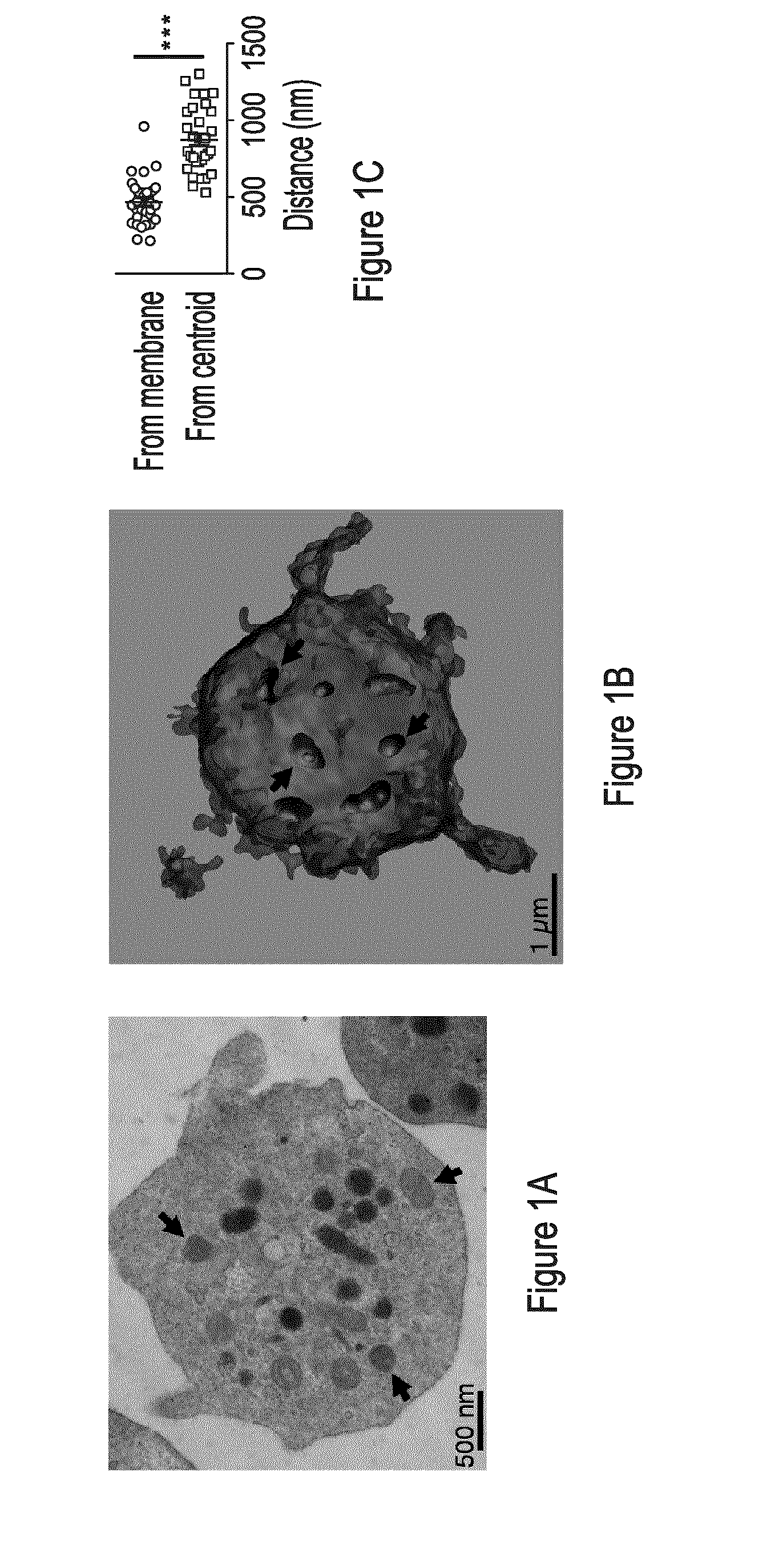
[0148]Using fluorescence and transmission electron microscopy (TEM), we found that unactivated platelets contain an average of ˜4 mitochondria, frequently located in the vicinity of the plasma membrane (FIGS. 1A-C; FIG. 8A and data not shown). Remarkably, a fraction of these mitochondria promptly localizes in pseudopodia on activation by thrombin, a serine protease that participates in blood coagulation (FIG. 8B and data not shown).

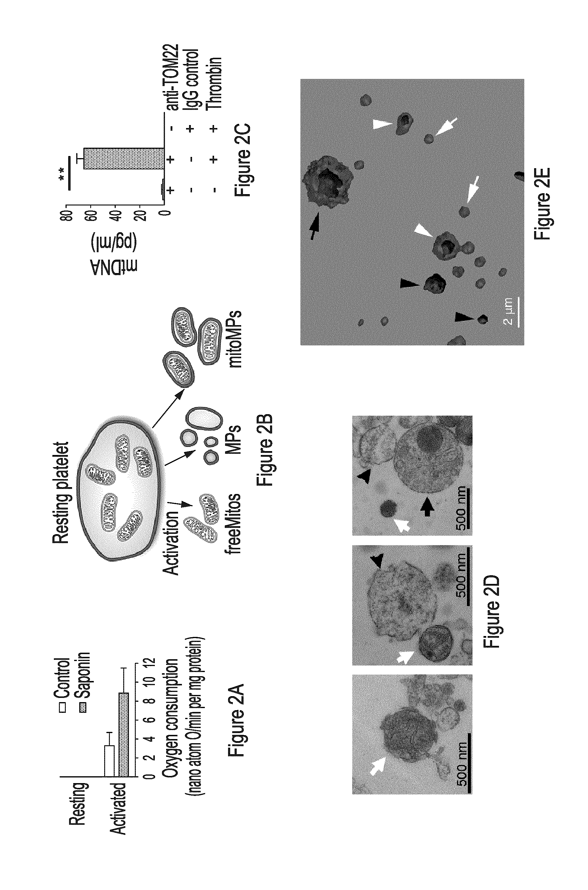
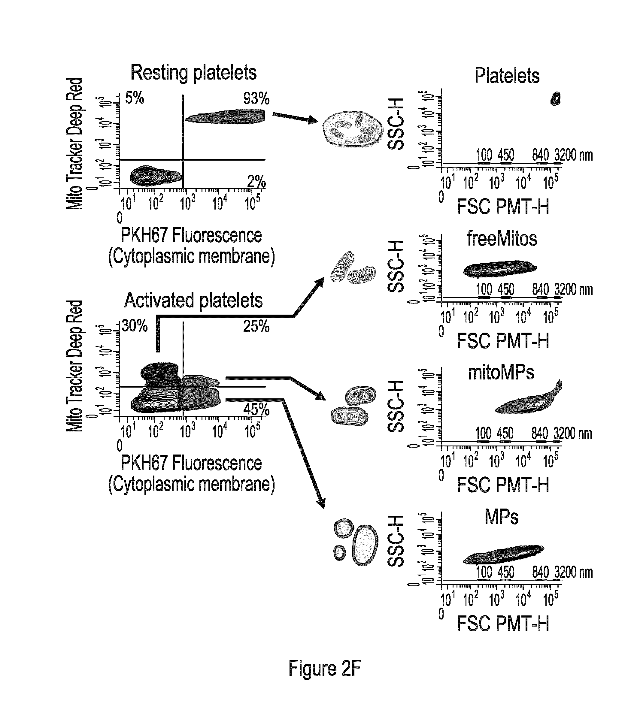
[0149]In addition to promoting release of granule contents, platelet activation triggers cytoplasmic membrane budding and the shedding of submicron vesicles called microparticles (MPs) (Boilard et al. 2012, György et al. 2011). Taking into account the localization of mitochondria in the vicinity of the cytoplasmic membrane, we hypothesized that mitochondria might be packaged within MPs and form mitochondria-containing micro...

example iii

Characterization of Mitochondria from Platelets in Lupus

[0168]The material an methods used in this Example are provided in Example I.

[0169]We verified the presence of mitochondrial immune complexes in the plasma of 192 patients from the established University of Toronto Lupus Clinic (Table 2).

[0170]Table 2. Demographics and disease characteristics of the 192 women that participated in the study of fatty acids in lupus. We have recruited and characterized 192 women with lupus from the University of Toronto Lupus Clinic. This longitudinal observational study includes more than 1 600 persons with SLE that are followed prospectively according to a standardized protocol. We recruited 192 consecutive female patients with SLE and collected demographic variables and disease characteristics as well as serum and plasma. Baseline demographic variables and disease characteristics of these 192 women demonstrate that this is a sample of middle-aged women (mean age (SD)=46.3 years (14.7)) with a l...


PUM


Abstract

The present disclosure highlights the relationship between extracellular mitochondrial components, optionally in combination with the secreted phospholipase A2-IIA and / or an auto-antibody, and in vivo as well as in vitro and inflammatory reactions / conditions, especially those released as a result of the degradation of a platelet. The present disclosure provides methods for determining the presence of inflammatory mediators, for limiting inflammatory reactions / conditions, for the diagnosis inflammatory reactions / conditions, for screening therapeutics for the treatment and / or the alleviation of symptoms of inflammatory reactions / conditions based on the detection or modulation of the level of these extracellular mitochondrial components.

Description

CROSS-REFERENCE TO RELATED APPLICATIONS[0001]This application claims priority from U.S. Provisional Patent Application Ser. No. 61 / 889,798 filed on Oct. 11, 2013 which is herewith incorporated in its entirety.TECHNOLOGICAL FIELD[0002]The present disclosure relates to the diagnosis, prevention, treatment, alleviation of symptoms of inflammatory conditions or reactions.BACKGROUND[0003]Platelets are small (2-4 μm) discoid cells released by megakaryocytes present in the bone marrow. During this process, megakaryocytes transfer components to daughter platelets, including messenger RNA and microRNA as well as cytoplasmic organelles. Mitochondria are among these organelles and although they can amplify platelet activation and thrombosis, very little is known regarding their number and distribution in platelets.[0004]Platelets circulate in blood to promote haemostasis and play pivotal roles in the prevention of bleeding. Structurally, platelets are small anucleated cells that contain a numb...

Claims


Application Information

Patent Timeline
IPC IPC(8): G01N33/569G01N15/14G01N33/564
CPCG01N33/56966G01N33/564G01N2015/1486G01N2015/0084G01N15/14G01N33/502G01N2500/10G01N33/5079G01N2800/104G01N2800/222G01N2800/7095G01N2015/018
Inventor BOILARD, ERICBOUDREAU, LUCTHIBAULT, LOUISGELB, MICHAEL HERMAN
Owner HEMA QUEBEC
Learn More